Phyre Home Retrieve Phyre Job Id
Emailmdejesus@rockefeller.edu
DescriptionRVBD0027_(-)_31189_31506
DateTue Jul 23 14:50:05 BST 2019
Unique Job ID758d4c8861938612
SequenceMTDRIHVQPA ... Download FASTA
Job Typenormal
Job Expiry

Summary 

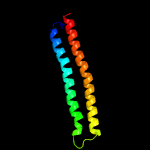
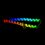
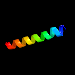
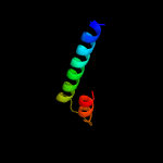
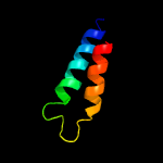
Top model
Image coloured by rainbow N → C terminus
Model dimensions (Å):X:22.198 Y:68.872 Z:27.608
Model (left) based on template c4iogD_
Top template information
PDB header:unknown function
Chain: D: PDB Molecule:secreted protein esxb;
PDBTitle: the crystal structure of a secreted protein esxb (wild-type, in p212 space group) from bacillus anthracis str. sterne
Confidence and coverage
Confidence: 89.4% Coverage: 81%
85 residues ( 81% of your sequence) have been modelled with 89.4% confidence by the single highest scoring template.
You may wish to submit your sequence to Phyrealarm. This will automatically scan your sequence every week for new potential templates as they appear in the Phyre2 library.
Please note: You must be registered and logged in to use Phyrealarm.
3D viewing
Interactive 3D view in JSmol
For other options to view your downloaded structure offline see the FAQ

Sequence analysis 

Download FASTA version

Secondary structure and disorder prediction [Show] 

Domain analysis [Show] 

Detailed template information [Hide] 


#
Template Alignment Coverage3D Model Confidence
% i.d. Template Information
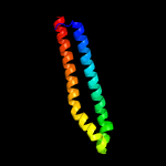
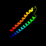
1c4iogD_


89.4 20
PDB header:unknown function
Chain: D: PDB Molecule:secreted protein esxb;
PDBTitle: the crystal structure of a secreted protein esxb (wild-type, in p212 space group) from bacillus anthracis str. sterne
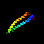
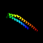
2c2vs0B_


87.3 16
PDB header:cell invasion
Chain: B: PDB Molecule:virulence factor esxa;
PDBTitle: structural analysis of homodimeric staphylococcal aureus2 virulence factor esxa
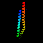
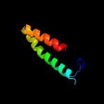
3d1wa8a1


82.0 15
Fold:Ferritin-like
Superfamily:EsxAB dimer-like
Family:ESAT-6 like
4c3gvmA_


49.4 17
PDB header:viral protein
Chain: A: PDB Molecule:putative uncharacterized protein sag1039;
PDBTitle: structure of the homodimeric wxg-100 family protein from streptococcus2 agalactiae
5c4lwsA_


31.7 21
PDB header:unknown function
Chain: A: PDB Molecule:uncharacterized protein;
PDBTitle: esxa : esxb (semet) hetero-dimer from thermomonospora curvata
6c4lwsB_


24.1 10
PDB header:unknown function
Chain: B: PDB Molecule:uncharacterized protein;
PDBTitle: esxa : esxb (semet) hetero-dimer from thermomonospora curvata
7c3zbhC_


18.6 14
PDB header:unknown function
Chain: C: PDB Molecule:esxa;
PDBTitle: geobacillus thermodenitrificans esxa crystal form i
8c3a98A_


16.9 22
PDB header:signaling protein
Chain: A: PDB Molecule:dedicator of cytokinesis protein 2;
PDBTitle: crystal structure of the complex of the interacting regions of dock22 and elmo1
9c6fueC_


15.6 50
PDB header:protein transport
Chain: C: PDB Molecule:fapf;
PDBTitle: periplasmic coiled coil domain of the fapf amyloid transporter
10c6fueA_


15.6 50
PDB header:protein transport
Chain: A: PDB Molecule:fapf;
PDBTitle: periplasmic coiled coil domain of the fapf amyloid transporter
11c3bboZ_


13.3 23
PDB header:ribosome
Chain: Z: PDB Molecule:ribosomal protein l29;
PDBTitle: homology model for the spinach chloroplast 50s subunit fitted to 9.4a2 cryo-em map of the 70s chlororibosome
12c5jzrA_


13.1 56
PDB header:viral protein
Chain: A: PDB Molecule:coat protein;
PDBTitle: solid-state mas nmr structure of acinetobacter phage 205 (ap205) coat2 protein in assembled capsid particles
13c3l8jA_


12.9 29
PDB header:protein binding
Chain: A: PDB Molecule:programmed cell death protein 10;
PDBTitle: crystal structure of ccm3, a cerebral cavernous malformation protein2 critical for vascular integrity
14c1pyvA_


11.4 73
PDB header:hydrolase
Chain: A: PDB Molecule:atp synthase beta chain, mitochondrial precursor;
PDBTitle: nmr solution structure of the mitochondrial f1b presequence2 peptide from nicotiana plumbaginifolia
15c2pl9D_


11.0 50
PDB header:signaling protien
Chain: D: PDB Molecule:chemotaxis protein chez;
PDBTitle: crystal structure of chey-mg(2+)-bef(3)(-) in complex with chez(c19)2 peptide solved from a p2(1)2(1)2 crystal
16c2e8pA_


10.9 16
PDB header:signaling protein
Chain: A: PDB Molecule:elf3 protein;
PDBTitle: solution structure of the n-terminal sam-domain of e74-like2 factor 3
17c5mlcZ_


10.5 20
PDB header:ribosome
Chain: Z: PDB Molecule:50s ribosomal protein l29, chloroplastic;
PDBTitle: cryo-em structure of the spinach chloroplast ribosome reveals the2 location of plastid-specific ribosomal proteins and extensions
18c4m70A_


8.7 69
PDB header:plant protein
Chain: A: PDB Molecule:rx protein;
PDBTitle: crystal structure of potato rx-cc domain in complex with rangap2-wpp2 domain
19c2odmA_


8.6 44
PDB header:unknown function
Chain: A: PDB Molecule:upf0358 protein mw0995;
PDBTitle: crystal structure of s. aureus ylan, an essential leucine rich protein2 involved in the control of cell shape
20c2lndA_


7.9 41
PDB header:de novo protein
Chain: A: PDB Molecule:de novo designed protein, pfk fold;
PDBTitle: solution nmr structure of de novo designed protein, pfk fold,2 northeast structural genomics consortium target or134
21c2lpeA_


not modelled 7.7 23
PDB header:signaling protein
Chain: A: PDB Molecule:kinase suppressor of ras 1;
PDBTitle: solution nmr structure of the ksr1 ca1-ca1a domain
22c3j3wX_


not modelled 7.6 27
PDB header:ribosome
Chain: X: PDB Molecule:50s ribosomal protein l29;
PDBTitle: atomic model of the immature 50s subunit from bacillus subtilis (state2 ii-a)
23c2pl9F_


not modelled 6.9 54
PDB header:signaling protien
Chain: F: PDB Molecule:chemotaxis protein chez;
PDBTitle: crystal structure of chey-mg(2+)-bef(3)(-) in complex with chez(c19)2 peptide solved from a p2(1)2(1)2 crystal
24c2pl9E_


not modelled 6.9 54
PDB header:signaling protien
Chain: E: PDB Molecule:chemotaxis protein chez;
PDBTitle: crystal structure of chey-mg(2+)-bef(3)(-) in complex with chez(c19)2 peptide solved from a p2(1)2(1)2 crystal
25c1i7wB_


not modelled 6.8 38
PDB header:cell adhesion
Chain: B: PDB Molecule:epithelial-cadherin;
PDBTitle: beta-catenin/phosphorylated e-cadherin complex
26c6dlmB_


not modelled 6.4 41
PDB header:de novo protein
Chain: B: PDB Molecule:dhd127_b;
PDBTitle: dhd127
27d1vqov1


not modelled 6.4 25
Fold:Long alpha-hairpin
Superfamily:Ribosomal protein L29 (L29p)
Family:Ribosomal protein L29 (L29p)
28c2w0cT_


not modelled 6.1 75
PDB header:virus
Chain: T: PDB Molecule:protein p6;
PDBTitle: x-ray structure of the entire lipid-containing bacteriophage pm2
29d1r73a_


not modelled 6.1 27
Fold:Long alpha-hairpin
Superfamily:Ribosomal protein L29 (L29p)
Family:Ribosomal protein L29 (L29p)
30c5o60Z_


not modelled 5.9 23
PDB header:ribosome
Chain: Z: PDB Molecule:50s ribosomal protein l29;
PDBTitle: structure of the 50s large ribosomal subunit from mycobacterium2 smegmatis
31c6fikC_


not modelled 5.6 50
PDB header:biosynthetic protein
Chain: C: PDB Molecule:polyketide synthase;
PDBTitle: acp2 crosslinked to the ks of the loading/condensing region of the2 ctb1 pks
32d1fnoa3


not modelled 5.5 33
Fold:Ferredoxin-like
Superfamily:Bacterial exopeptidase dimerisation domain
Family:Bacterial exopeptidase dimerisation domain

Binding site prediction 

Due to computational demand, binding site predictions are not run for batch jobs

If you want to predict binding sites, please manually submit your model of choice to 3DLigandSite