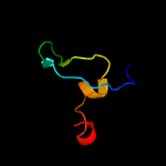
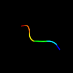
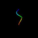
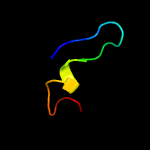
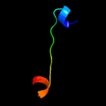
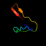
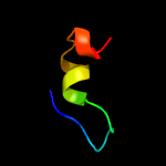
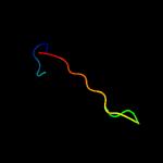
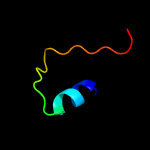
1 c2equA_
46.5
30
PDB header: protein bindingChain: A: PDB Molecule: phd finger protein 20-like 1;PDBTitle: solution structure of the tudor domain of phd finger2 protein 20-like 1
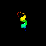
2 c2yf2C_
39.9
29
PDB header: immune systemChain: C: PDB Molecule: c4b binding protein;PDBTitle: crystal structure of the oligomerisation domain of c4b-binding2 protein from gallus gallus
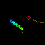
3 c3w0lD_
27.1
43
PDB header: transferase/transferase inhibitorChain: D: PDB Molecule: glucokinase regulatory protein;PDBTitle: the crystal structure of xenopus glucokinase and glucokinase2 regulatory protein complex
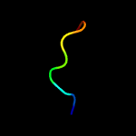
4 c1qfnB_
26.8
78
PDB header: electron transport/oxidoreductaseChain: B: PDB Molecule: protein (ribonucleoside-diphosphate reductase 1);PDBTitle: glutaredoxin-1-ribonucleotide reductase b1 mixed disulfide2 bond
5 c4bbaA_
26.2
24
PDB header: protein-binding proteinChain: A: PDB Molecule: glucokinase regulatory protein;PDBTitle: crystal structure of glucokinase regulatory protein complexed to2 phosphate
6 c2kzbA_
24.8
46
PDB header: protein transportChain: A: PDB Molecule: autophagy-related protein 19;PDBTitle: solution structure of alpha-mannosidase binding domain of atg19
7 d1npla_
22.6
29
Fold: beta-Prism IISuperfamily: alpha-D-mannose-specific plant lectinsFamily: alpha-D-mannose-specific plant lectins
8 c3r0eC_
22.0
24
PDB header: sugar binding proteinChain: C: PDB Molecule: lectin;PDBTitle: structure of remusatia vivipara lectin
9 d1v54k_
19.8
67
Fold: Single transmembrane helixSuperfamily: Mitochondrial cytochrome c oxidase subunit VIIbFamily: Mitochondrial cytochrome c oxidase subunit VIIb
10 c2y69X_
19.8
67
PDB header: electron transportChain: X: PDB Molecule: cytochrome c oxidase polypeptide 7b;PDBTitle: bovine heart cytochrome c oxidase re-refined with molecular oxygen
11 c2ov2O_
18.5
11
PDB header: protein binding/transferaseChain: O: PDB Molecule: serine/threonine-protein kinase pak 4;PDBTitle: the crystal structure of the human rac3 in complex with the crib2 domain of human p21-activated kinase 4 (pak4)
12 c4lc9A_
17.7
20
PDB header: transferase/transferase regulatorChain: A: PDB Molecule: glucokinase regulatory protein;PDBTitle: structural basis for regulation of human glucokinase by glucokinase2 regulatory protein
13 c2kzkA_
16.4
46
PDB header: protein transportChain: A: PDB Molecule: uncharacterized protein yol083w;PDBTitle: solution structure of alpha-mannosidase binding domain of atg34
14 d1qw1a1
14.3
22
Fold: SH3-like barrelSuperfamily: C-terminal domain of transcriptional repressorsFamily: FeoA-like
15 d1vqod1
14.1
31
Fold: RL5-likeSuperfamily: RL5-likeFamily: Ribosomal protein L5
16 c2ldmA_
13.8
24
PDB header: transcription/protein bindingChain: A: PDB Molecule: uncharacterized protein;PDBTitle: solution structure of human phf20 tudor2 domain bound to a p53 segment2 containing a dimethyllysine analog p53k370me2
17 d1b2pa_
13.4
29
Fold: beta-Prism IISuperfamily: alpha-D-mannose-specific plant lectinsFamily: alpha-D-mannose-specific plant lectins
18 c2odbB_
13.2
16
PDB header: protein bindingChain: B: PDB Molecule: serine/threonine-protein kinase pak 6;PDBTitle: the crystal structure of human cdc42 in complex with the crib domain2 of human p21-activated kinase 6 (pak6)
19 d1odha_
12.9
25
Fold: GCM domainSuperfamily: GCM domainFamily: GCM domain
20 c1vraB_
12.9
29
PDB header: transferaseChain: B: PDB Molecule: arginine biosynthesis bifunctional protein argj;PDBTitle: crystal structure of arginine biosynthesis bifunctional protein argj2 (10175521) from bacillus halodurans at 2.00 a resolution
21 c2w80E_
not modelled
12.8
21
PDB header: immune systemChain: E: PDB Molecule: complement factor h;PDBTitle: structure of a complex between neisseria meningitidis2 factor h binding protein and ccps 6-7 of human complement3 factor h
22 c4aydA_
not modelled
12.7
21
PDB header: immune systemChain: A: PDB Molecule: complement factor h;PDBTitle: structure of a complex between ccps 6 and 7 of human2 complement factor h and neisseria meningitidis fhbp3 variant 1 r106a mutant
23 c5gkeB_
not modelled
12.4
20
PDB header: hydrolase/dnaChain: B: PDB Molecule: endonuclease endoms;PDBTitle: structure of endoms-dsdna1 complex
24 c5d0bB_
not modelled
12.3
45
PDB header: oxidoreductase/rnaChain: B: PDB Molecule: epoxyqueuosine reductase;PDBTitle: crystal structure of epoxyqueuosine reductase with a trna-tyr2 epoxyqueuosine-modified trna stem loop
25 c5d6sB_
not modelled
12.2
54
PDB header: oxidoreductaseChain: B: PDB Molecule: epoxyqueuosine reductase;PDBTitle: structure of epoxyqueuosine reductase from streptococcus thermophilus.
26 c4jhnD_
not modelled
11.8
11
PDB header: unknown functionChain: D: PDB Molecule: x-linked retinitis pigmentosa gtpase regulator;PDBTitle: the crystal structure of the rpgr rcc1-like domain
27 c2kx2A_
not modelled
11.7
56
PDB header: structural genomics, unknown functionChain: A: PDB Molecule: putative uncharacterized protein;PDBTitle: the solution structure of mth1821
28 c3faoA_
not modelled
11.6
58
PDB header: hydrolaseChain: A: PDB Molecule: non-structural protein;PDBTitle: crystal structure of s118a mutant 3clsp of prrsv
29 c2ra9A_
not modelled
11.5
36
PDB header: unknown functionChain: A: PDB Molecule: uncharacterized protein duf1285;PDBTitle: crystal structure of a duf1285 family protein (sbal_2486) from2 shewanella baltica os155 at 1.40 a resolution
30 c1fllX_
not modelled
11.4
47
PDB header: apoptosisChain: X: PDB Molecule: b-cell surface antigen cd40;PDBTitle: molecular basis for cd40 signaling mediated by traf3
31 c3qiiA_
not modelled
11.4
22
PDB header: transcription regulatorChain: A: PDB Molecule: phd finger protein 20;PDBTitle: crystal structure of tudor domain 2 of human phd finger protein 20
32 c1fllY_
not modelled
11.1
47
PDB header: apoptosisChain: Y: PDB Molecule: b-cell surface antigen cd40;PDBTitle: molecular basis for cd40 signaling mediated by traf3
33 c3uaqB_
not modelled
10.5
44
PDB header: protein bindingChain: B: PDB Molecule: lbpb b-lobe;PDBTitle: crystal structure of the n-lobe domain of lactoferrin binding protein2 b (lbpb) of moraxella bovis
34 c1f3mB_
not modelled
10.4
42
PDB header: transferaseChain: B: PDB Molecule: serine/threonine-protein kinase pak-alpha;PDBTitle: crystal structure of human serine/threonine kinase pak1
35 c3en9B_
not modelled
10.4
22
PDB header: hydrolaseChain: B: PDB Molecule: o-sialoglycoprotein endopeptidase/protein kinase;PDBTitle: structure of the methanococcus jannaschii kae1-bud32 fusion2 protein
36 c1xniI_
not modelled
10.2
24
PDB header: cell cycleChain: I: PDB Molecule: tumor suppressor p53-binding protein 1;PDBTitle: tandem tudor domain of 53bp1
37 c4a1cD_
not modelled
10.1
25
PDB header: ribosomeChain: D: PDB Molecule: 60s ribosomal protein l11;PDBTitle: t.thermophila 60s ribosomal subunit in complex with2 initiation factor 6. this file contains 5s rrna,3 5.8s rrna and proteins of molecule 4.
38 d2ed6a1
not modelled
10.1
35
Fold: WSSV envelope protein-likeSuperfamily: WSSV envelope protein-likeFamily: WSSV envelope protein-like
39 d1mbma_
not modelled
10.0
40
Fold: Trypsin-like serine proteasesSuperfamily: Trypsin-like serine proteasesFamily: Viral proteases
40 c3p8dB_
not modelled
9.7
21
PDB header: protein bindingChain: B: PDB Molecule: medulloblastoma antigen mu-mb-50.72;PDBTitle: crystal structure of the second tudor domain of human phf20 (homodimer2 form)
41 d2rb6a1
not modelled
9.2
28
Fold: Sm-like foldSuperfamily: Sm-like ribonucleoproteinsFamily: YgdI/YgdR-like
42 d1i7na1
not modelled
9.1
20
Fold: PreATP-grasp domainSuperfamily: PreATP-grasp domainFamily: Synapsin domain
43 d2rd1a1
not modelled
8.8
22
Fold: Sm-like foldSuperfamily: Sm-like ribonucleoproteinsFamily: YgdI/YgdR-like
44 d1vr3a1
not modelled
8.6
26
Fold: Double-stranded beta-helixSuperfamily: RmlC-like cupinsFamily: Acireductone dioxygenase
45 c3c6fD_
not modelled
8.5
14
PDB header: structural genomics, unknown functionChain: D: PDB Molecule: yetf protein;PDBTitle: crystal structure of protein bsu07140 from bacillus subtilis
46 c1s1iJ_
not modelled
8.5
27
PDB header: ribosomeChain: J: PDB Molecule: 60s ribosomal protein l11;PDBTitle: structure of the ribosomal 80s-eef2-sordarin complex from yeast2 obtained by docking atomic models for rna and protein components into3 a 11.7 a cryo-em map. this file, 1s1i, contains 60s subunit. the 40s4 ribosomal subunit is in file 1s1h.
47 d3bdua1
not modelled
8.4
28
Fold: Sm-like foldSuperfamily: Sm-like ribonucleoproteinsFamily: YgdI/YgdR-like
48 c2re3A_
not modelled
8.4
50
PDB header: unknown functionChain: A: PDB Molecule: uncharacterized protein;PDBTitle: crystal structure of a duf1285 family protein (spo_0140) from2 silicibacter pomeroyi dss-3 at 2.50 a resolution
49 c1e0aB_
not modelled
8.2
37
PDB header: signalling protein/kinaseChain: B: PDB Molecule: serine/threonine-protein kinase pak-alpha;PDBTitle: cdc42 complexed with the gtpase binding domain of p212 activated kinase
50 c5gsmB_
not modelled
8.1
16
PDB header: hydrolaseChain: B: PDB Molecule: exo-beta-d-glucosaminidase;PDBTitle: glycoside hydrolase b with product
51 c4u9cA_
not modelled
8.1
47
PDB header: lactoferrin-binding proteinChain: A: PDB Molecule: lactoferrin-binding protein b;PDBTitle: structure of the lbpb n-lobe from neisseria meningitidis
52 c4aydB_
not modelled
8.0
21
PDB header: immune systemChain: B: PDB Molecule: complement factor h;PDBTitle: structure of a complex between ccps 6 and 7 of human2 complement factor h and neisseria meningitidis fhbp3 variant 1 r106a mutant
53 c3it4B_
not modelled
7.9
21
PDB header: transferaseChain: B: PDB Molecule: arginine biosynthesis bifunctional protein argjPDBTitle: the crystal structure of ornithine acetyltransferase from2 mycobacterium tuberculosis (rv1653) at 1.7 a
54 d2ra2a1
not modelled
7.5
29
Fold: Sm-like foldSuperfamily: Sm-like ribonucleoproteinsFamily: YgdI/YgdR-like
55 c3fetA_
not modelled
7.1
13
PDB header: electron transportChain: A: PDB Molecule: electron transfer flavoprotein subunit alpha relatedPDBTitle: crystal structure of the electron transfer flavoprotein subunit alpha2 related protein ta0212 from thermoplasma acidophilum
56 d1gesa3
not modelled
7.0
18
Fold: CO dehydrogenase flavoprotein C-domain-likeSuperfamily: FAD/NAD-linked reductases, dimerisation (C-terminal) domainFamily: FAD/NAD-linked reductases, dimerisation (C-terminal) domain
57 c2eoyA_
not modelled
6.8
86
PDB header: transcriptionChain: A: PDB Molecule: zinc finger protein 473;PDBTitle: solution structure of the c2h2 type zinc finger (region 557-2 589) of human zinc finger protein 473
58 c2ybyA_
not modelled
6.7
24
PDB header: immune systemChain: A: PDB Molecule: complement factor h;PDBTitle: structure of domains 6 and 7 of the mouse complement regulator2 factor h
59 d1l4db_
not modelled
6.7
16
Fold: beta-Grasp (ubiquitin-like)Superfamily: Staphylokinase/streptokinaseFamily: Staphylokinase/streptokinase
60 c3zf7L_
not modelled
6.6
29
PDB header: ribosomeChain: L: PDB Molecule: 60s ribosomal protein l11, putative;PDBTitle: high-resolution cryo-electron microscopy structure of the trypanosoma2 brucei ribosome
61 d2k57a1
not modelled
6.4
29
Fold: Sm-like foldSuperfamily: Sm-like ribonucleoproteinsFamily: YgdI/YgdR-like
62 d1vlva1
not modelled
6.4
29
Fold: ATC-likeSuperfamily: Aspartate/ornithine carbamoyltransferaseFamily: Aspartate/ornithine carbamoyltransferase
63 c3c00B_
not modelled
6.2
27
PDB header: membrane protein, protein transportChain: B: PDB Molecule: escu;PDBTitle: crystal structural of the mutated g247t escu/spas c-terminal domain
64 c4aqqA_
not modelled
6.2
64
PDB header: viral proteinChain: A: PDB Molecule: l2 protein iii (penton base);PDBTitle: dodecahedron formed of penton base protein from adenovirus ad3
65 c4d77A_
not modelled
6.0
33
PDB header: signaling proteinChain: A: PDB Molecule: gliomedin;PDBTitle: high-resolution structure of the extracellular olfactomedin2 domain from gliomedin
66 c1vgpA_
not modelled
5.9
39
PDB header: transferaseChain: A: PDB Molecule: 373aa long hypothetical citrate synthase;PDBTitle: crystal structure of an isozyme of citrate synthase from sulfolbus2 tokodaii strain7
67 d1k7ka_
not modelled
5.7
17
Fold: Anticodon-binding domain-likeSuperfamily: ITPase-likeFamily: ITPase (Ham1)
68 d1pk8a1
not modelled
5.7
23
Fold: PreATP-grasp domainSuperfamily: PreATP-grasp domainFamily: Synapsin domain
69 c4pdtA_
not modelled
5.6
26
PDB header: sugar binding proteinChain: A: PDB Molecule: mannose recognizing lectin;PDBTitle: japanese marasmius oreades lectin
70 c2c6xA_
not modelled
5.6
45
PDB header: transferaseChain: A: PDB Molecule: citrate synthase 1;PDBTitle: structure of bacillus subtilis citrate synthase
71 c5o7hE_
not modelled
5.6
35
PDB header: antiviral proteinChain: E: PDB Molecule: cas7fv;PDBTitle: structure of the cascade-i-fv complex from shewanella putrefaciens
72 c2qqpD_
not modelled
5.6
67
PDB header: virusChain: D: PDB Molecule: small capsid protein;PDBTitle: crystal structure of authentic providence virus
73 d1aj8a_
not modelled
5.3
33
Fold: Citrate synthaseSuperfamily: Citrate synthaseFamily: Citrate synthase
74 c1o94D_
not modelled
5.3
30
PDB header: electron transportChain: D: PDB Molecule: electron transfer flavoprotein alpha-subunit;PDBTitle: ternary complex between trimethylamine dehydrogenase and2 electron transferring flavoprotein
75 c4xi7A_
not modelled
5.1
33
PDB header: ligaseChain: A: PDB Molecule: e3 ubiquitin-protein ligase mib1;PDBTitle: crystal structure of the mzm-rep domains of mind bomb 1 in complex2 with jagged1 n-box peptide
76 c2x0dA_
not modelled
5.1
25
PDB header: transferaseChain: A: PDB Molecule: wsaf;PDBTitle: apo structure of wsaf
77 c2vzkD_
not modelled
5.1
21
PDB header: transferaseChain: D: PDB Molecule: glutamate n-acetyltransferase 2 beta chain;PDBTitle: structure of the acyl-enzyme complex of an n-terminal nucleophile2 (ntn) hydrolase, oat2
78 d1jida_
not modelled
5.0
29
Fold: SRP19Superfamily: SRP19Family: SRP19
79 c4d4pG_
not modelled
5.0
32
PDB header: translationChain: G: PDB Molecule: protein ats1, diphthamide biosynthesis protein 3;PDBTitle: crystal structure of the kti11 kti13 heterodimer spacegroup p65


