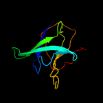
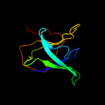
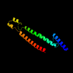
1 c6ic4C_
99.9
27
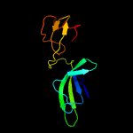
PDB header: protein transportChain: C: PDB Molecule: toluene tolerance efflux transporter (abc superfamily,PDBTitle: cryo-em structure of the a. baumannii mla complex at 8.7 a resolution
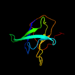
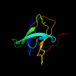
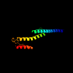
2 c5uw8C_
99.8
24
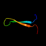
PDB header: transport proteinChain: C: PDB Molecule: probable phospholipid abc transporter-binding protein mlad;PDBTitle: structure of e. coli mce protein mlad, core mce domain
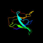
3 c5uvnA_
99.5
21
PDB header: transport proteinChain: A: PDB Molecule: paraquat-inducible protein b;PDBTitle: structure of e. coli mce protein pqib, periplasmic domain
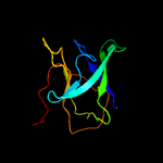
4 c5uvnF_
99.5
21
PDB header: transport proteinChain: F: PDB Molecule: paraquat-inducible protein b;PDBTitle: structure of e. coli mce protein pqib, periplasmic domain
5 c5uvnC_
99.5
21
PDB header: transport proteinChain: C: PDB Molecule: paraquat-inducible protein b;PDBTitle: structure of e. coli mce protein pqib, periplasmic domain
6 c5uvnD_
99.5
21
PDB header: transport proteinChain: D: PDB Molecule: paraquat-inducible protein b;PDBTitle: structure of e. coli mce protein pqib, periplasmic domain
7 c5uvnB_
99.5
21
PDB header: transport proteinChain: B: PDB Molecule: paraquat-inducible protein b;PDBTitle: structure of e. coli mce protein pqib, periplasmic domain
8 c5uvnE_
99.5
21
PDB header: transport proteinChain: E: PDB Molecule: paraquat-inducible protein b;PDBTitle: structure of e. coli mce protein pqib, periplasmic domain
9 c3g67A_
92.2
13
PDB header: signaling proteinChain: A: PDB Molecule: methyl-accepting chemotaxis protein;PDBTitle: crystal structure of a soluble chemoreceptor from thermotoga2 maritima
10 c1qu7A_
89.5
13
PDB header: signaling proteinChain: A: PDB Molecule: methyl-accepting chemotaxis protein i;PDBTitle: four helical-bundle structure of the cytoplasmic domain of a serine2 chemotaxis receptor
11 c6b7nC_
87.3
9
PDB header: viral proteinChain: C: PDB Molecule: spike protein;PDBTitle: cryo-electron microscopy structure of porcine delta coronavirus spike2 protein in the pre-fusion state
12 c3lnrA_
85.0
8
PDB header: signaling proteinChain: A: PDB Molecule: aerotaxis transducer aer2;PDBTitle: crystal structure of poly-hamp domains from the p. aeruginosa soluble2 receptor aer2
13 c3zx6A_
77.9
12
PDB header: signalingChain: A: PDB Molecule: hamp, methyl-accepting chemotaxis protein i;PDBTitle: structure of hamp(af1503)-tsr fusion - hamp (a291v) mutant
14 c2ch7A_
75.8
9
PDB header: chemotaxisChain: A: PDB Molecule: methyl-accepting chemotaxis protein;PDBTitle: crystal structure of the cytoplasmic domain of a bacterial2 chemoreceptor from thermotoga maritima
15 c2qf4A_
75.3
17
PDB header: structural proteinChain: A: PDB Molecule: cell shape determining protein mrec;PDBTitle: high resolution structure of the major periplasmic domain from the2 cell shape-determining filament mrec (orthorhombic form)
16 c3ojaB_
68.5
12
PDB header: protein bindingChain: B: PDB Molecule: anopheles plasmodium-responsive leucine-rich repeat proteinPDBTitle: crystal structure of lrim1/apl1c complex
17 c2j5uB_
67.3
13
PDB header: cell shape regulationChain: B: PDB Molecule: mrec protein;PDBTitle: mrec lysteria monocytogenes
18 c4rh7A_
66.8
7
PDB header: motor proteinChain: A: PDB Molecule: green fluorescent protein/cytoplasmic dynein 2 heavy chainPDBTitle: crystal structure of human cytoplasmic dynein 2 motor domain in2 complex with adp.vi
19 c6e6aB_
65.2
17
PDB header: protein bindingChain: B: PDB Molecule: inclusion membrane protein a;PDBTitle: triclinic crystal form of inca g144a point mutant
20 c2d4yA_
64.9
8
PDB header: structural proteinChain: A: PDB Molecule: flagellar hook-associated protein 1;PDBTitle: crystal structure of a 49k fragment of hap1 (flgk)
21 c2wpqA_
not modelled
64.1
9
PDB header: membrane proteinChain: A: PDB Molecule: trimeric autotransporter adhesin fragment;PDBTitle: salmonella enterica sada 479-519 fused to gcn4 adaptors (sadak3, in-2 register fusion)
22 c1deqF_
not modelled
63.4
12
PDB header: blood clottingChain: F: PDB Molecule: fibrinogen (gamma chain);PDBTitle: the crystal structure of modified bovine fibrinogen (at ~42 angstrom resolution)
23 c3vkhA_
not modelled
63.2
7
PDB header: motor proteinChain: A: PDB Molecule: dynein heavy chain, cytoplasmic;PDBTitle: x-ray structure of a functional full-length dynein motor domain
24 c3cwgA_
not modelled
62.9
11
PDB header: transcriptionChain: A: PDB Molecule: signal transducer and activator of transcriptionPDBTitle: unphosphorylated mouse stat3 core fragment
25 c5lp5F_
not modelled
61.6
13
PDB header: hydrolase/antibioticChain: F: PDB Molecule: rod shape-determining protein (mrec);PDBTitle: complex between penicillin-binding protein (pbp2) and mrec from2 helicobacter pylori
26 c5zuvB_
not modelled
59.6
8
PDB header: viral protein, inhibitorChain: B: PDB Molecule: spike glycoprotein,spike glycoprotein,inhibitor ek1;PDBTitle: crystal structure of the human coronavirus 229e hr1 motif in complex2 with pan-covs inhibitor ek1
27 c6gajA_
not modelled
55.9
13
PDB header: viral proteinChain: A: PDB Molecule: outer capsid protein sigma-1;PDBTitle: crystal structure of the t1l reovirus sigma1 coiled coil tail (iodide)
28 c6nb3B_
not modelled
55.2
9
PDB header: virusChain: B: PDB Molecule: spike glycoprotein;PDBTitle: mers-cov complex with human neutralizing lca60 antibody fab fragment2 (state 1)
29 c3j99M_
not modelled
54.7
10
PDB header: hydrolaseChain: M: PDB Molecule: synaptosomal-associated protein 25;PDBTitle: structure of 20s supercomplex determined by single particle2 cryoelectron microscopy (state iiib)
30 c2ieqC_
not modelled
53.2
10
PDB header: viral proteinChain: C: PDB Molecule: spike glycoprotein;PDBTitle: core structure of s2 from the human coronavirus nl63 spike2 glycoprotein
31 c5szsC_
not modelled
52.8
15
PDB header: viral proteinChain: C: PDB Molecule: spike glycoprotein;PDBTitle: glycan shield and epitope masking of a coronavirus spike protein2 observed by cryo-electron microscopy
32 c4iogD_
not modelled
51.2
10
PDB header: unknown functionChain: D: PDB Molecule: secreted protein esxb;PDBTitle: the crystal structure of a secreted protein esxb (wild-type, in p212 space group) from bacillus anthracis str. sterne
33 c3zbhC_
not modelled
41.5
16
PDB header: unknown functionChain: C: PDB Molecule: esxa;PDBTitle: geobacillus thermodenitrificans esxa crystal form i
34 c6grjG_
not modelled
40.9
10
PDB header: toxinChain: G: PDB Molecule: ahlb;PDBTitle: structure of the ahlb pore of the tripartite alpha-pore forming toxin,2 ahl, from aeromonas hydrophila.
35 c1deqO_
not modelled
40.1
11
PDB header: blood clottingChain: O: PDB Molecule: fibrinogen (beta chain);PDBTitle: the crystal structure of modified bovine fibrinogen (at ~42 angstrom resolution)
36 c1ei3E_
not modelled
39.0
7
PDB header: blood clottingChain: E: PDB Molecule: fibrinogen;PDBTitle: crystal structure of native chicken fibrinogen
37 c4wsrA_
not modelled
37.3
15
PDB header: viral proteinChain: A: PDB Molecule: hemagglutinin;PDBTitle: the crystal structure of hemagglutinin form a/chicken/new york/14677-2 13/1998
38 c6gapB_
not modelled
36.8
12
PDB header: viral proteinChain: B: PDB Molecule: outer capsid protein sigma-1;PDBTitle: crystal structure of the t3d reovirus sigma1 coiled coil tail and body
39 c6f0kA_
not modelled
36.8
17
PDB header: membrane proteinChain: A: PDB Molecule: cytochrome c family protein;PDBTitle: alternative complex iii
40 c6ezvX_
not modelled
36.7
13
PDB header: toxinChain: X: PDB Molecule: non-hemolytic enterotoxin lytic component l1;PDBTitle: the cytotoxin maka from vibrio cholerae
41 c5xbjA_
not modelled
36.6
10
PDB header: biosynthetic proteinChain: A: PDB Molecule: flagellar hook-associated protein flgk;PDBTitle: the structure of the flagellar hook junction protein hap1 (flgk) from2 campylobacter jejuni
42 d1v5va1
not modelled
36.5
15
Fold: Elongation factor/aminomethyltransferase common domainSuperfamily: Aminomethyltransferase beta-barrel domainFamily: Aminomethyltransferase beta-barrel domain
43 d1pj5a1
not modelled
35.2
10
Fold: Elongation factor/aminomethyltransferase common domainSuperfamily: Aminomethyltransferase beta-barrel domainFamily: Aminomethyltransferase beta-barrel domain
44 c3gvmA_
not modelled
33.4
8
PDB header: viral proteinChain: A: PDB Molecule: putative uncharacterized protein sag1039;PDBTitle: structure of the homodimeric wxg-100 family protein from streptococcus2 agalactiae
45 c5u0pU_
not modelled
32.7
10
PDB header: transcriptionChain: U: PDB Molecule: mediator complex subunit 21;PDBTitle: cryo-em structure of the transcriptional mediator
46 c1ei3C_
not modelled
32.4
5
PDB header: blood clottingChain: C: PDB Molecule: fibrinogen;PDBTitle: crystal structure of native chicken fibrinogen
47 c4njlA_
not modelled
32.3
10
PDB header: viral proteinChain: A: PDB Molecule: s protein;PDBTitle: crystal structure of middle east respiratory syndrome coronavirus s22 protein fusion core
48 c2vs0B_
not modelled
30.9
14
PDB header: cell invasionChain: B: PDB Molecule: virulence factor esxa;PDBTitle: structural analysis of homodimeric staphylococcal aureus2 virulence factor esxa
49 c1t98B_
not modelled
30.1
11
PDB header: cell cycleChain: B: PDB Molecule: chromosome partition protein mukf;PDBTitle: crystal structure of mukf(1-287)
50 c1kmiZ_
not modelled
29.4
12
PDB header: signaling proteinChain: Z: PDB Molecule: chemotaxis protein chez;PDBTitle: crystal structure of an e.coli chemotaxis protein, chez
51 c2wr2B_
not modelled
29.3
15
PDB header: viral proteinChain: B: PDB Molecule: hemagglutinin;PDBTitle: structure of influenza h2 avian hemagglutinin with avian2 receptor
52 c4ut1A_
not modelled
29.3
8
PDB header: motor proteinChain: A: PDB Molecule: flagellar hook-associated protein;PDBTitle: the structure of the flagellar hook junction protein flgk2 from burkholderia pseudomallei
53 c1v5vA_
not modelled
29.0
14
PDB header: transferaseChain: A: PDB Molecule: aminomethyltransferase;PDBTitle: crystal structure of a component of glycine cleavage system: t-protein2 from pyrococcus horikoshii ot3 at 1.5 a resolution
54 c4fiuC_
not modelled
27.3
11
PDB header: viral proteinChain: C: PDB Molecule: hemagglutinin;PDBTitle: the structure of hemagglutinin of h16 subtype influenza virus with2 v327g mutation
55 c4abxB_
not modelled
26.0
8
PDB header: dna binding proteinChain: B: PDB Molecule: dna repair protein recn;PDBTitle: crystal structure of deinococcus radiodurans recn coiled-2 coil domain
56 c4mc5C_
not modelled
25.1
9
PDB header: viral proteinChain: C: PDB Molecule: hemagglutinin;PDBTitle: crystal structure of a subtype h18 hemagglutinin homologue from2 a/flat-faced bat/peru/033/2010 (h18n11)
57 c1worA_
not modelled
25.0
14
PDB header: transferaseChain: A: PDB Molecule: aminomethyltransferase;PDBTitle: crystal structure of t-protein of the glycine cleavage2 system
58 c5dmaA_
not modelled
24.8
18
PDB header: hydrolaseChain: A: PDB Molecule: atp-dependent dna helicase pcra;PDBTitle: crystal structure of c-terminal tudor domain in pcra/uvrd helicase
59 d1wa8a1
not modelled
24.5
11
Fold: Ferritin-likeSuperfamily: EsxAB dimer-likeFamily: ESAT-6 like
60 c1ha0A_
not modelled
24.5
9
PDB header: viral proteinChain: A: PDB Molecule: protein (hemagglutinin precursor);PDBTitle: hemagglutinin precursor ha0
61 c5xl9B_
not modelled
24.4
9
PDB header: viral proteinChain: B: PDB Molecule: hemagglutinin;PDBTitle: the structure of hemagglutinin g228s mutant from an avian-origin h4n62 influenza virus in complex with avian receptor analog lsta
62 c4lwsB_
not modelled
23.8
8
PDB header: unknown functionChain: B: PDB Molecule: uncharacterized protein;PDBTitle: esxa : esxb (semet) hetero-dimer from thermomonospora curvata
63 c3bt6B_
not modelled
23.4
10
PDB header: viral proteinChain: B: PDB Molecule: influenza b hemagglutinin (ha);PDBTitle: crystal structure of influenza b virus hemagglutinin
64 c5zhyA_
not modelled
23.3
13
PDB header: viral proteinChain: A: PDB Molecule: spike glycoprotein, spike glycoprotein;PDBTitle: structural characterization of the hcov-229e fusion core
65 c1mqlB_
not modelled
22.9
8
PDB header: viral proteinChain: B: PDB Molecule: hemagglutinin ha2 chain;PDBTitle: bha of ukr/63
66 c2gl2B_
not modelled
22.4
10
PDB header: cell adhesionChain: B: PDB Molecule: adhesion a;PDBTitle: crystal structure of the tetra muntant (t66g,r67g,f68g,y69g) of2 bacterial adhesin fada
67 d1szia_
not modelled
21.7
12
Fold: Four-helical up-and-down bundleSuperfamily: Mannose-6-phosphate receptor binding protein 1 (Tip47), C-terminal domainFamily: Mannose-6-phosphate receptor binding protein 1 (Tip47), C-terminal domain
68 c1zvaA_
not modelled
20.8
15
PDB header: viral proteinChain: A: PDB Molecule: e2 glycoprotein;PDBTitle: a structure-based mechanism of sars virus membrane fusion
69 c1jsdB_
not modelled
19.8
12
PDB header: viral proteinChain: B: PDB Molecule: haemagglutinin (ha2 chain);PDBTitle: crystal structure of swine h9 haemagglutinin
70 d1jjcb3
not modelled
19.7
28
Fold: OB-foldSuperfamily: Nucleic acid-binding proteinsFamily: Myf domain
71 d1st6a4
not modelled
19.0
13
Fold: Four-helical up-and-down bundleSuperfamily: alpha-catenin/vinculin-likeFamily: alpha-catenin/vinculin
72 c2wrhI_
not modelled
17.8
15
PDB header: viral proteinChain: I: PDB Molecule: hemagglutinin ha2 chain;PDBTitle: structure of h1 duck albert hemagglutinin with human2 receptor
73 c1ru7B_
not modelled
17.5
13
PDB header: viral proteinChain: B: PDB Molecule: hemagglutinin;PDBTitle: 1934 human h1 hemagglutinin
74 c2qihA_
not modelled
17.2
9
PDB header: cell adhesionChain: A: PDB Molecule: protein uspa1;PDBTitle: crystal structure of 527-665 fragment of uspa1 protein from moraxella2 catarrhalis
75 c1hgeD_
not modelled
17.1
9
PDB header: viral proteinChain: D: PDB Molecule: hemagglutinin, (g135r), ha1 chain;PDBTitle: binding of influenza virus hemagglutinin to analogs of its cell-2 surface receptor, sialic acid: analysis by proton nuclear magnetic3 resonance spectroscopy and x-ray crystallography
76 c6cv0C_
not modelled
17.0
8
PDB header: viral proteinChain: C: PDB Molecule: spike glycoprotein;PDBTitle: cryo-electron microscopy structure of infectious bronchitis2 coronavirus spike protein
77 c1yx2B_
not modelled
16.8
19
PDB header: transferaseChain: B: PDB Molecule: aminomethyltransferase;PDBTitle: crystal structure of the probable aminomethyltransferase2 from bacillus subtilis
78 d1wosa1
not modelled
15.7
13
Fold: Elongation factor/aminomethyltransferase common domainSuperfamily: Aminomethyltransferase beta-barrel domainFamily: Aminomethyltransferase beta-barrel domain
79 c2iakA_
not modelled
15.3
13
PDB header: cell adhesionChain: A: PDB Molecule: bullous pemphigoid antigen 1, isoform 5;PDBTitle: crystal structure of a protease resistant fragment of the plakin2 domain of bullous pemphigoid antigen1 (bpag1)
80 c1wyyB_
not modelled
15.2
16
PDB header: viral proteinChain: B: PDB Molecule: e2 glycoprotein;PDBTitle: post-fusion hairpin conformation of the sars coronavirus spike2 glycoprotein
81 c3izcN_
not modelled
15.0
20
PDB header: ribosomeChain: N: PDB Molecule: 60s ribosomal protein rpl14 (l14e);PDBTitle: localization of the large subunit ribosomal proteins into a 6.1 a2 cryo-em map of saccharomyces cerevisiae translating 80s ribosome
82 d1eq1a_
not modelled
14.9
13
Fold: Apolipophorin-IIISuperfamily: Apolipophorin-IIIFamily: Apolipophorin-III
83 c4lwsA_
not modelled
14.7
6
PDB header: unknown functionChain: A: PDB Molecule: uncharacterized protein;PDBTitle: esxa : esxb (semet) hetero-dimer from thermomonospora curvata
84 c6nzkB_
not modelled
14.1
10
PDB header: viral proteinChain: B: PDB Molecule: spike surface glycoprotein;PDBTitle: structural basis for human coronavirus attachment to sialic acid2 receptors
85 c3pe0B_
not modelled
13.7
7
PDB header: structural proteinChain: B: PDB Molecule: plectin;PDBTitle: structure of the central region of the plakin domain of plectin
86 c1wncE_
not modelled
13.5
14
PDB header: viral proteinChain: E: PDB Molecule: e2 glycoprotein;PDBTitle: crystal structure of the sars-cov spike protein fusion core
87 c5x5bB_
not modelled
13.5
8
PDB header: viral proteinChain: B: PDB Molecule: spike glycoprotein;PDBTitle: prefusion structure of sars-cov spike glycoprotein, conformation 2
88 c5x5fC_
not modelled
13.3
11
PDB header: viral proteinChain: C: PDB Molecule: s protein;PDBTitle: prefusion structure of mers-cov spike glycoprotein, conformation 2
89 c4a19F_
not modelled
12.8
25
PDB header: ribosomeChain: F: PDB Molecule: rpl14;PDBTitle: t.thermophila 60s ribosomal subunit in complex with2 initiation factor 6. this file contains 26s rrna and3 proteins of molecule 2.
90 d1xmec1
not modelled
12.8
21
Fold: Single transmembrane helixSuperfamily: Bacterial ba3 type cytochrome c oxidase subunit IIaFamily: Bacterial ba3 type cytochrome c oxidase subunit IIa
91 c3bvdC_
not modelled
12.8
21
PDB header: oxidoreductaseChain: C: PDB Molecule: cytochrome c oxidase polypeptide 2a;PDBTitle: structure of surface-engineered cytochrome ba3 oxidase from thermus2 thermophilus under xenon pressure, 100psi 5min
92 c5wrgB_
not modelled
12.7
8
PDB header: virus like particleChain: B: PDB Molecule: spike glycoprotein;PDBTitle: sars-cov spike glycoprotein
93 c3iz5N_
not modelled
12.6
29
PDB header: ribosomeChain: N: PDB Molecule: 60s ribosomal protein l14 (l14e);PDBTitle: localization of the large subunit ribosomal proteins into a 5.5 a2 cryo-em map of triticum aestivum translating 80s ribosome
94 c5b0oB_
not modelled
12.5
35
PDB header: hydrolase/motor proteinChain: B: PDB Molecule: flagellum-specific atp synthase;PDBTitle: structure of the flih-flii complex
95 d1wa8b1
not modelled
12.5
8
Fold: Ferritin-likeSuperfamily: EsxAB dimer-likeFamily: ESAT-6 like
96 c3j3bM_
not modelled
12.5
17
PDB header: ribosomeChain: M: PDB Molecule: 60s ribosomal protein l14;PDBTitle: structure of the human 60s ribosomal proteins
97 c3zf7P_
not modelled
12.5
21
PDB header: ribosomeChain: P: PDB Molecule: probable 60s ribosomal protein l14;PDBTitle: high-resolution cryo-electron microscopy structure of the trypanosoma2 brucei ribosome
98 c6gaoC_
not modelled
12.4
9
PDB header: viral proteinChain: C: PDB Molecule: outer capsid protein sigma-1;PDBTitle: crystal structure of the t1l reovirus sigma1 coiled coil tail and body
99 c3ghgK_
not modelled
12.4
10
PDB header: blood clottingChain: K: PDB Molecule: fibrinogen beta chain;PDBTitle: crystal structure of human fibrinogen