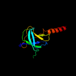
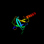
1 c6ic4C_
99.9
24
PDB header: protein transportChain: C: PDB Molecule: toluene tolerance efflux transporter (abc superfamily,PDBTitle: cryo-em structure of the a. baumannii mla complex at 8.7 a resolution
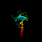
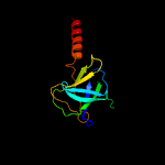
2 c5uw8C_
99.7
28
PDB header: transport proteinChain: C: PDB Molecule: probable phospholipid abc transporter-binding protein mlad;PDBTitle: structure of e. coli mce protein mlad, core mce domain
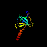
3 c5uvnF_
99.3
17
PDB header: transport proteinChain: F: PDB Molecule: paraquat-inducible protein b;PDBTitle: structure of e. coli mce protein pqib, periplasmic domain
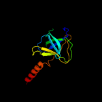
4 c5uvnA_
99.3
17
PDB header: transport proteinChain: A: PDB Molecule: paraquat-inducible protein b;PDBTitle: structure of e. coli mce protein pqib, periplasmic domain
5 c5uvnC_
99.3
17
PDB header: transport proteinChain: C: PDB Molecule: paraquat-inducible protein b;PDBTitle: structure of e. coli mce protein pqib, periplasmic domain
6 c5uvnE_
99.3
17
PDB header: transport proteinChain: E: PDB Molecule: paraquat-inducible protein b;PDBTitle: structure of e. coli mce protein pqib, periplasmic domain
7 c5uvnD_
99.3
17
PDB header: transport proteinChain: D: PDB Molecule: paraquat-inducible protein b;PDBTitle: structure of e. coli mce protein pqib, periplasmic domain
8 c5uvnB_
99.3
17
PDB header: transport proteinChain: B: PDB Molecule: paraquat-inducible protein b;PDBTitle: structure of e. coli mce protein pqib, periplasmic domain
9 c3g67A_
96.5
10
PDB header: signaling proteinChain: A: PDB Molecule: methyl-accepting chemotaxis protein;PDBTitle: crystal structure of a soluble chemoreceptor from thermotoga2 maritima
10 c1qu7A_
96.2
8
PDB header: signaling proteinChain: A: PDB Molecule: methyl-accepting chemotaxis protein i;PDBTitle: four helical-bundle structure of the cytoplasmic domain of a serine2 chemotaxis receptor
11 c2ch7A_
95.8
13
PDB header: chemotaxisChain: A: PDB Molecule: methyl-accepting chemotaxis protein;PDBTitle: crystal structure of the cytoplasmic domain of a bacterial2 chemoreceptor from thermotoga maritima
12 c5szsC_
95.0
17
PDB header: viral proteinChain: C: PDB Molecule: spike glycoprotein;PDBTitle: glycan shield and epitope masking of a coronavirus spike protein2 observed by cryo-electron microscopy
13 c2d4yA_
94.7
13
PDB header: structural proteinChain: A: PDB Molecule: flagellar hook-associated protein 1;PDBTitle: crystal structure of a 49k fragment of hap1 (flgk)
14 c6e6aB_
94.7
13
PDB header: protein bindingChain: B: PDB Molecule: inclusion membrane protein a;PDBTitle: triclinic crystal form of inca g144a point mutant
15 c2wpqA_
94.5
5
PDB header: membrane proteinChain: A: PDB Molecule: trimeric autotransporter adhesin fragment;PDBTitle: salmonella enterica sada 479-519 fused to gcn4 adaptors (sadak3, in-2 register fusion)
16 c6nzkB_
94.4
16
PDB header: viral proteinChain: B: PDB Molecule: spike surface glycoprotein;PDBTitle: structural basis for human coronavirus attachment to sialic acid2 receptors
17 c6b7nC_
94.2
17
PDB header: viral proteinChain: C: PDB Molecule: spike protein;PDBTitle: cryo-electron microscopy structure of porcine delta coronavirus spike2 protein in the pre-fusion state
18 c1ei3C_
94.0
10
PDB header: blood clottingChain: C: PDB Molecule: fibrinogen;PDBTitle: crystal structure of native chicken fibrinogen
19 c6gajA_
94.0
19
PDB header: viral proteinChain: A: PDB Molecule: outer capsid protein sigma-1;PDBTitle: crystal structure of the t1l reovirus sigma1 coiled coil tail (iodide)
20 c3lnrA_
93.9
11
PDB header: signaling proteinChain: A: PDB Molecule: aerotaxis transducer aer2;PDBTitle: crystal structure of poly-hamp domains from the p. aeruginosa soluble2 receptor aer2
21 c2ieqC_
not modelled
93.5
14
PDB header: viral proteinChain: C: PDB Molecule: spike glycoprotein;PDBTitle: core structure of s2 from the human coronavirus nl63 spike2 glycoprotein
22 c5xbjA_
not modelled
93.4
6
PDB header: biosynthetic proteinChain: A: PDB Molecule: flagellar hook-associated protein flgk;PDBTitle: the structure of the flagellar hook junction protein hap1 (flgk) from2 campylobacter jejuni
23 c4abxB_
not modelled
93.1
14
PDB header: dna binding proteinChain: B: PDB Molecule: dna repair protein recn;PDBTitle: crystal structure of deinococcus radiodurans recn coiled-2 coil domain
24 c1ei3E_
not modelled
92.8
16
PDB header: blood clottingChain: E: PDB Molecule: fibrinogen;PDBTitle: crystal structure of native chicken fibrinogen
25 c1deqO_
not modelled
92.8
14
PDB header: blood clottingChain: O: PDB Molecule: fibrinogen (beta chain);PDBTitle: the crystal structure of modified bovine fibrinogen (at ~42 angstrom resolution)
26 c2dq3A_
not modelled
92.8
13
PDB header: ligaseChain: A: PDB Molecule: seryl-trna synthetase;PDBTitle: crystal structure of aq_298
27 c6ezvX_
not modelled
92.7
10
PDB header: toxinChain: X: PDB Molecule: non-hemolytic enterotoxin lytic component l1;PDBTitle: the cytotoxin maka from vibrio cholerae
28 c6nb3B_
not modelled
92.7
15
PDB header: virusChain: B: PDB Molecule: spike glycoprotein;PDBTitle: mers-cov complex with human neutralizing lca60 antibody fab fragment2 (state 1)
29 c5i08A_
not modelled
92.6
15
PDB header: viral proteinChain: A: PDB Molecule: spike glycoprotein, envelope glycoprotein chimera;PDBTitle: prefusion structure of a human coronavirus spike protein
30 c5x5bB_
not modelled
92.6
11
PDB header: viral proteinChain: B: PDB Molecule: spike glycoprotein;PDBTitle: prefusion structure of sars-cov spike glycoprotein, conformation 2
31 c2p22A_
not modelled
92.4
10
PDB header: transport proteinChain: A: PDB Molecule: suppressor protein stp22 of temperature-sensitive alpha-PDBTitle: structure of the yeast escrt-i heterotetramer core
32 c6gapB_
not modelled
92.2
12
PDB header: viral proteinChain: B: PDB Molecule: outer capsid protein sigma-1;PDBTitle: crystal structure of the t3d reovirus sigma1 coiled coil tail and body
33 c3cwgA_
not modelled
92.1
7
PDB header: transcriptionChain: A: PDB Molecule: signal transducer and activator of transcriptionPDBTitle: unphosphorylated mouse stat3 core fragment
34 c6cs2A_
not modelled
91.7
11
PDB header: viral protein/hydrolaseChain: A: PDB Molecule: spike glycoprotein,fibritin;PDBTitle: sars spike glycoprotein - human ace2 complex, stabilized variant, all2 ace2-bound particles
35 c3jclC_
not modelled
91.6
15
PDB header: viral proteinChain: C: PDB Molecule: spike glycoprotein;PDBTitle: cryo-electron microscopy structure of a coronavirus spike glycoprotein2 trimer
36 c1deqF_
not modelled
91.0
13
PDB header: blood clottingChain: F: PDB Molecule: fibrinogen (gamma chain);PDBTitle: the crystal structure of modified bovine fibrinogen (at ~42 angstrom resolution)
37 c2bbjB_
not modelled
91.0
13
PDB header: metal transport/membrane proteinChain: B: PDB Molecule: divalent cation transport-related protein;PDBTitle: crystal structure of the cora mg2+ transporter
38 c4ut1A_
not modelled
91.0
15
PDB header: motor proteinChain: A: PDB Molecule: flagellar hook-associated protein;PDBTitle: the structure of the flagellar hook junction protein flgk2 from burkholderia pseudomallei
39 c3zx6A_
not modelled
90.8
7
PDB header: signalingChain: A: PDB Molecule: hamp, methyl-accepting chemotaxis protein i;PDBTitle: structure of hamp(af1503)-tsr fusion - hamp (a291v) mutant
40 c5xlrC_
not modelled
89.9
11
PDB header: viral proteinChain: C: PDB Molecule: spike glycoprotein;PDBTitle: structure of sars-cov spike glycoprotein
41 c6gy8B_
not modelled
89.1
11
PDB header: toxinChain: B: PDB Molecule: xaxa;PDBTitle: crystal structure of xaxa from xenorhabdus nematophila
42 c2qihA_
not modelled
89.0
12
PDB header: cell adhesionChain: A: PDB Molecule: protein uspa1;PDBTitle: crystal structure of 527-665 fragment of uspa1 protein from moraxella2 catarrhalis
43 c5dfzA_
not modelled
88.7
13
PDB header: transferaseChain: A: PDB Molecule: vacuolar protein sorting-associated protein 38;PDBTitle: structure of vps34 complex ii from s. cerevisiae.
44 c1wyyB_
not modelled
88.7
22
PDB header: viral proteinChain: B: PDB Molecule: e2 glycoprotein;PDBTitle: post-fusion hairpin conformation of the sars coronavirus spike2 glycoprotein
45 c6b3oB_
not modelled
88.5
20
PDB header: viral proteinChain: B: PDB Molecule: spike glycoprotein;PDBTitle: tectonic conformational changes of a coronavirus spike glycoprotein2 promote membrane fusion
46 c5u0pU_
not modelled
87.8
16
PDB header: transcriptionChain: U: PDB Molecule: mediator complex subunit 21;PDBTitle: cryo-em structure of the transcriptional mediator
47 d1ykhb1
not modelled
87.8
13
Fold: Mediator hinge subcomplex-likeSuperfamily: Mediator hinge subcomplex-likeFamily: CSE2-like
48 c5zhyA_
not modelled
87.8
9
PDB header: viral proteinChain: A: PDB Molecule: spike glycoprotein, spike glycoprotein;PDBTitle: structural characterization of the hcov-229e fusion core
49 c4rh7A_
not modelled
87.7
11
PDB header: motor proteinChain: A: PDB Molecule: green fluorescent protein/cytoplasmic dynein 2 heavy chainPDBTitle: crystal structure of human cytoplasmic dynein 2 motor domain in2 complex with adp.vi
50 c3ojaB_
not modelled
87.3
13
PDB header: protein bindingChain: B: PDB Molecule: anopheles plasmodium-responsive leucine-rich repeat proteinPDBTitle: crystal structure of lrim1/apl1c complex
51 c5wrgB_
not modelled
87.1
11
PDB header: virus like particleChain: B: PDB Molecule: spike glycoprotein;PDBTitle: sars-cov spike glycoprotein
52 c1kmiZ_
not modelled
87.1
13
PDB header: signaling proteinChain: Z: PDB Molecule: chemotaxis protein chez;PDBTitle: crystal structure of an e.coli chemotaxis protein, chez
53 c4nl6C_
not modelled
86.9
15
PDB header: splicingChain: C: PDB Molecule: survival motor neuron protein;PDBTitle: structure of the full-length form of the protein smn found in healthy2 patients
54 c5nmoA_
not modelled
86.8
15
PDB header: cell cycleChain: A: PDB Molecule: chromosome partition protein smc,chromosome partitionPDBTitle: structure of the bacillus subtilis smc joint domain
55 c1quuA_
not modelled
85.7
14
PDB header: contractile proteinChain: A: PDB Molecule: human skeletal muscle alpha-actinin 2;PDBTitle: crystal structure of two central spectrin-like repeats from alpha-2 actinin
56 c5lskD_
not modelled
85.4
17
PDB header: cell cycleChain: D: PDB Molecule: kinetochore-associated protein dsn1 homolog;PDBTitle: crystal structure of the human kinetochore mis12-cenp-c complex
57 c2nrjA_
not modelled
85.3
15
PDB header: toxinChain: A: PDB Molecule: hbl b protein;PDBTitle: crystal structure of hemolysin binding component from2 bacillus cereus
58 c4e40A_
not modelled
85.3
8
PDB header: transport proteinChain: A: PDB Molecule: putative uncharacterized protein;PDBTitle: the haptoglobin-hemoglobin receptor of trypanosoma congolense
59 c6grjG_
not modelled
85.0
10
PDB header: toxinChain: G: PDB Molecule: ahlb;PDBTitle: structure of the ahlb pore of the tripartite alpha-pore forming toxin,2 ahl, from aeromonas hydrophila.
60 c5j65A_
not modelled
84.5
11
PDB header: toxinChain: A: PDB Molecule: pesticidal crystal protein cry6aa;PDBTitle: crystal structure of trypsin activated cry6aa
61 c3ghgI_
not modelled
83.8
7
PDB header: blood clottingChain: I: PDB Molecule: fibrinogen gamma chain;PDBTitle: crystal structure of human fibrinogen
62 c3ghgK_
not modelled
83.3
14
PDB header: blood clottingChain: K: PDB Molecule: fibrinogen beta chain;PDBTitle: crystal structure of human fibrinogen
63 c3j99M_
not modelled
82.1
12
PDB header: hydrolaseChain: M: PDB Molecule: synaptosomal-associated protein 25;PDBTitle: structure of 20s supercomplex determined by single particle2 cryoelectron microscopy (state iiib)
64 c5cwsC_
not modelled
81.9
10
PDB header: protein transportChain: C: PDB Molecule: nucleoporin nsp1;PDBTitle: crystal structure of the intact chaetomium thermophilum nsp1-nup49-2 nup57 channel nucleoporin heterotrimer bound to its nic96 nuclear3 pore complex attachment site
65 c2gl2B_
not modelled
81.0
12
PDB header: cell adhesionChain: B: PDB Molecule: adhesion a;PDBTitle: crystal structure of the tetra muntant (t66g,r67g,f68g,y69g) of2 bacterial adhesin fada
66 d1eq1a_
not modelled
80.9
13
Fold: Apolipophorin-IIISuperfamily: Apolipophorin-IIIFamily: Apolipophorin-III
67 c5yfpG_
not modelled
78.4
13
PDB header: exocytosisChain: G: PDB Molecule: exocyst complex component exo70;PDBTitle: cryo-em structure of the exocyst complex
68 c6gaoC_
not modelled
78.1
11
PDB header: viral proteinChain: C: PDB Molecule: outer capsid protein sigma-1;PDBTitle: crystal structure of the t1l reovirus sigma1 coiled coil tail and body
69 d1g4us1
not modelled
78.0
15
Fold: Four-helical up-and-down bundleSuperfamily: Bacterial GAP domainFamily: Bacterial GAP domain
70 c5cwsJ_
not modelled
78.0
8
PDB header: protein transportChain: J: PDB Molecule: nucleoporin nup49;PDBTitle: crystal structure of the intact chaetomium thermophilum nsp1-nup49-2 nup57 channel nucleoporin heterotrimer bound to its nic96 nuclear3 pore complex attachment site
71 d1st6a4
not modelled
77.1
17
Fold: Four-helical up-and-down bundleSuperfamily: alpha-catenin/vinculin-likeFamily: alpha-catenin/vinculin
72 c6el1F_
not modelled
76.5
14
PDB header: membrane proteinChain: F: PDB Molecule: yaxa;PDBTitle: yaxab pore complex
73 d1quua1
not modelled
74.6
15
Fold: Spectrin repeat-likeSuperfamily: Spectrin repeatFamily: Spectrin repeat
74 c2efrB_
not modelled
73.6
13
PDB header: contractile proteinChain: B: PDB Molecule: general control protein gcn4 and tropomyosin 1 alpha chain;PDBTitle: crystal structure of the c-terminal tropomyosin fragment with n- and2 c-terminal extensions of the leucine zipper at 1.8 angstroms3 resolution
75 c4a7fB_
not modelled
73.4
10
PDB header: structural protein/hydrolaseChain: B: PDB Molecule: tropomyosin 1 alpha;PDBTitle: structure of the actin-tropomyosin-myosin complex (rigor atm 3)
76 c5gasN_
not modelled
73.2
12
PDB header: hydrolaseChain: N: PDB Molecule: archaeal/vacuolar-type h+-atpase subunit i;PDBTitle: thermus thermophilus v/a-atpase, conformation 2
77 c2xzrA_
not modelled
73.1
8
PDB header: cell adhesionChain: A: PDB Molecule: immunoglobulin-binding protein eibd;PDBTitle: escherichia coli immunoglobulin-binding protein eibd 391-438 fused2 to gcn4 adaptors
78 c3lbxB_
not modelled
72.9
9
PDB header: structural proteinChain: B: PDB Molecule: spectrin beta chain, erythrocyte;PDBTitle: crystal structure of the erythrocyte spectrin tetramerization domain2 complex
79 c1u5pA_
not modelled
72.4
11
PDB header: structural proteinChain: A: PDB Molecule: spectrin alpha chain, brain;PDBTitle: crystal structure of repeats 15 and 16 of chicken brain2 alpha spectrin
80 c4fiuC_
not modelled
71.3
13
PDB header: viral proteinChain: C: PDB Molecule: hemagglutinin;PDBTitle: the structure of hemagglutinin of h16 subtype influenza virus with2 v327g mutation
81 c2fxmB_
not modelled
70.2
14
PDB header: contractile proteinChain: B: PDB Molecule: myosin heavy chain, cardiac muscle beta isoform;PDBTitle: structure of the human beta-myosin s2 fragment
82 c2yfaA_
not modelled
70.1
9
PDB header: receptorChain: A: PDB Molecule: methyl-accepting chemotaxis transducer;PDBTitle: x-ray structure of mcps ligand binding domain in complex with malate
83 c2bezC_
not modelled
69.9
25
PDB header: viral proteinChain: C: PDB Molecule: e2 glycoprotein;PDBTitle: structure of a proteolitically resistant core from the severe acute2 respiratory syndrome coronavirus s2 fusion protein
84 c5zuvB_
not modelled
68.9
18
PDB header: viral protein, inhibitorChain: B: PDB Molecule: spike glycoprotein,spike glycoprotein,inhibitor ek1;PDBTitle: crystal structure of the human coronavirus 229e hr1 motif in complex2 with pan-covs inhibitor ek1
85 c1z23A_
not modelled
68.4
14
PDB header: cell adhesionChain: A: PDB Molecule: crk-associated substrate;PDBTitle: the serine-rich domain from crk-associated substrate2 (p130cas)
86 c2dq0A_
not modelled
68.3
9
PDB header: ligaseChain: A: PDB Molecule: seryl-trna synthetase;PDBTitle: crystal structure of seryl-trna synthetase from pyrococcus2 horikoshii complexed with a seryl-adenylate analog
87 c5j4oA_
not modelled
67.8
13
PDB header: structural proteinChain: A: PDB Molecule: spectrin alpha chain, erythrocytic 1;PDBTitle: structure of human erythrocytic spectrin alpha chain repeats 16-17
88 c4wsrA_
not modelled
66.9
17
PDB header: viral proteinChain: A: PDB Molecule: hemagglutinin;PDBTitle: the crystal structure of hemagglutinin form a/chicken/new york/14677-2 13/1998
89 d1hcia4
not modelled
66.2
7
Fold: Spectrin repeat-likeSuperfamily: Spectrin repeatFamily: Spectrin repeat
90 c2d3eD_
not modelled
66.1
10
PDB header: contractile proteinChain: D: PDB Molecule: general control protein gcn4 and tropomyosin 1 alpha chain;PDBTitle: crystal structure of the c-terminal fragment of rabbit skeletal alpha-2 tropomyosin
91 c3dtpA_
not modelled
65.6
13
PDB header: contractile proteinChain: A: PDB Molecule: myosin 2 heavy chain chimera of smooth and cardiac muscle;PDBTitle: tarantula heavy meromyosin obtained by flexible docking to tarantula2 muscle thick filament cryo-em 3d-map
92 c2wr2B_
not modelled
64.4
13
PDB header: viral proteinChain: B: PDB Molecule: hemagglutinin;PDBTitle: structure of influenza h2 avian hemagglutinin with avian2 receptor
93 c3vkhA_
not modelled
64.3
14
PDB header: motor proteinChain: A: PDB Molecule: dynein heavy chain, cytoplasmic;PDBTitle: x-ray structure of a functional full-length dynein motor domain
94 c3gvmA_
not modelled
61.8
9
PDB header: viral proteinChain: A: PDB Molecule: putative uncharacterized protein sag1039;PDBTitle: structure of the homodimeric wxg-100 family protein from streptococcus2 agalactiae
95 c3bt6B_
not modelled
61.1
7
PDB header: viral proteinChain: B: PDB Molecule: influenza b hemagglutinin (ha);PDBTitle: crystal structure of influenza b virus hemagglutinin
96 c4n21E_
not modelled
60.2
11
PDB header: viral proteinChain: E: PDB Molecule: gp2 ectodomain;PDBTitle: crystal structure of the gp2 core domain from the california academy2 of science virus
97 c1ha0A_
not modelled
59.7
10
PDB header: viral proteinChain: A: PDB Molecule: protein (hemagglutinin precursor);PDBTitle: hemagglutinin precursor ha0
98 c4l1bB_
not modelled
59.5
7
PDB header: signaling protein/transferase/inhibitorChain: B: PDB Molecule: phosphatidylinositol 3-kinase regulatory subunit alpha;PDBTitle: crystal structure of p110alpha complexed with nish2 of p85alpha
99 c4mc5C_
not modelled
58.8
16
PDB header: viral proteinChain: C: PDB Molecule: hemagglutinin;PDBTitle: crystal structure of a subtype h18 hemagglutinin homologue from2 a/flat-faced bat/peru/033/2010 (h18n11)
100 c2q13A_
not modelled
58.6
9
PDB header: protein transportChain: A: PDB Molecule: dcc-interacting protein 13 alpha;PDBTitle: crystal structure of bar-ph domain of appl1
101 c5wwlN_
not modelled
58.6
12
PDB header: cell cycleChain: N: PDB Molecule: kinetochore protein nnf1;PDBTitle: crystal structure of the schizogenesis pombe kinetochore mis12c2 subcomplex
102 c2dnxA_
not modelled
58.5
17
PDB header: transport proteinChain: A: PDB Molecule: syntaxin-12;PDBTitle: solution structure of rsgi ruh-063, an n-terminal domain of2 syntaxin 12 from human cdna
103 c6j9rA_
not modelled
58.2
10
PDB header: translationChain: A: PDB Molecule: brain tumor protein;PDBTitle: coiled-coil domain of drosophila trim protein brat
104 c4lwsA_
not modelled
58.0
11
PDB header: unknown functionChain: A: PDB Molecule: uncharacterized protein;PDBTitle: esxa : esxb (semet) hetero-dimer from thermomonospora curvata
105 c5tvbB_
not modelled
57.9
10
PDB header: transferaseChain: B: PDB Molecule: nucleoprotein tpr;PDBTitle: structure of the tpr oligomerization domain
106 c5dmaA_
not modelled
57.8
28
PDB header: hydrolaseChain: A: PDB Molecule: atp-dependent dna helicase pcra;PDBTitle: crystal structure of c-terminal tudor domain in pcra/uvrd helicase
107 c6gy6Q_
not modelled
57.1
10
PDB header: toxinChain: Q: PDB Molecule: xaxa;PDBTitle: xaxab pore complex from xenorhabdus nematophila
108 c5ijnF_
not modelled
57.1
12
PDB header: transport proteinChain: F: PDB Molecule: nuclear pore complex protein nup54;PDBTitle: composite structure of the inner ring of the human nuclear pore2 complex (32 copies of nup205)
109 c2oevA_
not modelled
56.8
14
PDB header: protein transportChain: A: PDB Molecule: programmed cell death 6-interacting protein;PDBTitle: crystal structure of alix/aip1
110 c2j5uB_
not modelled
56.8
17
PDB header: cell shape regulationChain: B: PDB Molecule: mrec protein;PDBTitle: mrec lysteria monocytogenes
111 c3o0zD_
not modelled
55.7
9
PDB header: transferaseChain: D: PDB Molecule: rho-associated protein kinase 1;PDBTitle: crystal structure of a coiled-coil domain from human rock i
112 c6h9xA_
not modelled
55.1
15
PDB header: ligaseChain: A: PDB Molecule: serine--trna ligase;PDBTitle: klebsiella pneumoniae seryl-trna synthetase in complex with the2 intermediate analog 5'-o-(n-(l-seryl)-sulfamoyl)adenosine
113 d1u5pa1
not modelled
55.0
14
Fold: Spectrin repeat-likeSuperfamily: Spectrin repeatFamily: Spectrin repeat
114 c2ld3A_
not modelled
54.8
18
PDB header: motor proteinChain: A: PDB Molecule: myosin vi;PDBTitle: solution structure of myosin vi lever arm extension
115 c5yfpD_
not modelled
54.8
14
PDB header: exocytosisChain: D: PDB Molecule: exocyst complex component sec8;PDBTitle: cryo-em structure of the exocyst complex
116 c5lm2B_
not modelled
54.7
12
PDB header: hydrolaseChain: B: PDB Molecule: tyrosine-protein phosphatase non-receptor type 23;PDBTitle: crystal structure of hd-ptp phosphatase
117 c4lwsB_
not modelled
54.6
6
PDB header: unknown functionChain: B: PDB Molecule: uncharacterized protein;PDBTitle: esxa : esxb (semet) hetero-dimer from thermomonospora curvata
118 d1s35a1
not modelled
54.2
13
Fold: Spectrin repeat-likeSuperfamily: Spectrin repeatFamily: Spectrin repeat
119 c1y8oA_
not modelled
53.6
12
PDB header: transferaseChain: A: PDB Molecule: [pyruvate dehydrogenase [lipoamide]] kinase isozyme 3;PDBTitle: crystal structure of the pdk3-l2 complex
120 c1hgeD_
not modelled
53.3
10
PDB header: viral proteinChain: D: PDB Molecule: hemagglutinin, (g135r), ha1 chain;PDBTitle: binding of influenza virus hemagglutinin to analogs of its cell-2 surface receptor, sialic acid: analysis by proton nuclear magnetic3 resonance spectroscopy and x-ray crystallography