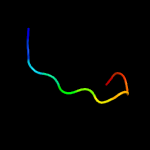
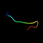
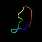
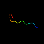
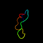
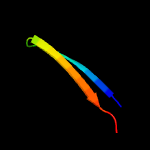
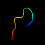
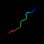
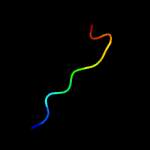
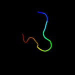
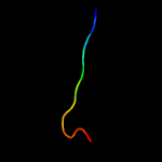
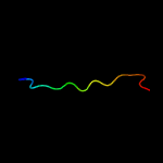
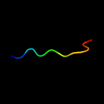
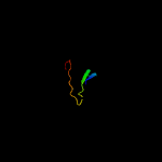
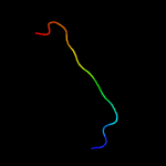
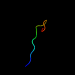
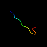
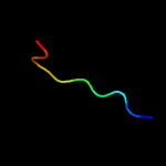
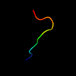
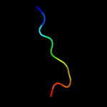
1 c5xknE_
60.7
55
PDB header: transferase/signaling proteinChain: E: PDB Molecule: epidermal patterning factor-like protein 4;PDBTitle: crystal structure of plant receptor erl2 in complexe with epfl4
2 c5xknF_
60.7
55
PDB header: transferase/signaling proteinChain: F: PDB Molecule: epidermal patterning factor-like protein 4;PDBTitle: crystal structure of plant receptor erl2 in complexe with epfl4
3 c2mw0A_
27.8
60
PDB header: protein bindingChain: A: PDB Molecule: kalata b7;PDBTitle: kalata b7 ser mutant
4 c2elnA_
27.4
50
PDB header: transcriptionChain: A: PDB Molecule: zinc finger protein 406;PDBTitle: solution structure of the 11th c2h2 zinc finger of human2 zinc finger protein 406
5 d1bx7a_
22.4
28
Fold: Knottins (small inhibitors, toxins, lectins)Superfamily: Leech antihemostatic proteinsFamily: Huristasin-like
6 c3zxqA_
19.5
20
PDB header: transferaseChain: A: PDB Molecule: hypoxia sensor histidine kinase response regulator dost;PDBTitle: crystal structure of the atp-binding domain of mycobacterium2 tuberculosis dost
7 d2j7ja2
19.0
60
Fold: beta-beta-alpha zinc fingersSuperfamily: beta-beta-alpha zinc fingersFamily: Classic zinc finger, C2H2
8 d1ddza2
16.9
40
Fold: Resolvase-likeSuperfamily: beta-carbonic anhydrase, cabFamily: beta-carbonic anhydrase, cab
9 d1ddza1
16.4
60
Fold: Resolvase-likeSuperfamily: beta-carbonic anhydrase, cabFamily: beta-carbonic anhydrase, cab
10 d1ejab_
16.2
45
Fold: Knottins (small inhibitors, toxins, lectins)Superfamily: Leech antihemostatic proteinsFamily: Huristasin-like
11 c5ztpB_
15.4
27
PDB header: lyaseChain: B: PDB Molecule: carbonic anhydrase;PDBTitle: carbonic anhydrase from glaciozyma antarctica
12 c3lasA_
15.4
20
PDB header: lyaseChain: A: PDB Molecule: putative carbonic anhydrase;PDBTitle: crystal structure of carbonic anhydrase from streptococcus mutans to2 1.4 angstrom resolution
13 c2a8cE_
15.1
40
PDB header: lyaseChain: E: PDB Molecule: carbonic anhydrase 2;PDBTitle: haemophilus influenzae beta-carbonic anhydrase
14 c3j0cH_
15.0
32
PDB header: virusChain: H: PDB Molecule: e2 envelope glycoprotein;PDBTitle: models of e1, e2 and cp of venezuelan equine encephalitis virus tc-832 strain restrained by a near atomic resolution cryo-em map
15 c1ylkA_
14.9
20
PDB header: unknown functionChain: A: PDB Molecule: hypothetical protein rv1284/mt1322;PDBTitle: crystal structure of rv1284 from mycobacterium tuberculosis in complex2 with thiocyanate
16 d1dx5i1
14.8
36
Fold: Knottins (small inhibitors, toxins, lectins)Superfamily: EGF/LamininFamily: EGF-type module
17 c4o1jB_
14.8
40
PDB header: lyaseChain: B: PDB Molecule: carbonic anhydrase;PDBTitle: crystal structures of two tetrameric beta-carbonic anhydrases from the2 filamentous ascomycete sordaria macrospora.
18 d1i6pa_
14.3
50
Fold: Resolvase-likeSuperfamily: beta-carbonic anhydrase, cabFamily: beta-carbonic anhydrase, cab
19 c1ddzA_
14.1
60
PDB header: lyaseChain: A: PDB Molecule: carbonic anhydrase;PDBTitle: x-ray structure of a beta-carbonic anhydrase from the red2 alga, porphyridium purpureum r-1
20 c2w3nA_
13.8
40
PDB header: lyaseChain: A: PDB Molecule: carbonic anhydrase 2;PDBTitle: structure and inhibition of the co2-sensing carbonic anhydrase can22 from the pathogenic fungus cryptococcus neoformans
21 c2a5vB_
not modelled
13.5
30
PDB header: lyaseChain: B: PDB Molecule: carbonic anhydrase (carbonate dehydratase) (carbonicPDBTitle: crystal structure of m. tuberculosis beta carbonic anhydrase, rv3588c,2 tetrameric form
22 c2fynO_
not modelled
13.3
35
PDB header: oxidoreductaseChain: O: PDB Molecule: ubiquinol-cytochrome c reductase iron-sulfurPDBTitle: crystal structure analysis of the double mutant rhodobacter2 sphaeroides bc1 complex
23 c5cxkG_
not modelled
13.3
50
PDB header: lyaseChain: G: PDB Molecule: carbonic anhydrase;PDBTitle: crystal structure of beta carbonic anhydrase from vibrio cholerae
24 c3eyxB_
not modelled
13.1
40
PDB header: lyaseChain: B: PDB Molecule: carbonic anhydrase;PDBTitle: crystal structure of carbonic anhydrase nce103 from2 saccharomyces cerevisiae
25 c4o1kA_
not modelled
12.6
50
PDB header: lyaseChain: A: PDB Molecule: carbonic anhydrase;PDBTitle: crystal structures of two tetrameric beta-carbonic anhydrases from the2 filamentous ascomycete sordaria macrospora.
26 d1g5ca_
not modelled
12.5
27
Fold: Resolvase-likeSuperfamily: beta-carbonic anhydrase, cabFamily: beta-carbonic anhydrase, cab
27 c3tenD_
not modelled
11.9
30
PDB header: hydrolaseChain: D: PDB Molecule: cs2 hydrolase;PDBTitle: holo form of carbon disulfide hydrolase
28 c4biyD_
not modelled
11.8
25
PDB header: transferaseChain: D: PDB Molecule: sensor protein cpxa;PDBTitle: crystal structure of cpxahdc (monoclinic form 2)
29 c3ucoB_
not modelled
11.8
50
PDB header: lyase/lyase inhibitorChain: B: PDB Molecule: carbonic anhydrase;PDBTitle: coccomyxa beta-carbonic anhydrase in complex with iodide
30 d1ekja_
not modelled
11.6
27
Fold: Resolvase-likeSuperfamily: beta-carbonic anhydrase, cabFamily: beta-carbonic anhydrase, cab
31 c5swcE_
not modelled
11.5
40
PDB header: lyaseChain: E: PDB Molecule: carbonic anhydrase;PDBTitle: the structure of the beta-carbonic anhydrase ccaa
32 c3vrkA_
not modelled
11.1
27
PDB header: hydrolaseChain: A: PDB Molecule: carbonyl sulfide hydrolase;PDBTitle: crystal structutre of thiobacillus thioparus thi115 carbonyl sulfide2 hydrolase / thiocyanate complex
33 c2xfbI_
not modelled
11.0
32
PDB header: virusChain: I: PDB Molecule: e2 envelope glycoprotein;PDBTitle: the chikungunya e1 e2 envelope glycoprotein complex fit into2 the sindbis virus cryo-em map
34 c3n43B_
not modelled
10.9
32
PDB header: viral proteinChain: B: PDB Molecule: e2 envelope glycoprotein;PDBTitle: crystal structures of the mature envelope glycoprotein complex2 (trypsin cleavage) of chikungunya virus.
35 c6gwuB_
not modelled
10.9
36
PDB header: lyaseChain: B: PDB Molecule: carbonic anhydrase;PDBTitle: carbonic anhydrase cance103p from candida albicans
36 c2e76D_
not modelled
10.9
35
PDB header: photosynthesisChain: D: PDB Molecule: cytochrome b6-f complex iron-sulfur subunit;PDBTitle: crystal structure of the cytochrome b6f complex with tridecyl-2 stigmatellin (tds) from m.laminosus
37 c6avjB_
not modelled
10.5
70
PDB header: metal binding proteinChain: B: PDB Molecule: cdgsh iron-sulfur domain-containing protein 3,PDBTitle: crystal structure of human mitochondrial inner neet protein (mint)2 /cisd3
38 d2i9wa3
not modelled
10.5
47
Fold: Sec-C motifSuperfamily: Sec-C motifFamily: Sec-C motif
39 c4rxyA_
not modelled
10.5
50
PDB header: lyaseChain: A: PDB Molecule: carbonic anhydrase;PDBTitle: crystal structure of the beta carbonic anhydrase psca3 isolated from2 pseudomonas aeruginosa
40 c3q7tB_
not modelled
10.1
42
PDB header: transcriptionChain: B: PDB Molecule: transcriptional regulatory protein;PDBTitle: 2.15a resolution structure (i41 form) of the chxr receiver domain from2 chlamydia trachomatis
41 c6atyA_
not modelled
9.6
80
PDB header: toxinChain: A: PDB Molecule: venom protein 51.1;PDBTitle: exploring cystine dense peptide space to open a unique molecular2 toolbox
42 c6hwhB_
not modelled
9.3
53
PDB header: electron transportChain: B: PDB Molecule: ubiquinol-cytochrome c reductase iron-sulfur subunit;PDBTitle: structure of a functional obligate respiratory supercomplex from2 mycobacterium smegmatis
43 c5xjoF_
not modelled
9.2
46
PDB header: transferase/membrane proteinChain: F: PDB Molecule: protein epidermal patterning factor 1;PDBTitle: plant receptor erl1-tmm in complex with peptide epf1
44 d2akla2
not modelled
9.0
55
Fold: Rubredoxin-likeSuperfamily: Zinc beta-ribbonFamily: PhnA zinc-binding domain
45 c2xpoB_
not modelled
8.9
57
PDB header: transcriptionChain: B: PDB Molecule: chromatin structure modulator;PDBTitle: crystal structure of a spt6-iws1(spn1) complex from2 encephalitozoon cuniculi, form ii
46 c2xpoD_
not modelled
8.8
57
PDB header: transcriptionChain: D: PDB Molecule: chromatin structure modulator;PDBTitle: crystal structure of a spt6-iws1(spn1) complex from2 encephalitozoon cuniculi, form ii
47 c5xkjF_
not modelled
8.6
46
PDB header: transferase/membrane protein/hormoneChain: F: PDB Molecule: protein epidermal patterning factor 2;PDBTitle: crystal structure of plant receptor erl1-tmm in complexe with epf2
48 d1omba_
not modelled
8.4
43
Fold: Knottins (small inhibitors, toxins, lectins)Superfamily: omega toxin-likeFamily: Spider toxins
49 d2bz1a1
not modelled
8.0
33
Fold: RibA-likeSuperfamily: RibA-likeFamily: RibA-like
50 d2g45a1
not modelled
7.9
44
Fold: RING/U-boxSuperfamily: RING/U-boxFamily: Zf-UBP
51 c1g9iI_
not modelled
7.8
44
PDB header: hydrolase/hydrolase inhibitorChain: I: PDB Molecule: bowman-birk type trypsin inhibitor;PDBTitle: crystal structure of beta-trysin complex in cyclohexane
52 c1p84E_
not modelled
7.6
35
PDB header: oxidoreductaseChain: E: PDB Molecule: ubiquinol-cytochrome c reductase iron-sulfur subunit;PDBTitle: hdbt inhibited yeast cytochrome bc1 complex
53 c2jr7A_
not modelled
7.5
50
PDB header: metal binding proteinChain: A: PDB Molecule: dph3 homolog;PDBTitle: solution structure of human desr1
54 d1agga_
not modelled
7.3
43
Fold: Knottins (small inhibitors, toxins, lectins)Superfamily: omega toxin-likeFamily: Spider toxins
55 d1ywsa1
not modelled
7.3
71
Fold: Rubredoxin-likeSuperfamily: CSL zinc fingerFamily: CSL zinc finger
56 c3n40P_
not modelled
7.1
32
PDB header: viral proteinChain: P: PDB Molecule: p62 envelope glycoprotein;PDBTitle: crystal structure of the immature envelope glycoprotein complex of2 chikungunya virus.
57 d3cx5e1
not modelled
7.1
27
Fold: ISP domainSuperfamily: ISP domainFamily: Rieske iron-sulfur protein (ISP)
58 c3zxoB_
not modelled
7.0
30
PDB header: transferaseChain: B: PDB Molecule: redox sensor histidine kinase response regulator devs;PDBTitle: crystal structure of the mutant atp-binding domain of2 mycobacterium tuberculosis doss
59 c2xppB_
not modelled
6.9
57
PDB header: transcriptionChain: B: PDB Molecule: chromatin structure modulator;PDBTitle: crystal structure of a spt6-iws1(spn1) complex from2 encephalitozoon cuniculi, form iii
60 c2xpnB_
not modelled
6.8
57
PDB header: transcriptionChain: B: PDB Molecule: chromatin structure modulator;PDBTitle: crystal structure of a spt6-iws1(spn1) complex from2 encephalitozoon cuniculi, form i
61 d2glia3
not modelled
6.8
57
Fold: beta-beta-alpha zinc fingersSuperfamily: beta-beta-alpha zinc fingersFamily: Classic zinc finger, C2H2
62 c4rl4B_
not modelled
6.6
33
PDB header: hydrolaseChain: B: PDB Molecule: gtp cyclohydrolase-2;PDBTitle: crystal structure of gtp cyclohydrolase ii from helicobacter pylori2 26695
63 d1wgea1
not modelled
6.5
71
Fold: Rubredoxin-likeSuperfamily: CSL zinc fingerFamily: CSL zinc finger
64 c2yrtA_
not modelled
6.3
46
PDB header: transcriptionChain: A: PDB Molecule: chord containing protein-1;PDBTitle: solution structure of the chord domain of human chord-2 containing protein 1
65 c1bi6H_
not modelled
6.2
44
PDB header: cysteine protease inhibitorChain: H: PDB Molecule: bromelain inhibitor vi;PDBTitle: nmr structure of bromelain inhibitor vi from pineapple stem
66 c2bi6H_
not modelled
6.0
44
PDB header: cysteine protease inhibitorChain: H: PDB Molecule: bromelain inhibitor vi;PDBTitle: nmr study of bromelain inhibitor vi from pineapple stem
67 d1x3ca1
not modelled
6.0
67
Fold: beta-beta-alpha zinc fingersSuperfamily: beta-beta-alpha zinc fingersFamily: Classic zinc finger, C2H2
68 d1a1ia1
not modelled
5.8
33
Fold: beta-beta-alpha zinc fingersSuperfamily: beta-beta-alpha zinc fingersFamily: Classic zinc finger, C2H2
69 d1jm1a_
not modelled
5.8
45
Fold: ISP domainSuperfamily: ISP domainFamily: Rieske iron-sulfur protein (ISP)
70 c1sx0A_
not modelled
5.8
47
PDB header: protein transportChain: A: PDB Molecule: seca;PDBTitle: solution nmr structure and x-ray absorption analysis of the2 c-terminal zinc-binding domain of the seca atpase
71 c1sx1A_
not modelled
5.7
47
PDB header: protein transportChain: A: PDB Molecule: seca;PDBTitle: solution nmr structure and x-ray absorption analysis of the2 c-terminal zinc-binding domain of the seca atpase
72 d1bdsa_
not modelled
5.6
67
Fold: Defensin-likeSuperfamily: Defensin-likeFamily: Defensin
73 c1bdsA_
not modelled
5.6
67
PDB header: anti-hypertensive, anti-viral proteinChain: A: PDB Molecule: bds-i;PDBTitle: determination of the three-dimensional solution structure of the2 antihypertensive and antiviral protein bds-i from the sea anemone3 anemonia sulcata. a study using nuclear magnetic resonance and hybrid4 distance geometry-dynamical simulated annealing
74 c4mi0A_
not modelled
5.6
25
PDB header: transferaseChain: A: PDB Molecule: histone-lysine n-methyltransferase ezh2;PDBTitle: human enhancer of zeste (drosophila) homolog 2(ezh2)
75 d1gtra2
not modelled
5.5
33
Fold: Adenine nucleotide alpha hydrolase-likeSuperfamily: Nucleotidylyl transferaseFamily: Class I aminoacyl-tRNA synthetases (RS), catalytic domain
76 d1nzja_
not modelled
5.5
42
Fold: Adenine nucleotide alpha hydrolase-likeSuperfamily: Nucleotidylyl transferaseFamily: Class I aminoacyl-tRNA synthetases (RS), catalytic domain
77 c1sbwI_
not modelled
5.3
30
PDB header: hydrolase/hydrolase inhibitorChain: I: PDB Molecule: protein (mung bean inhibitor lysin activePDBTitle: crystal structure of mung bean inhibitor lysine active2 fragment complex with bovine beta-trypsin at 1.8a3 resolution
78 d1tf3a2
not modelled
5.3
44
Fold: beta-beta-alpha zinc fingersSuperfamily: beta-beta-alpha zinc fingersFamily: Classic zinc finger, C2H2
79 c2odxA_
not modelled
5.3
44
PDB header: oxidoreductaseChain: A: PDB Molecule: cytochrome c oxidase polypeptide iv;PDBTitle: solution structure of zn(ii)cox4
80 c6mwcN_
not modelled
5.3
46
PDB header: virus/immune systemChain: N: PDB Molecule: e2;PDBTitle: cryoem structure of chimeric eastern equine encephalitis virus with2 fab of eeev-5 antibody
81 d1ozbi_
not modelled
5.3
38
Fold: Sec-C motifSuperfamily: Sec-C motifFamily: Sec-C motif
82 c1ozbI_
not modelled
5.3
38
PDB header: protein transportChain: I: PDB Molecule: preprotein translocase seca subunit;PDBTitle: crystal structure of secb complexed with seca c-terminus
83 c1ozbJ_
not modelled
5.2
38
PDB header: protein transportChain: J: PDB Molecule: preprotein translocase seca subunit;PDBTitle: crystal structure of secb complexed with seca c-terminus
84 d1iarb1
not modelled
5.1
23
Fold: Immunoglobulin-like beta-sandwichSuperfamily: Fibronectin type IIIFamily: Fibronectin type III
85 c4g6zA_
not modelled
5.1
25
PDB header: ligaseChain: A: PDB Molecule: glutamate-trna ligase;PDBTitle: crystal structure of a glutamyl-trna synthetase glurs from2 burkholderia thailandensis bound to l-glutamate
86 c4kp4B_
not modelled
5.1
30
PDB header: transferase/signaling proteinChain: B: PDB Molecule: osmolarity sensor protein envz, histidine kinase;PDBTitle: deciphering cis-trans directionality and visualizing2 autophosphorylation in histidine kinases.


