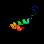
| 1 |
|
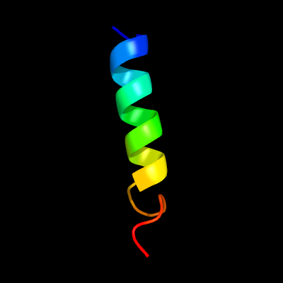
PDB 6gcs chain J
Region: 108 - 129
Aligned: 20
Modelled: 22
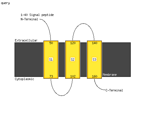
Confidence: 24.9%
Identity: 35%
PDB header:oxidoreductase
Chain: J: PDB Molecule:nujm subunit;
PDBTitle: cryo-em structure of respiratory complex i from yarrowia lipolytica
Phyre2
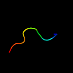
| 2 |
|
PDB 3wmm chain P
Region: 49 - 65
Aligned: 17
Modelled: 17
Confidence: 17.8%
Identity: 47%
PDB header:photosynthesis
Chain: P: PDB Molecule:lh1 beta polypeptide;
PDBTitle: crystal structure of the lh1-rc complex from thermochromatium tepidum2 in c2 form
Phyre2
| 3 |
|
PDB 4dzo chain A
Region: 103 - 115
Aligned: 13
Modelled: 13
Confidence: 16.0%
Identity: 54%
PDB header:cell cycle
Chain: A: PDB Molecule:mitotic spindle assembly checkpoint protein mad1;
PDBTitle: structure of human mad1 c-terminal domain reveals its involvement in2 kinetochore targeting
Phyre2
| 4 |
|
PDB 4rta chain B
Region: 95 - 144
Aligned: 34
Modelled: 34
Confidence: 14.1%
Identity: 47%
PDB header:protein binding
Chain: B: PDB Molecule:protein dpy-30 homolog;
PDBTitle: cystal structure of the dpy30 for mll/set1 compass h3k4 trimethylation
Phyre2
| 5 |
|
PDB 5m1h chain A
Region: 4 - 20
Aligned: 17
Modelled: 17
Confidence: 11.6%
Identity: 41%
PDB header:viral protein
Chain: A: PDB Molecule:gag protein;
PDBTitle: structure of a spumaretrovirus gag central domain reveals an ancient2 retroviral capsid
Phyre2
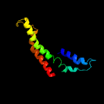
| 6 |
|
PDB 6hcy chain A
Region: 64 - 164
Aligned: 98
Modelled: 101
Confidence: 10.0%
Identity: 18%
PDB header:membrane protein
Chain: A: PDB Molecule:metalloreductase steap4;
PDBTitle: human steap4 bound to nadp, fad, heme and fe(iii)-nta.
Phyre2
| 7 |
|
PDB 2eqf chain A
Region: 8 - 16
Aligned: 9
Modelled: 9
Confidence: 9.8%
Identity: 78%
PDB header:hydrolase
Chain: A: PDB Molecule:tumor necrosis factor, alpha-induced protein 3;
PDBTitle: solution structure of the 7th a20-type zinc finger domain2 from human tumor necrosis factor, alpha-induced protein3
Phyre2
| 8 |
|
PDB 5d2m chain G
Region: 90 - 101
Aligned: 12
Modelled: 12
Confidence: 7.5%
Identity: 42%
PDB header:ligase,protein binding
Chain: G: PDB Molecule:zinc finger protein 451;
PDBTitle: complex between human sumo2-rangap1, ubc9 and znf451
Phyre2
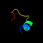
| 9 |
|
PDB 6j8l chain B
Region: 134 - 143
Aligned: 10
Modelled: 10
Confidence: 7.4%
Identity: 50%
PDB header:immune system
Chain: B: PDB Molecule:avh240;
PDBTitle: phytophthora sojae effector psavh240 inhibits a host aspartic protease2 secretion to promote infection
Phyre2
| 10 |
|
PDB 6bo5 chain D
Region: 1 - 22
Aligned: 22
Modelled: 22
Confidence: 6.2%
Identity: 23%
PDB header:membrane protein
Chain: D: PDB Molecule:transient receptor potential cation channel subfamily v
PDBTitle: trpv2 ion channel in partially closed state
Phyre2