1 c4kk7A_
100.0
37
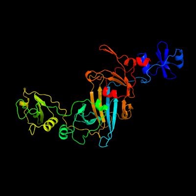
PDB header: protein transportChain: A: PDB Molecule: esx-1 secretion system protein eccb1;PDBTitle: structure of eccb1 from the type vii (esx-1) secretion system of2 mycobacterium tuberculosis.
2 c5cyuA_
100.0
37
PDB header: membrane proteinChain: A: PDB Molecule: conserved membrane protein;PDBTitle: structure of the soluble domain of eccb1 from the mycobacterium2 smegmatis esx-1 secretion system.
3 d2fb5a1
49.7
10
Fold: YojJ-likeSuperfamily: YojJ-likeFamily: YojJ-like
4 c6navI_
42.4
20
PDB header: structural proteinChain: I: PDB Molecule: m9ud72;PDBTitle: cryo-em reconstruction of sulfolobus islandicus lal14/1 pilus
5 c2k9yA_
32.7
20
PDB header: transferaseChain: A: PDB Molecule: ephrin type-a receptor 2;PDBTitle: epha2 dimeric structure in the lipidic bicelle at ph 5.0
6 c2k9yB_
32.7
20
PDB header: transferaseChain: B: PDB Molecule: ephrin type-a receptor 2;PDBTitle: epha2 dimeric structure in the lipidic bicelle at ph 5.0
7 d1vh6a_
18.7
24
Fold: Four-helical up-and-down bundleSuperfamily: Flagellar export chaperone FliSFamily: Flagellar export chaperone FliS
8 c1vh6A_
18.7
24
PDB header: structural genomics, unknown functionChain: A: PDB Molecule: flagellar protein flis;PDBTitle: crystal structure of a flagellar protein
9 c5xefA_
18.6
5
PDB header: chaperoneChain: A: PDB Molecule: flagellar protein flis;PDBTitle: crystal structure of flagellar chaperone from bacteria
10 c5gzbA_
18.4
40
PDB header: transcription/dnaChain: A: PDB Molecule: transcriptional enhancer factor tef-3;PDBTitle: crystal structure of transcription factor tead4 in complex with m-cat2 dna
11 c2hzdA_
17.4
40
PDB header: gene regulationChain: A: PDB Molecule: transcriptional enhancer factor tef-1;PDBTitle: nmr structure of the dna-binding tea domain and insights2 into tef-1 function
12 c3k1iA_
17.2
5
PDB header: chaperoneChain: A: PDB Molecule: flagellar protein;PDBTitle: crystal strcture of flis-hp1076 complex in h. pylori
13 c6f0kA_
16.3
14
PDB header: membrane proteinChain: A: PDB Molecule: cytochrome c family protein;PDBTitle: alternative complex iii
14 c2ciuA_
14.8
30
PDB header: protein transportChain: A: PDB Molecule: import inner membrane translocase subunit tim21PDBTitle: structure of the ims domain of the mitochondrial import2 protein tim21 from s. cerevisiae
15 c3c1zA_
14.4
8
PDB header: dna binding proteinChain: A: PDB Molecule: dna integrity scanning protein disa;PDBTitle: structure of the ligand-free form of a bacterial dna damage sensor2 protein
16 c6gyyB_
14.3
17
PDB header: transferaseChain: B: PDB Molecule: diadenylate cyclase;PDBTitle: crystal structure of daca from staphylococcus aureus, n166c/t172c2 double mutant
17 c2lxfA_
14.1
16
PDB header: structural genomics, unknown functionChain: A: PDB Molecule: uncharacterized protein;PDBTitle: solution nmr structure of a potential acylphosphatase from giardia2 lamblia, seattle structural genomics center for infectious disease3 target gilaa.01396.a
18 c6cfwE_
13.8
18
PDB header: membrane proteinChain: E: PDB Molecule: mbh subunit;PDBTitle: cryoem structure of a respiratory membrane-bound hydrogenase
19 c3hshA_
13.0
18
PDB header: protein bindingChain: A: PDB Molecule: collagen alpha-1(xviii) chain;PDBTitle: crystal structure of human collagen xviii trimerization domain2 (tetragonal crystal form)
20 d2zjru1
13.0
20
Fold: L28p-likeSuperfamily: L28p-likeFamily: Ribosomal protein L28
21 c2a7oA_
not modelled
11.2
15
PDB header: transcriptionChain: A: PDB Molecule: huntingtin interacting protein b;PDBTitle: solution structure of the hset2/hypb sri domain
22 c3n3fB_
not modelled
10.3
9
PDB header: protein bindingChain: B: PDB Molecule: collagen alpha-1(xv) chain;PDBTitle: crystal structure of the human collagen xv trimerization domain: a2 potent trimerizing unit common to multiplexin collagens
23 c6ch3B_
not modelled
10.2
14
PDB header: structural proteinChain: B: PDB Molecule: flagellar secretion chaperone flis,flagellin;PDBTitle: crystal structure of the cytoplasmic domain of flha and flis-flic2 complex
24 d1gado2
not modelled
10.0
21
Fold: FwdE/GAPDH domain-likeSuperfamily: Glyceraldehyde-3-phosphate dehydrogenase-like, C-terminal domainFamily: GAPDH-like
25 c1zzaA_
not modelled
9.9
14
PDB header: membrane proteinChain: A: PDB Molecule: stannin;PDBTitle: solution nmr structure of the membrane protein stannin
26 d1iioa_
not modelled
9.5
24
Fold: EF Hand-likeSuperfamily: Hypothetical protein MTH865Family: Hypothetical protein MTH865
27 c2l02B_
not modelled
8.9
17
PDB header: structural genomics, unknown functionChain: B: PDB Molecule: uncharacterized protein;PDBTitle: solution nmr structure of protein bt2368 from bacteroides2 thetaiotaomicron, northeast structural genomics consortium target3 btr375
28 d2i5nh1
not modelled
8.8
26
Fold: PRC-barrel domainSuperfamily: PRC-barrel domainFamily: Photosynthetic reaction centre, H-chain, cytoplasmic domain
29 d1y7ma1
not modelled
8.7
7
Fold: L,D-transpeptidase catalytic domain-likeSuperfamily: L,D-transpeptidase catalytic domain-likeFamily: L,D-transpeptidase catalytic domain-like
30 c1k6nH_
not modelled
8.7
20
PDB header: photosynthesisChain: H: PDB Molecule: photosynthetic reaction center h subunit;PDBTitle: e(l212)a,d(l213)a double mutant structure of photosynthetic reaction2 center from rhodobacter sphaeroides
31 c4rv7C_
not modelled
8.4
10
PDB header: transferaseChain: C: PDB Molecule: diadenylate cyclase;PDBTitle: characterization of an essential diadenylate cyclase
32 c6hraD_
not modelled
8.0
42
PDB header: membrane proteinChain: D: PDB Molecule: potassium-transporting atpase kdpf subunit;PDBTitle: cryo-em structure of the kdpfabc complex in an e1 outward-facing state2 (state 1)
33 c5mrwD_
not modelled
8.0
42
PDB header: hydrolaseChain: D: PDB Molecule: potassium-transporting atpase kdpf subunit;PDBTitle: structure of the kdpfabc complex
34 c5mrwH_
not modelled
8.0
42
PDB header: hydrolaseChain: H: PDB Molecule: potassium-transporting atpase kdpf subunit;PDBTitle: structure of the kdpfabc complex
35 c6hrbD_
not modelled
8.0
42
PDB header: membrane proteinChain: D: PDB Molecule: potassium-transporting atpase kdpf subunit;PDBTitle: cryo-em structure of the kdpfabc complex in an e2 inward-facing state2 (state 2)
36 c5mrwL_
not modelled
8.0
42
PDB header: hydrolaseChain: L: PDB Molecule: potassium-transporting atpase kdpf subunit;PDBTitle: structure of the kdpfabc complex
37 c2kncA_
not modelled
7.5
17
PDB header: cell adhesionChain: A: PDB Molecule: integrin alpha-iib;PDBTitle: platelet integrin alfaiib-beta3 transmembrane-cytoplasmic2 heterocomplex
38 c2lf3A_
not modelled
7.4
56
PDB header: signaling proteinChain: A: PDB Molecule: effector protein hopab3;PDBTitle: solution nmr structure of hoppmal_281_385 from pseudomonas syringae2 pv. maculicola str. es4326, midwest center for structural genomics3 target apc40104.5 and northeast structural genomics consortium target4 pst2a
39 c2i5nH_
not modelled
7.2
26
PDB header: photosynthesisChain: H: PDB Molecule: reaction center protein h chain;PDBTitle: 1.96 a x-ray structure of photosynthetic reaction center from2 rhodopseudomonas viridis:crystals grown by microfluidic technique
40 c2dzqA_
not modelled
7.1
27
PDB header: transcriptionChain: A: PDB Molecule: general transcription factor ii-i repeat domain-PDBTitle: solution structure of rsgi ruh-066, a gtf2i domain in human2 cdna
41 c2dn5A_
not modelled
7.0
41
PDB header: transcriptionChain: A: PDB Molecule: general transcription factor ii-i repeat domain-PDBTitle: solution structure of rsgi ruh-057, a gtf2i domain in human2 cdna
42 d1ixsa_
not modelled
6.7
29
Fold: RuvA C-terminal domain-likeSuperfamily: DNA helicase RuvA subunit, C-terminal domainFamily: DNA helicase RuvA subunit, C-terminal domain
43 c2d99A_
not modelled
6.6
36
PDB header: transcriptionChain: A: PDB Molecule: general transcription factor ii-i repeat domain-PDBTitle: solution structure of rsgi ruh-048, a gtf2i domain in human2 cdna
44 d1q60a_
not modelled
6.4
36
Fold: GTF2I-like repeatSuperfamily: GTF2I-like repeatFamily: GTF2I-like repeat
45 c2ed2A_
not modelled
6.3
23
PDB header: transcriptionChain: A: PDB Molecule: general transcription factor ii-i;PDBTitle: solution structure of rsgi ruh-069, a gtf2i domain in human2 cdna
46 c2e3lA_
not modelled
6.3
27
PDB header: transcriptionChain: A: PDB Molecule: transcription factor gtf2ird2 beta;PDBTitle: solution structure of rsgi ruh-068, a gtf2i domain in human2 cdna
47 c5zjiO_
not modelled
6.3
22
PDB header: membrane proteinChain: O: PDB Molecule: 16kda membrane protein;PDBTitle: structure of photosystem i supercomplex with light-harvesting2 complexes i and ii
48 c2ejeA_
not modelled
5.9
27
PDB header: transcriptionChain: A: PDB Molecule: general transcription factor ii-i;PDBTitle: solution structure of rsgi ruh-071, a gtf2i domain in human2 cdna
49 c2l8sA_
not modelled
5.5
9
PDB header: cell adhesionChain: A: PDB Molecule: integrin alpha-1;PDBTitle: solution nmr structure of transmembrane and cytosolic regions of2 integrin alpha1 in detergent micelles
50 c2dn4A_
not modelled
5.4
27
PDB header: transcriptionChain: A: PDB Molecule: general transcription factor ii-i;PDBTitle: solution structure of rsgi ruh-060, a gtf2i domain in human2 cdna
51 c4jonA_
not modelled
5.4
40
PDB header: structural genomics, unknown functionChain: A: PDB Molecule: centrosomal protein of 170 kda;PDBTitle: crystal structure of a centrosomal protein 170kda, transcript variant2 beta (cep170) from homo sapiens at 2.15 a resolution (psi community3 target, sundstrom)
52 c2dzrA_
not modelled
5.1
32
PDB header: transcriptionChain: A: PDB Molecule: general transcription factor ii-i repeat domain-PDBTitle: solution structure of rsgi ruh-067, a gtf2i domain in human2 cdna
53 c1ohfB_
not modelled
5.0
30
PDB header: virusChain: B: PDB Molecule: nudaurelia capensis omega virus capsid protein;PDBTitle: the refined structure of nudaurelia capensis omega virus
54 c2k1kB_
not modelled
5.0
42
PDB header: signaling proteinChain: B: PDB Molecule: ephrin type-a receptor 1;PDBTitle: nmr structures of dimeric transmembrane domain of the2 receptor tyrosine kinase epha1 in lipid bicelles at ph 4.3
55 c2k1kA_
not modelled
5.0
42
PDB header: signaling proteinChain: A: PDB Molecule: ephrin type-a receptor 1;PDBTitle: nmr structures of dimeric transmembrane domain of the2 receptor tyrosine kinase epha1 in lipid bicelles at ph 4.3
56 c2k1lB_
not modelled
5.0
42
PDB header: signaling proteinChain: B: PDB Molecule: ephrin type-a receptor 1;PDBTitle: nmr structures of dimeric transmembrane domain of the2 receptor tyrosine kinase epha1 in lipid bicelles at ph 6.3