Phyre Home Retrieve Phyre Job Id
Emailmdejesus@rockefeller.edu
DescriptionRVBD0287_(esxG)_351525_351818
DateTue Jul 23 14:50:35 BST 2019
Unique Job ID6318e440e8d82b4b
SequenceMSLLDAHIPQ ... Download FASTA
Job Typenormal
Job Expiry

Summary 

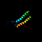
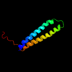
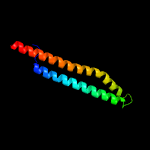
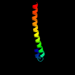
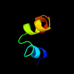
Top model
Image coloured by rainbow N → C terminus
Model dimensions (Å):X:33.928 Y:76.493 Z:45.469
Model (left) based on template c2kg7A_
Top template information
PDB header:unknown function
Chain: A: PDB Molecule:uncharacterized protein esxg (pe family protein);
PDBTitle: structure and features of the complex formed by the tuberculosis2 virulence factors rv0287 and rv0288
Confidence and coverage
Confidence:100.0% Coverage:100%
97 residues (100% of your sequence) have been modelled with 100.0% confidence by the single highest scoring template.
3D viewing
Interactive 3D view in JSmol
For other options to view your downloaded structure offline see the FAQ

Sequence analysis 

Download FASTA version

Secondary structure and disorder prediction [Show] 

Domain analysis [Show] 

Detailed template information [Hide] 


#
Template Alignment Coverage3D Model Confidence
% i.d. Template Information
1c2kg7A_


100.0 100
PDB header:unknown function
Chain: A: PDB Molecule:uncharacterized protein esxg (pe family protein);
PDBTitle: structure and features of the complex formed by the tuberculosis2 virulence factors rv0287 and rv0288
2c3h6pB_


100.0 91
PDB header:structural genomics, unknown function
Chain: B: PDB Molecule:esat-6 like protein esxs;
PDBTitle: crystal structure of rv3019c-rv3020c from mycobacterium tuberculosis
3c2kg7B_


95.0 19
PDB header:unknown function
Chain: B: PDB Molecule:esat-6-like protein esxh;
PDBTitle: structure and features of the complex formed by the tuberculosis2 virulence factors rv0287 and rv0288
4d1wa8b1


94.7 17
Fold:Ferritin-like
Superfamily:EsxAB dimer-like
Family:ESAT-6 like
5d1wa8a1


94.3 31
Fold:Ferritin-like
Superfamily:EsxAB dimer-like
Family:ESAT-6 like
6c3zbhC_


92.5 21
PDB header:unknown function
Chain: C: PDB Molecule:esxa;
PDBTitle: geobacillus thermodenitrificans esxa crystal form i
7c4lwsA_


92.1 24
PDB header:unknown function
Chain: A: PDB Molecule:uncharacterized protein;
PDBTitle: esxa : esxb (semet) hetero-dimer from thermomonospora curvata
8c4lwsB_


89.1 17
PDB header:unknown function
Chain: B: PDB Molecule:uncharacterized protein;
PDBTitle: esxa : esxb (semet) hetero-dimer from thermomonospora curvata
9c3gvmA_


89.0 18
PDB header:viral protein
Chain: A: PDB Molecule:putative uncharacterized protein sag1039;
PDBTitle: structure of the homodimeric wxg-100 family protein from streptococcus2 agalactiae
10c2vs0B_


73.0 15
PDB header:cell invasion
Chain: B: PDB Molecule:virulence factor esxa;
PDBTitle: structural analysis of homodimeric staphylococcal aureus2 virulence factor esxa
11c4iogD_


65.6 17
PDB header:unknown function
Chain: D: PDB Molecule:secreted protein esxb;
PDBTitle: the crystal structure of a secreted protein esxb (wild-type, in p212 space group) from bacillus anthracis str. sterne
12d2i9fa1


13.0 38
Fold:Nucleocapsid protein dimerization domain
Superfamily:Nucleocapsid protein dimerization domain
Family:Arterivirus nucleocapsid protein
13c3u0cA_


12.5 20
PDB header:cell invasion
Chain: A: PDB Molecule:invasin ipab;
PDBTitle: crystal structure of n-terminal region of type iii secretion first2 translocator ipab (residues 74-224)
14c2i9fB_


12.2 38
PDB header:viral protein
Chain: B: PDB Molecule:nucleocapsid;
PDBTitle: structure of the equine arterivirus nucleocapsid protein
15c5j9iH_


11.2 29
PDB header:antitoxin
Chain: H: PDB Molecule:antitoxin iga-2;
PDBTitle: crystal structure of the higa2 antitoxin c-terminal domain
16d1e6va1


11.1 41
Fold:Methyl-coenzyme M reductase alpha and beta chain C-terminal domain
Superfamily:Methyl-coenzyme M reductase alpha and beta chain C-terminal domain
Family:Methyl-coenzyme M reductase alpha and beta chain C-terminal domain
17c3bd1B_


10.6 37
PDB header:transcription
Chain: B: PDB Molecule:cro protein;
PDBTitle: structure of the cro protein from putative prophage element xfaso 1 in2 xylella fastidiosa strain ann-1
18d1x57a1


10.0 24
Fold:lambda repressor-like DNA-binding domains
Superfamily:lambda repressor-like DNA-binding domains
Family:EDF1-like
19d1hbna1


9.6 41
Fold:Methyl-coenzyme M reductase alpha and beta chain C-terminal domain
Superfamily:Methyl-coenzyme M reductase alpha and beta chain C-terminal domain
Family:Methyl-coenzyme M reductase alpha and beta chain C-terminal domain
20c2rilA_


9.4 28
PDB header:oxidoreductase
Chain: A: PDB Molecule:antibiotic biosynthesis monooxygenase;
PDBTitle: crystal structure of a putative monooxygenase (yp_001095275.1) from2 shewanella loihica pv-4 at 1.26 a resolution
21d2ppxa1


not modelled 9.0 29
Fold:lambda repressor-like DNA-binding domains
Superfamily:lambda repressor-like DNA-binding domains
Family:SinR domain-like
22c2ppxA_


not modelled 9.0 29
PDB header:structural genomics, unknown function
Chain: A: PDB Molecule:uncharacterized protein atu1735;
PDBTitle: crystal structure of a hth xre-family like protein from agrobacterium2 tumefaciens
23d1xbwa_


not modelled 8.6 45
Fold:Ferredoxin-like
Superfamily:Dimeric alpha+beta barrel
Family:PG130-like
24c2oszA_


not modelled 8.4 20
PDB header:structural protein
Chain: A: PDB Molecule:nucleoporin p58/p45;
PDBTitle: structure of nup58/45 suggests flexible nuclear pore diameter by2 intermolecular sliding
25c2jvlA_


not modelled 8.3 19
PDB header:transcription
Chain: A: PDB Molecule:trmbf1;
PDBTitle: nmr structure of the c-terminal domain of mbf1 of trichoderma reesei
26c5v6hC_


not modelled 7.7 42
PDB header:protein binding
Chain: C: PDB Molecule:pdz domain-containing protein gipc2;
PDBTitle: crystal structure of myosin vi in complex with gh2 domain of gipc2
27c1wd6B_


not modelled 7.5 39
PDB header:structural genomics, unknown function
Chain: B: PDB Molecule:protein ydhr;
PDBTitle: crystal structure of jw1657 from escherichia coli
28c5d4zF_


not modelled 7.4 24
PDB header:dna binding protein
Chain: F: PDB Molecule:repressor;
PDBTitle: crystal structure of repressor from salmonella-temperate phage
29c4xy3A_


not modelled 7.3 15
PDB header:protein transport
Chain: A: PDB Molecule:esx-1 secretion-associated protein espb;
PDBTitle: structure of esx-1 secreted protein espb
30c4ghjA_


not modelled 7.2 43
PDB header:transcription
Chain: A: PDB Molecule:probable transcriptional regulator;
PDBTitle: 1.75 angstrom crystal structure of transcriptional regulator ftom2 vibrio vulnificus.
31c4ybaA_


not modelled 7.1 24
PDB header:gene regulation
Chain: A: PDB Molecule:regulatory protein c;
PDBTitle: the structure of the c.kpn2i controller protein
32d1xrxa1


not modelled 7.0 67
Fold:Ribbon-helix-helix
Superfamily:Ribbon-helix-helix
Family:SeqA N-terminal domain-like
33c1xrxD_


not modelled 7.0 67
PDB header:replication inhibitor
Chain: D: PDB Molecule:seqa protein;
PDBTitle: crystal structure of a dna-binding protein
34c6csuA_


not modelled 6.9 75
PDB header:cell cycle
Chain: A: PDB Molecule:centrosomal protein of 63 kda;
PDBTitle: the structure of the cep63-cep152 heterotetrameric complex
35c3fmyA_


not modelled 6.8 14
PDB header:dna binding protein
Chain: A: PDB Molecule:hth-type transcriptional regulator mqsa
PDBTitle: structure of the c-terminal domain of the e. coli protein2 mqsa (ygit/b3021)
36d1sq8a_


not modelled 6.5 33
Fold:lambda repressor-like DNA-binding domains
Superfamily:lambda repressor-like DNA-binding domains
Family:Phage repressors
37c4pu4C_


not modelled 6.2 25
PDB header:toxin/antitoxin/dna
Chain: C: PDB Molecule:toxin-antitoxin system antidote transcriptional repressor
PDBTitle: shewanella oneidensis mr-1 toxin antitoxin system hipa, hipb and its2 operator dna complex (space group p21)
38c5jaaB_


not modelled 6.0 29
PDB header:toxin
Chain: B: PDB Molecule:antitoxin iga-2;
PDBTitle: crystal structure of the higba2 toxin-antitoxin complex
39c5bu3A_


not modelled 5.7 35
PDB header:lyase
Chain: A: PDB Molecule:pyri4;
PDBTitle: crystal structure of diels-alderase pyri4 in complex with its product
40c2eouA_


not modelled 5.6 75
PDB header:transcription
Chain: A: PDB Molecule:zinc finger protein 473;
PDBTitle: solution structure of the c2h2 type zinc finger (region 370-2 400) of human zinc finger protein 473
41c2ebyA_


not modelled 5.4 10
PDB header:transcription
Chain: A: PDB Molecule:putative hth-type transcriptional regulator ybaq;
PDBTitle: crystal structure of a hypothetical protein from e. coli
42c5n1qD_


not modelled 5.3 44
PDB header:transferase
Chain: D: PDB Molecule:methyl-coenzyme m reductase iii from methanothermococcus
PDBTitle: methyl-coenzyme m reductase iii from methanothermococcus2 thermolithotrophicus at 1.9 a resolution
43c4e1pA_


not modelled 5.3 20
PDB header:dna binding protein
Chain: A: PDB Molecule:protein lsr2;
PDBTitle: crystal structure of the dimerization domain of lsr2 from2 mycobacterium tuberculosis in the p 1 21 1 space group
44c4e1rA_


not modelled 5.3 20
PDB header:dna binding protein
Chain: A: PDB Molecule:protein lsr2;
PDBTitle: crystal structure of the dimerization domain of lsr2 from2 mycobacterium tuberculosis in the p 31 2 1 space group
45c5z1yA_


not modelled 5.2 83
PDB header:antibiotic
Chain: A: PDB Molecule:mbjamp1 peptide;
PDBTitle: mbjamp1 structure

Binding site prediction 

Due to computational demand, binding site predictions are not run for batch jobs

If you want to predict binding sites, please manually submit your model of choice to 3DLigandSite