| 1 |
|
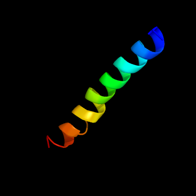
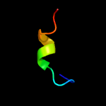
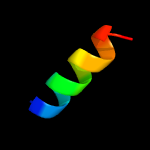
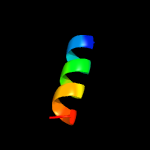
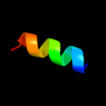
PDB 1iij chain A
Region: 91 - 120
Aligned: 30
Modelled: 30
Confidence: 11.5%
Identity: 13%
PDB header:signaling protein
Chain: A: PDB Molecule:erbb-2 receptor protein-tyrosine kinase;
PDBTitle: solution structure of the neu/erbb-2 membrane spanning2 segment
Phyre2
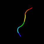
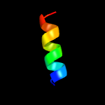
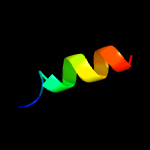
| 2 |
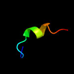
|
PDB 2low chain A
Region: 91 - 108
Aligned: 18
Modelled: 18
Confidence: 10.3%
Identity: 28%
PDB header:membrane protein
Chain: A: PDB Molecule:apelin receptor;
PDBTitle: solution structure of ar55 in 50% hfip
Phyre2
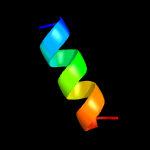
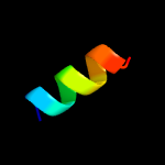
| 3 |
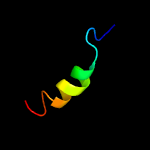
|
PDB 1n93 chain X
Region: 21 - 34
Aligned: 14
Modelled: 14
Confidence: 9.4%
Identity: 36%
Fold: P40 nucleoprotein
Superfamily: P40 nucleoprotein
Family: P40 nucleoprotein
Phyre2
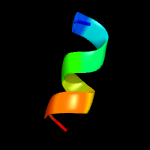
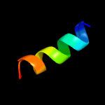
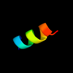
| 4 |
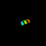
|
PDB 1n93 chain X
Region: 21 - 34
Aligned: 14
Modelled: 14
Confidence: 9.4%
Identity: 36%
PDB header:viral protein
Chain: X: PDB Molecule:p40 nucleoprotein;
PDBTitle: crystal structure of the borna disease virus nucleoprotein
Phyre2
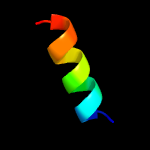
| 5 |
|
PDB 2lx0 chain A
Region: 4 - 24
Aligned: 21
Modelled: 21
Confidence: 7.9%
Identity: 33%
PDB header:membrane protein
Chain: A: PDB Molecule:membrane fusion protein p14;
PDBTitle: arced helix (arch) nmr structure of the reovirus p14 fusion-associated2 small transmembrane (fast) protein transmembrane domain (tmd) in3 dodecyl phosphocholine (dpc) micelles
Phyre2
| 6 |
|
PDB 5tfy chain J
Region: 94 - 106
Aligned: 13
Modelled: 13
Confidence: 7.4%
Identity: 54%
PDB header:cell adhesion
Chain: J: PDB Molecule:flagellin;
PDBTitle: the archaeal flagellum of methanospirillum hungatei strain jf1.
Phyre2
| 7 |
|
PDB 1g5t chain A
Region: 75 - 88
Aligned: 14
Modelled: 14
Confidence: 6.8%
Identity: 29%
Fold: P-loop containing nucleoside triphosphate hydrolases
Superfamily: P-loop containing nucleoside triphosphate hydrolases
Family: RecA protein-like (ATPase-domain)
Phyre2
| 8 |
|
PDB 5tp6 chain B
Region: 1 - 6
Aligned: 6
Modelled: 6
Confidence: 6.7%
Identity: 67%
PDB header:oxidoreductase
Chain: B: PDB Molecule:nitric oxide synthase, inducible;
PDBTitle: solution structure of the cam34 with the inos cam binding domain2 peptide
Phyre2
| 9 |
|
PDB 1ldd chain A
Region: 38 - 47
Aligned: 10
Modelled: 10
Confidence: 6.5%
Identity: 30%
Fold: DNA/RNA-binding 3-helical bundle
Superfamily: "Winged helix" DNA-binding domain
Family: SCF ubiquitin ligase complex WHB domain
Phyre2
| 10 |
|
PDB 1yt3 chain A domain 2
Region: 105 - 111
Aligned: 7
Modelled: 7
Confidence: 6.2%
Identity: 57%
Fold: SAM domain-like
Superfamily: HRDC-like
Family: RNase D C-terminal domains
Phyre2
| 11 |
|
PDB 1g64 chain B
Region: 75 - 88
Aligned: 14
Modelled: 14
Confidence: 6.2%
Identity: 29%
Fold: P-loop containing nucleoside triphosphate hydrolases
Superfamily: P-loop containing nucleoside triphosphate hydrolases
Family: RecA protein-like (ATPase-domain)
Phyre2
| 12 |
|
PDB 6b2z chain S
Region: 42 - 55
Aligned: 14
Modelled: 14
Confidence: 5.9%
Identity: 21%
PDB header:membrane protein
Chain: S: PDB Molecule:atp synthase subunit j, mitochondrial;
PDBTitle: cryo-em structure of the dimeric fo region of yeast mitochondrial atp2 synthase
Phyre2
| 13 |
|
PDB 6b2z chain I
Region: 42 - 55
Aligned: 14
Modelled: 14
Confidence: 5.9%
Identity: 21%
PDB header:membrane protein
Chain: I: PDB Molecule:atp synthase subunit c, mitochondrial;
PDBTitle: cryo-em structure of the dimeric fo region of yeast mitochondrial atp2 synthase
Phyre2
| 14 |
|
PDB 5o4u chain K
Region: 94 - 106
Aligned: 13
Modelled: 13
Confidence: 5.6%
Identity: 46%
PDB header:cell adhesion
Chain: K: PDB Molecule:flagellin;
PDBTitle: the flagellin of pyrococcus furiosus
Phyre2
| 15 |
|
PDB 3n1h chain A
Region: 34 - 48
Aligned: 15
Modelled: 15
Confidence: 5.6%
Identity: 20%
PDB header:dna binding protein
Chain: A: PDB Molecule:stwhy2;
PDBTitle: crystal structure of stwhy2
Phyre2
| 16 |
|
PDB 5z1l chain L
Region: 94 - 106
Aligned: 13
Modelled: 13
Confidence: 5.4%
Identity: 38%
PDB header:protein fibril
Chain: L: PDB Molecule:flagellin;
PDBTitle: cryo-em structure of methanoccus maripaludis archaellum
Phyre2
| 17 |
|
PDB 5j6v chain A
Region: 36 - 49
Aligned: 14
Modelled: 14
Confidence: 5.2%
Identity: 21%
PDB header:antimicrobial protein
Chain: A: PDB Molecule:hylin-d;
PDBTitle: nmr structures of hylin-a1 analogs: hylin-d
Phyre2
| 18 |
|
PDB 4ogq chain F
Region: 45 - 56
Aligned: 12
Modelled: 12
Confidence: 5.2%
Identity: 25%
PDB header:electron transport
Chain: F: PDB Molecule:cytochrome b6-f complex subunit 7;
PDBTitle: internal lipid architecture of the hetero-oligomeric cytochrome b6f2 complex
Phyre2
| 19 |
|
PDB 4h44 chain F
Region: 45 - 56
Aligned: 12
Modelled: 12
Confidence: 5.2%
Identity: 25%
PDB header:photosynthesis
Chain: F: PDB Molecule:cytochrome b6-f complex subunit 7;
PDBTitle: 2.70 a cytochrome b6f complex structure from nostoc pcc 7120
Phyre2