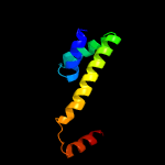
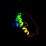
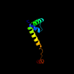
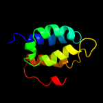
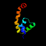
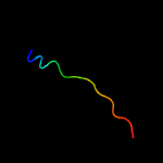
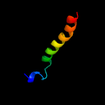
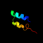
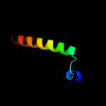
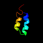
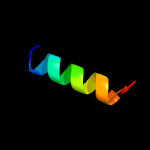
1 c3vdoB_
99.6
100
PDB header: dna binding protein/protein bindingChain: B: PDB Molecule: anti-sigma-k factor rska;PDBTitle: structure of extra-cytoplasmic function(ecf) sigma factor sigk in2 complex with its negative regulator rska from mycobacterium3 tuberculosis
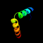
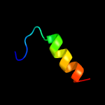
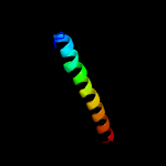
2 c2z2sD_
99.3
12
PDB header: transcriptionChain: D: PDB Molecule: anti-sigma factor chrr, transcriptional activator chrr;PDBTitle: crystal structure of rhodobacter sphaeroides sige in complex with the2 anti-sigma chrr
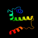
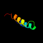
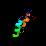
3 c5wuqD_
99.1
7
PDB header: metal binding proteinChain: D: PDB Molecule: anti-sigma-w factor rsiw;PDBTitle: crystal structure of sigw in complex with its anti-sigma rsiw, a zinc2 binding form
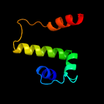
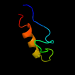
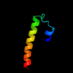
4 c5frhA_
98.9
7
PDB header: transcriptionChain: A: PDB Molecule: anti-sigma factor rsra;PDBTitle: solution structure of oxidised rsra
5 c3hugJ_
98.8
16
PDB header: transcription/membrane proteinChain: J: PDB Molecule: probable conserved membrane protein;PDBTitle: crystal structure of mycobacterium tuberculosis anti-sigma factor rsla2 in complex with -35 promoter binding domain of sigl
6 c5camC_
67.7
29
PDB header: transcriptionChain: C: PDB Molecule: pupr protein;PDBTitle: crystal structure of the cytoplasmic domain of the pseudomonas putida2 anti-sigma factor pupr (semet)
7 d1or7c_
34.4
13
Fold: N-terminal, cytoplasmic domain of anti-sigmaE factor RseASuperfamily: N-terminal, cytoplasmic domain of anti-sigmaE factor RseAFamily: N-terminal, cytoplasmic domain of anti-sigmaE factor RseA
8 c1or7C_
34.4
13
PDB header: transcriptionChain: C: PDB Molecule: sigma-e factor negative regulatory protein;PDBTitle: crystal structure of escherichia coli sigmae with the cytoplasmic2 domain of its anti-sigma rsea
9 c5ee2A_
30.5
38
PDB header: metal transportChain: A: PDB Molecule: hemoglobin-haptoglobin-utilization protein;PDBTitle: the crystal structure of the c-terminal beta-barrel of hpua from2 neisseria gonorrhoeae
10 c2mjlA_
22.5
15
PDB header: hydrolaseChain: A: PDB Molecule: peptidyl-trna hydrolase;PDBTitle: solution structure of peptidyl-trna hyrolase from vibrio cholerae
11 c5j84A_
14.6
9
PDB header: lyaseChain: A: PDB Molecule: dihydroxy-acid dehydratase;PDBTitle: crystal structure of l-arabinonate dehydratase in holo-form
12 d2ptha_
14.6
18
Fold: Phosphorylase/hydrolase-likeSuperfamily: Peptidyl-tRNA hydrolase-likeFamily: Peptidyl-tRNA hydrolase-like
13 c5ze4A_
13.5
12
PDB header: lyaseChain: A: PDB Molecule: dihydroxy-acid dehydratase, chloroplastic;PDBTitle: the structure of holo- structure of dhad complex with [2fe-2s] cluster
14 c4jd9B_
12.8
5
PDB header: protein bindingChain: B: PDB Molecule: 14.5 kda salivary protein;PDBTitle: contact pathway inhibitor from a sand fly
15 c5oynB_
11.4
18
PDB header: lyaseChain: B: PDB Molecule: dehydratase, ilvd/edd family;PDBTitle: crystal structure of d-xylonate dehydratase in holo-form
16 d1j2na_
11.4
22
Fold: Myosin phosphatase inhibitor 17kDa protein, CPI-17Superfamily: Myosin phosphatase inhibitor 17kDa protein, CPI-17Family: Myosin phosphatase inhibitor 17kDa protein, CPI-17
17 c5ja9D_
11.2
22
PDB header: toxinChain: D: PDB Molecule: toxin higb-2;PDBTitle: crystal structure of the higb2 toxin in complex with nb6
18 c5lbmD_
11.2
6
PDB header: transcriptionChain: D: PDB Molecule: transcriptional repressor frmr;PDBTitle: the asymmetric tetrameric structure of the formaldehyde sensing2 transcriptional repressor frmr from escherichia coli
19 d1k5oa_
11.0
22
Fold: Myosin phosphatase inhibitor 17kDa protein, CPI-17Superfamily: Myosin phosphatase inhibitor 17kDa protein, CPI-17Family: Myosin phosphatase inhibitor 17kDa protein, CPI-17
20 d1wrda1
10.6
5
Fold: Spectrin repeat-likeSuperfamily: GAT-like domainFamily: GAT domain
21 c4fopA_
not modelled
10.0
18
PDB header: hydrolaseChain: A: PDB Molecule: peptidyl-trna hydrolase;PDBTitle: crystal structure of peptidyl-trna hydrolase from acinetobacter2 baumannii at 1.86 a resolution
22 d1sq4a_
not modelled
8.7
15
Fold: Double-stranded beta-helixSuperfamily: RmlC-like cupinsFamily: YlbA-like
23 c4dhwA_
not modelled
8.6
15
PDB header: hydrolaseChain: A: PDB Molecule: peptidyl-trna hydrolase;PDBTitle: crystal structure of peptidyl-trna hydrolase from pseudomonas2 aeruginosa with adipic acid at 2.4 angstrom resolution
24 d2oqea3
not modelled
8.2
4
Fold: Cystatin-likeSuperfamily: Amine oxidase N-terminal regionFamily: Amine oxidase N-terminal region
25 c4ynhA_
not modelled
7.9
15
PDB header: structural proteinChain: A: PDB Molecule: spindle assembly abnormal protein 5;PDBTitle: structure of the c. elegans sas-5 implico dimerization domain
26 c1soxB_
not modelled
7.6
32
PDB header: oxidoreductaseChain: B: PDB Molecule: sulfite oxidase;PDBTitle: sulfite oxidase from chicken liver
27 c5lcyD_
not modelled
7.2
6
PDB header: transcriptionChain: D: PDB Molecule: frmr;PDBTitle: formaldehyde-responsive regulator frmr e64h variant from salmonella2 enterica serovar typhimurium
28 c5zx8A_
not modelled
7.1
21
PDB header: hydrolaseChain: A: PDB Molecule: peptidyl-trna hydrolase;PDBTitle: crystal structure of peptidyl-trna hydrolase from thermus thermophilus
29 c2zxkB_
not modelled
6.9
10
PDB header: oxidoreductaseChain: B: PDB Molecule: red chlorophyll catabolite reductase,PDBTitle: crystal structure of semet-red chlorophyll catabolite2 reductase
30 c5n1tM_
not modelled
6.4
14
PDB header: oxidoreductaseChain: M: PDB Molecule: copc;PDBTitle: crystal structure of complex between flavocytochrome c and copper2 chaperone copc from t. paradoxus
31 c4adzA_
not modelled
6.3
6
PDB header: transcriptionChain: A: PDB Molecule: csor;PDBTitle: crystal structure of the apo form of a copper-sensitive operon2 regulator (csor) protein from streptomyces lividans
32 d1rc6a_
not modelled
5.8
14
Fold: Double-stranded beta-helixSuperfamily: RmlC-like cupinsFamily: YlbA-like
33 c5u1cA_
not modelled
5.6
13
PDB header: viral proteinChain: A: PDB Molecule: hiv-1 integrase, sso7d chimera;PDBTitle: structure of tetrameric hiv-1 strand transfer complex intasome
34 c2hh7A_
not modelled
5.5
3
PDB header: unknown functionChain: A: PDB Molecule: hypothetical protein csor;PDBTitle: crystal structure of cu(i) bound csor from mycobacterium tuberculosis.
35 c2yreA_
not modelled
5.2
15
PDB header: protein bindingChain: A: PDB Molecule: f-box only protein 30;PDBTitle: solution structure of the zinc finger domains (1-87) from2 human f-box only protein
36 d1aqaa_
not modelled
5.1
21
Fold: Cytochrome b5-like heme/steroid binding domainSuperfamily: Cytochrome b5-like heme/steroid binding domainFamily: Cytochrome b5


