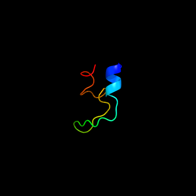
1 c4oo2D_
28.4
27
PDB header: oxidoreductaseChain: D: PDB Molecule: chlorophenol-4-monooxygenase;PDBTitle: streptomyces globisporus c-1027 fad dependent (s)-3-chloro-β-2 tyrosine-s-sgcc2 c-5 hydroxylase sgcc apo form
2 c1r8jB_
19.3
25
PDB header: circadian clock proteinChain: B: PDB Molecule: kaia;PDBTitle: crystal structure of circadian clock protein kaia from2 synechococcus elongatus
3 c4h9kA_
15.3
75
PDB header: hydrolaseChain: A: PDB Molecule: hog cholera virus;PDBTitle: crystal structure of cleavage site mutant of npro of classical swine2 fever virus.
4 c3hwcD_
15.1
21
PDB header: oxidoreductaseChain: D: PDB Molecule: chlorophenol-4-monooxygenase component 2;PDBTitle: crystal structure of chlorophenol 4-monooxygenase (tftd) of2 burkholderia cepacia ac1100
5 c6eb0A_
14.9
24
PDB header: oxidoreductaseChain: A: PDB Molecule: 4-hydroxyphenylacetate 3-monooxygenase, oxygenase subunit;PDBTitle: structure of 4-hydroxyphenylacetate 3-monooxygenase (hpab), oxygenase2 component from escherichia coli
6 c1eq8C_
11.3
53
PDB header: signaling proteinChain: C: PDB Molecule: acetylcholine receptor protein;PDBTitle: three-dimensional structure of the pentameric helical2 bundle of the acetylcholine receptor m2 transmembrane3 segment
7 c1eq8A_
11.3
53
PDB header: signaling proteinChain: A: PDB Molecule: acetylcholine receptor protein;PDBTitle: three-dimensional structure of the pentameric helical2 bundle of the acetylcholine receptor m2 transmembrane3 segment
8 c1eq8E_
11.3
53
PDB header: signaling proteinChain: E: PDB Molecule: acetylcholine receptor protein;PDBTitle: three-dimensional structure of the pentameric helical2 bundle of the acetylcholine receptor m2 transmembrane3 segment
9 c1eq8B_
11.3
53
PDB header: signaling proteinChain: B: PDB Molecule: acetylcholine receptor protein;PDBTitle: three-dimensional structure of the pentameric helical2 bundle of the acetylcholine receptor m2 transmembrane3 segment
10 c1eq8D_
11.3
53
PDB header: signaling proteinChain: D: PDB Molecule: acetylcholine receptor protein;PDBTitle: three-dimensional structure of the pentameric helical2 bundle of the acetylcholine receptor m2 transmembrane3 segment
11 c1a11A_
11.1
53
PDB header: acetylcholine receptorChain: A: PDB Molecule: acetylcholine receptor m2;PDBTitle: nmr structure of membrane spanning segment 2 of the2 acetylcholine receptor in dpc micelles, 10 structures
12 c4rv2A_
10.9
10
PDB header: lyaseChain: A: PDB Molecule: upf0336 protein msmeg_1340/msmei_1302;PDBTitle: crystal structure of (3r)-hydroxyacyl-acp dehydratase hadab hetero-2 dimer from mycobacterium smegmatis
13 c5n6yC_
10.3
24
PDB header: oxidoreductaseChain: C: PDB Molecule: nitrogenase vanadium-iron protein delta chain;PDBTitle: azotobacter vinelandii vanadium nitrogenase
14 d1fi6a_
9.9
20
Fold: EF Hand-likeSuperfamily: EF-handFamily: Eps15 homology domain (EH domain)
15 d2ghvc1
9.1
22
Fold: SARS receptor-binding domain-likeSuperfamily: SARS receptor-binding domain-likeFamily: SARS receptor-binding domain-like
16 c3p4hA_
8.9
22
PDB header: hydrolaseChain: A: PDB Molecule: atp-dependent dna ligase, n-terminal domain protein;PDBTitle: structures of archaeal members of the ligd 3'-phosphoesterase dna2 repair enzyme superfamily
17 d1l5ia_
8.2
29
Fold: Origin of replication-binding domain, RBD-likeSuperfamily: Origin of replication-binding domain, RBD-likeFamily: DNA-binding domain of REP protein
18 c1ii0A_
8.1
35
PDB header: hydrolaseChain: A: PDB Molecule: arsenical pump-driving atpase;PDBTitle: crystal structure of the escherichia coli arsenite-translocating2 atpase
19 d1rh2a_
7.5
13
Fold: 4-helical cytokinesSuperfamily: 4-helical cytokinesFamily: Interferons/interleukin-10 (IL-10)
20 c5l0lB_
7.5
41
PDB header: unknown functionChain: B: PDB Molecule: uncharacterized protein;PDBTitle: crystal structure of uncharacterized protein lpg0439
21 c1u8vA_
not modelled
7.2
24
PDB header: lyase, isomeraseChain: A: PDB Molecule: gamma-aminobutyrate metabolism dehydratase/isomerase;PDBTitle: crystal structure of 4-hydroxybutyryl-coa dehydratase from clostridium2 aminobutyricum: radical catalysis involving a [4fe-4s] cluster and3 flavin
22 c3p43A_
not modelled
7.2
25
PDB header: hydrolaseChain: A: PDB Molecule: putative uncharacterized protein;PDBTitle: structure and activities of archaeal members of the ligd 3'2 phosphoesterase dna repair enzyme superfamily
23 c6f0kA_
not modelled
7.2
16
PDB header: membrane proteinChain: A: PDB Molecule: cytochrome c family protein;PDBTitle: alternative complex iii
24 c5t42A_
not modelled
7.2
22
PDB header: viral proteinChain: A: PDB Molecule: envelope glycoprotein;PDBTitle: structure of the ebola virus envelope protein mper/tm domain and its2 interaction with the fusion loop explains their fusion activity
25 c4g5eD_
not modelled
7.1
22
PDB header: oxidoreductaseChain: D: PDB Molecule: 2,4,6-trichlorophenol 4-monooxygenase;PDBTitle: 2,4,6-trichlorophenol 4-monooxygenase
26 c2khnA_
not modelled
6.9
20
PDB header: signaling proteinChain: A: PDB Molecule: intersectin-1;PDBTitle: nmr solution structure of the eh 1 domain from human2 intersectin-1 protein. northeast structural genomics3 consortium target hr3646e.
27 c3zfnA_
not modelled
6.8
71
PDB header: hydrolaseChain: A: PDB Molecule: n-terminal protease npro;PDBTitle: crystal structure of product-like, processed n-terminal protease npro
28 c3sciE_
not modelled
6.7
22
PDB header: hydrolase/viral proteinChain: E: PDB Molecule: spike glycoprotein;PDBTitle: crystal structure of spike protein receptor-binding domain from a2 predicted sars coronavirus human strain complexed with human receptor3 ace2
29 c2kgrA_
not modelled
6.6
23
PDB header: protein bindingChain: A: PDB Molecule: intersectin-1;PDBTitle: solution structure of protein itsn1 from homo sapiens.2 northeast structural genomics consortium target hr5524a
30 c3ez6B_
not modelled
6.3
31
PDB header: dna binding proteinChain: B: PDB Molecule: plasmid partition protein a;PDBTitle: structure of para-adp complex:tetragonal form
31 d2ezla_
not modelled
6.3
13
Fold: DNA/RNA-binding 3-helical bundleSuperfamily: Homeodomain-likeFamily: Recombinase DNA-binding domain
32 c4qzvB_
not modelled
6.2
25
PDB header: hydrolase/viral proteinChain: B: PDB Molecule: spike protein s1;PDBTitle: bat-derived coronavirus hku4 uses mers-cov receptor human cd26 for2 cell entry
33 c5u3mA_
not modelled
6.0
80
PDB header: immune system/viral proteinChain: A: PDB Molecule: gp41 mper peptide;PDBTitle: crystal structure of dh511.11p fab in complex with hiv-1 gp41 mper2 peptide
34 c3fiaA_
not modelled
5.8
20
PDB header: protein bindingChain: A: PDB Molecule: intersectin-1;PDBTitle: crystal structure of the eh 1 domain from human intersectin-1 protein.2 northeast structural genomics consortium target hr3646e.
35 d2jxca1
not modelled
5.8
16
Fold: EF Hand-likeSuperfamily: EF-handFamily: Eps15 homology domain (EH domain)
36 c2m4mA_
not modelled
5.8
18
PDB header: unknown functionChain: A: PDB Molecule: hypothetical protein;PDBTitle: solution structure of the rrm domain of the hypothetical protein2 cagl0m09691g from candida glabrata
37 d1ihua2
not modelled
5.8
32
Fold: P-loop containing nucleoside triphosphate hydrolasesSuperfamily: P-loop containing nucleoside triphosphate hydrolasesFamily: Nitrogenase iron protein-like
38 c4zv4C_
not modelled
5.7
32
PDB header: translationChain: C: PDB Molecule: tse6;PDBTitle: structure of tse6 in complex with ef-tu
39 d2dk8a1
not modelled
5.3
44
Fold: DNA/RNA-binding 3-helical bundleSuperfamily: "Winged helix" DNA-binding domainFamily: RPO3F domain-like
40 c1javA_
not modelled
5.3
80
PDB header: viral proteinChain: A: PDB Molecule: transmembrane glycoprotein (gp41);PDBTitle: average nmr solution structure of the trp-rich peptide of2 hiv gp41 bound to dpc micelles
41 c2aenH_
not modelled
5.2
33
PDB header: viral proteinChain: H: PDB Molecule: outer capsid protein vp4, vp8* core;PDBTitle: crystal structure of the rotavirus strain ds-1 vp8* core
42 c2q01A_
not modelled
5.1
30
PDB header: isomeraseChain: A: PDB Molecule: uronate isomerase;PDBTitle: crystal structure of glucuronate isomerase from caulobacter crescentus
43 d1t61a2
not modelled
5.1
47
Fold: C-type lectin-likeSuperfamily: C-type lectin-likeFamily: Noncollagenous (NC1) domain of collagen IV