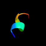
| 1 |
|
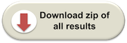
PDB 5m2u chain A
Region: 1 - 39
Aligned: 37
Modelled: 39
Confidence: 24.0%
Identity: 35%
PDB header:photosynthesis
Chain: A: PDB Molecule:ycf54;
PDBTitle: the structure of the ycf54 protein from synechocystis sp. pcc6803
Phyre2
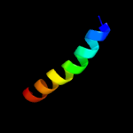
| 2 |
|
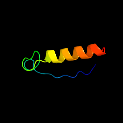
PDB 1qgt chain A
Region: 8 - 26
Aligned: 19
Modelled: 19
Confidence: 21.4%
Identity: 53%
Fold: Hepatitis B viral capsid (hbcag)
Superfamily: Hepatitis B viral capsid (hbcag)
Family: Hepatitis B viral capsid (hbcag)
Phyre2
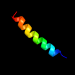
| 3 |
|
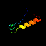
PDB 2k44 chain A
Region: 11 - 18
Aligned: 8
Modelled: 8
Confidence: 10.5%
Identity: 50%
PDB header:membrane protein
Chain: A: PDB Molecule:k+-channel voltage-sensor paddle domain of
PDBTitle: solution structure of a k+-channel voltage-sensor paddle2 domain
Phyre2
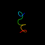
| 4 |
|
PDB 2jzi chain B
Region: 21 - 44
Aligned: 24
Modelled: 24
Confidence: 10.0%
Identity: 33%
PDB header:metal binding protein
Chain: B: PDB Molecule:serine/threonine-protein phosphatase 2b
PDBTitle: structure of calmodulin complexed with the calmodulin2 binding domain of calcineurin
Phyre2
| 5 |
|
PDB 4q5u chain C
Region: 21 - 44
Aligned: 24
Modelled: 24
Confidence: 8.9%
Identity: 33%
PDB header:calcium binding protein/protein binding
Chain: C: PDB Molecule:serine/threonine-protein phosphatase 2b catalytic subunit
PDBTitle: structure of calmodulin bound to its recognition site from calcineurin
Phyre2
| 6 |
|
PDB 2dco chain A
Region: 29 - 43
Aligned: 15
Modelled: 15
Confidence: 8.7%
Identity: 40%
PDB header:membrane protein
Chain: A: PDB Molecule:s1p4 first extracellular loop peptidomimetic;
PDBTitle: s1p4 first extracellular loop peptidomimetic
Phyre2
| 7 |
|
PDB 1m3v chain A
Region: 45 - 55
Aligned: 11
Modelled: 11
Confidence: 7.8%
Identity: 45%
PDB header:metal binding protein
Chain: A: PDB Molecule:fusion of the lim interacting domain of ldb1 and
PDBTitle: flin4: fusion of the lim binding domain of ldb1 and the n-2 terminal lim domain of lmo4
Phyre2
| 8 |
|
PDB 5vk2 chain A
Region: 2 - 14
Aligned: 13
Modelled: 13
Confidence: 7.0%
Identity: 46%
PDB header:viral protein/immune system
Chain: A: PDB Molecule:pre-glycoprotein polyprotein gp complex;
PDBTitle: structural basis for antibody-mediated neutralization of lassa virus
Phyre2
| 9 |
|
PDB 1mww chain A
Region: 6 - 28
Aligned: 23
Modelled: 23
Confidence: 6.1%
Identity: 17%
Fold: Tautomerase/MIF
Superfamily: Tautomerase/MIF
Family: Hypothetical protein HI1388.1
Phyre2