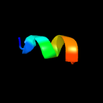
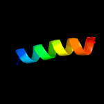
| 1 |
|
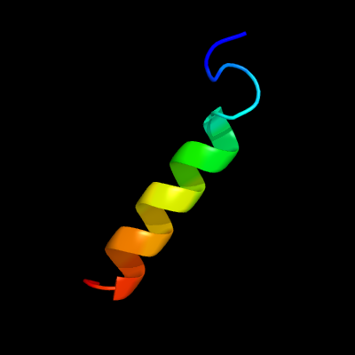
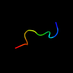
PDB 2n1p chain A
Region: 33 - 54
Aligned: 22
Modelled: 22
Confidence: 18.1%
Identity: 23%
PDB header:viral protein
Chain: A: PDB Molecule:non-structural protein 5b, ns5b;
PDBTitle: structure of the c-terminal membrane domain of hcv ns5b protein
Phyre2
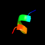
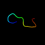
| 2 |
|
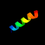
PDB 6d79 chain A
Region: 19 - 74
Aligned: 56
Modelled: 56
Confidence: 17.4%
Identity: 14%
PDB header:transport protein
Chain: A: PDB Molecule:sulfate transporter cysz;
PDBTitle: structure of cysz, a sulfate permease from pseudomonas fragi
Phyre2
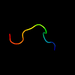
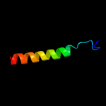
| 3 |
|
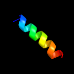
PDB 2c4f chain L domain 3
Region: 31 - 39
Aligned: 9
Modelled: 9
Confidence: 17.3%
Identity: 44%
Fold: GLA-domain
Superfamily: GLA-domain
Family: GLA-domain
Phyre2
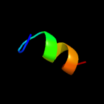
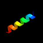
| 4 |
|
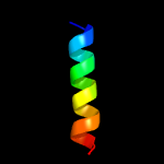
PDB 1dan chain L domain 3
Region: 31 - 39
Aligned: 9
Modelled: 9
Confidence: 15.9%
Identity: 44%
Fold: GLA-domain
Superfamily: GLA-domain
Family: GLA-domain
Phyre2
| 5 |
|
PDB 3ixs chain F
Region: 76 - 86
Aligned: 11
Modelled: 11
Confidence: 13.4%
Identity: 27%
PDB header:protein binding
Chain: F: PDB Molecule:ring1 and yy1-binding protein;
PDBTitle: ring1b c-terminal domain/rybp c-terminal domain complex
Phyre2
| 6 |
|
PDB 2zfd chain B
Region: 71 - 82
Aligned: 12
Modelled: 12
Confidence: 8.0%
Identity: 25%
PDB header:signaling protein/transferase
Chain: B: PDB Molecule:putative uncharacterized protein t20l15_90;
PDBTitle: the crystal structure of plant specific calcium binding protein atcbl22 in complex with the regulatory domain of atcipk14
Phyre2
| 7 |
|
PDB 5u89 chain B
Region: 72 - 81
Aligned: 10
Modelled: 10
Confidence: 7.4%
Identity: 40%
PDB header:hydrolase/inhibitor
Chain: B: PDB Molecule:mbth domain protein;
PDBTitle: crystal structure of a cross-module fragment from the dimodular nrps2 dhbf
Phyre2
| 8 |
|
PDB 3a0h chain X
Region: 35 - 52
Aligned: 18
Modelled: 18
Confidence: 7.0%
Identity: 28%
PDB header:electron transport
Chain: X: PDB Molecule:photosystem ii reaction center protein x;
PDBTitle: crystal structure of i-substituted photosystem ii complex
Phyre2
| 9 |
|
PDB 3a0b chain X
Region: 35 - 52
Aligned: 18
Modelled: 18
Confidence: 7.0%
Identity: 28%
PDB header:electron transport
Chain: X: PDB Molecule:photosystem ii reaction center protein x;
PDBTitle: crystal structure of br-substituted photosystem ii complex
Phyre2
| 10 |
|
PDB 3a0h chain X
Region: 35 - 52
Aligned: 18
Modelled: 18
Confidence: 7.0%
Identity: 28%
PDB header:electron transport
Chain: X: PDB Molecule:photosystem ii reaction center protein x;
PDBTitle: crystal structure of i-substituted photosystem ii complex
Phyre2
| 11 |
|
PDB 3a0b chain X
Region: 35 - 52
Aligned: 18
Modelled: 18
Confidence: 7.0%
Identity: 28%
PDB header:electron transport
Chain: X: PDB Molecule:photosystem ii reaction center protein x;
PDBTitle: crystal structure of br-substituted photosystem ii complex
Phyre2
| 12 |
|
PDB 1pjc chain A domain 2
Region: 70 - 81
Aligned: 12
Modelled: 12
Confidence: 6.7%
Identity: 8%
Fold: Flavodoxin-like
Superfamily: Formate/glycerate dehydrogenase catalytic domain-like
Family: L-alanine dehydrogenase-like
Phyre2
| 13 |
|
PDB 2knc chain A
Region: 28 - 58
Aligned: 31
Modelled: 31
Confidence: 5.9%
Identity: 23%
PDB header:cell adhesion
Chain: A: PDB Molecule:integrin alpha-iib;
PDBTitle: platelet integrin alfaiib-beta3 transmembrane-cytoplasmic2 heterocomplex
Phyre2
| 14 |
|
PDB 1s5l chain X
Region: 35 - 52
Aligned: 18
Modelled: 18
Confidence: 5.9%
Identity: 28%
PDB header:photosynthesis
Chain: X: PDB Molecule:photosystem ii psbx protein;
PDBTitle: architecture of the photosynthetic oxygen evolving center
Phyre2