Phyre Home Retrieve Phyre Job Id
Emailmdejesus@rockefeller.edu
DescriptionRVBD0544c_(-)_635938_636216
DateFri Jul 26 01:50:09 BST 2019
Unique Job IDc765c3a3323d7df9
SequenceVSAWFNYTAT ... Download FASTA
Job Typenormal
Job Expiry

Summary 

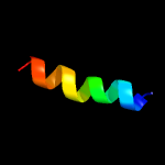
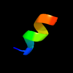
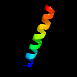
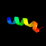
Top model
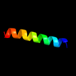
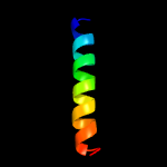
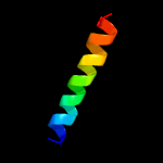
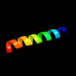
Image coloured by rainbow N → C terminus
Model dimensions (Å):X:11.648 Y:14.421 Z:27.221
Model (left) based on template c1ifpA_
Top template information
PDB header:virus
Chain: A: PDB Molecule:major coat protein assembly;
PDBTitle: inovirus (filamentous bacteriophage) strain pf3 major coat2 protein assembly
Confidence and coverage
Confidence: 55.5% Coverage: 18%
17 residues ( 18% of your sequence) have been modelled with 55.5% confidence by the single highest scoring template.
You may wish to submit your sequence to Phyrealarm. This will automatically scan your sequence every week for new potential templates as they appear in the Phyre2 library.
Please note: You must be registered and logged in to use Phyrealarm.
3D viewing
Interactive 3D view in JSmol
For other options to view your downloaded structure offline see the FAQ

Sequence analysis 

Download FASTA version

Secondary structure and disorder prediction [Show] 

Domain analysis [Show] 

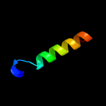
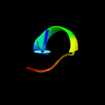
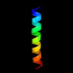
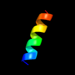
Detailed template information [Hide] 


#
Template Alignment Coverage3D Model Confidence
% i.d. Template Information
1c1ifpA_


55.5 41
PDB header:virus
Chain: A: PDB Molecule:major coat protein assembly;
PDBTitle: inovirus (filamentous bacteriophage) strain pf3 major coat2 protein assembly
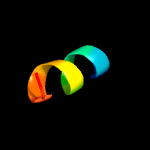
2c6bzfG_


24.9 20
PDB header:dna binding protein
Chain: G: PDB Molecule:sporulation-specific protein 16;
PDBTitle: structure of s. cerevisiae zip2:spo16 complex, c2 form
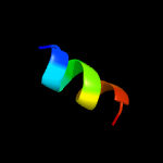
3c2k1kB_


20.5 64
PDB header:signaling protein
Chain: B: PDB Molecule:ephrin type-a receptor 1;
PDBTitle: nmr structures of dimeric transmembrane domain of the2 receptor tyrosine kinase epha1 in lipid bicelles at ph 4.3
4c2k1lB_


20.5 64
PDB header:signaling protein
Chain: B: PDB Molecule:ephrin type-a receptor 1;
PDBTitle: nmr structures of dimeric transmembrane domain of the2 receptor tyrosine kinase epha1 in lipid bicelles at ph 6.3
5c2k1kA_


20.5 64
PDB header:signaling protein
Chain: A: PDB Molecule:ephrin type-a receptor 1;
PDBTitle: nmr structures of dimeric transmembrane domain of the2 receptor tyrosine kinase epha1 in lipid bicelles at ph 4.3
6c2k1lA_


20.5 64
PDB header:signaling protein
Chain: A: PDB Molecule:ephrin type-a receptor 1;
PDBTitle: nmr structures of dimeric transmembrane domain of the2 receptor tyrosine kinase epha1 in lipid bicelles at ph 6.3
7d3e11a1


15.7 63
Fold:Zincin-like
Superfamily:Metalloproteases ("zincins"), catalytic domain
Family:TTHA0227-like
8c4pv1E_


15.4 25
PDB header:electron transport/inhibitor
Chain: E: PDB Molecule:cytochrome b6-f complex subunit 6;
PDBTitle: cytochrome b6f structure from m. laminosus with the quinone analog2 inhibitor stigmatellin
9c4i7zE_


15.4 25
PDB header:photosynthesis
Chain: E: PDB Molecule:cytochrome b6-f complex subunit 6;
PDBTitle: crystal structure of cytochrome b6f in dopg, with disordered rieske2 iron-sulfur protein soluble domain
10c4av3A_


14.0 31
PDB header:hydrolase
Chain: A: PDB Molecule:k(+)-stimulated pyrophosphate-energized sodium pump;
PDBTitle: crystal structure of thermotoga maritima sodium pumping membrane2 integral pyrophosphatase with metal ions in active site
11c2e74E_


12.9 25
PDB header:photosynthesis
Chain: E: PDB Molecule:cytochrome b6-f complex subunit 6;
PDBTitle: crystal structure of the cytochrome b6f complex from m.laminosus
12c4h0lE_


12.9 25
PDB header:photosynthesis
Chain: E: PDB Molecule:cytochrome b6-f complex subunit 6;
PDBTitle: cytochrome b6f complex crystal structure from mastigocladus laminosus2 with n-side inhibitor nqno
13c2e76E_


12.9 25
PDB header:photosynthesis
Chain: E: PDB Molecule:cytochrome b6-f complex subunit 6;
PDBTitle: crystal structure of the cytochrome b6f complex with tridecyl-2 stigmatellin (tds) from m.laminosus
14c1vf5R_


12.9 25
PDB header:photosynthesis
Chain: R: PDB Molecule:protein pet l;
PDBTitle: crystal structure of cytochrome b6f complex from m.laminosus
15c4h13E_


12.9 25
PDB header:photosynthesis
Chain: E: PDB Molecule:cytochrome b6-f complex subunit 6;
PDBTitle: crystal structure of the cytochrome b6f complex from mastigocladus2 laminosus with tds
16c2e75E_


12.9 25
PDB header:photosynthesis
Chain: E: PDB Molecule:cytochrome b6-f complex subunit 6;
PDBTitle: crystal structure of the cytochrome b6f complex with 2-nonyl-4-2 hydroxyquinoline n-oxide (nqno) from m.laminosus
17c1vf5E_


12.9 25
PDB header:photosynthesis
Chain: E: PDB Molecule:protein pet l;
PDBTitle: crystal structure of cytochrome b6f complex from m.laminosus
18d2e74e1


12.9 25
Fold:Single transmembrane helix
Superfamily:PetL subunit of the cytochrome b6f complex
Family:PetL subunit of the cytochrome b6f complex
19c4a01B_


12.5 38
PDB header:hydrolase
Chain: B: PDB Molecule:proton pyrophosphatase;
PDBTitle: crystal structure of the h-translocating pyrophosphatase
20c2zp2B_


12.4 50
PDB header:transferase inhibitor
Chain: B: PDB Molecule:kinase a inhibitor;
PDBTitle: c-terminal domain of kipi from bacillus subtilis
21d1v54d_


not modelled 11.3 10
Fold:Single transmembrane helix
Superfamily:Mitochondrial cytochrome c oxidase subunit IV
Family:Mitochondrial cytochrome c oxidase subunit IV
22c3cjsA_


not modelled 9.9 47
PDB header:transferase/ribosomal protein
Chain: A: PDB Molecule:ribosomal protein l11 methyltransferase;
PDBTitle: minimal recognition complex between prma and ribosomal protein l11
23d1bm8a_


not modelled 9.7 42
Fold:DNA-binding domain of Mlu1-box binding protein MBP1
Superfamily:DNA-binding domain of Mlu1-box binding protein MBP1
Family:DNA-binding domain of Mlu1-box binding protein MBP1
24d2phcb1


not modelled 9.5 44
Fold:Cyclophilin-like
Superfamily:Cyclophilin-like
Family:PH0987 C-terminal domain-like
25c3i71B_


not modelled 9.1 44
PDB header:unknown function
Chain: B: PDB Molecule:ethanolamine utilization protein eutk;
PDBTitle: ethanolamine utilization microcompartment shell subunit, eutk c-2 terminal domain
26c5dudB_


not modelled 9.0 44
PDB header:unknown function
Chain: B: PDB Molecule:ybgj;
PDBTitle: crystal structure of e. coli ybgjk
27c3mmlD_


not modelled 8.2 44
PDB header:hydrolase
Chain: D: PDB Molecule:allophanate hydrolase subunit 1;
PDBTitle: allophanate hydrolase complex from mycobacterium smegmatis, msmeg0435-2 msmeg0436
28d1nekc_


not modelled 8.1 14
Fold:Heme-binding four-helical bundle
Superfamily:Fumarate reductase respiratory complex transmembrane subunits
Family:Succinate dehydrogenase/Fumarate reductase transmembrane subunits (SdhC/FrdC and SdhD/FrdD)
29c2uwjG_


not modelled 7.2 40
PDB header:chaperone
Chain: G: PDB Molecule:type iii export protein pscg;
PDBTitle: structure of the heterotrimeric complex which regulates type iii2 secretion needle formation
30c2phcB_


not modelled 7.0 44
PDB header:structural genomics, unknown function
Chain: B: PDB Molecule:uncharacterized protein ph0987;
PDBTitle: crystal structure of conserved uncharacterized protein ph0987 from2 pyrococcus horikoshii
31c3kziE_


not modelled 7.0 31
PDB header:electron transport
Chain: E: PDB Molecule:cytochrome b559 subunit alpha;
PDBTitle: crystal structure of monomeric form of cyanobacterial photosystem ii
32c2y69Q_


not modelled 7.0 10
PDB header:electron transport
Chain: Q: PDB Molecule:cytochrome c oxidase subunit 4 isoform 1;
PDBTitle: bovine heart cytochrome c oxidase re-refined with molecular oxygen
33c5ncnB_


not modelled 6.8 33
PDB header:signaling protein
Chain: B: PDB Molecule:cell cycle protein kinase dbf2;
PDBTitle: crystal structure dbf2(ntr)-mob1 complex
34c4ux5B_


not modelled 6.7 42
PDB header:transcription
Chain: B: PDB Molecule:transcription factor mbp1;
PDBTitle: structure of dna complex of pcg2
35c2n28A_


not modelled 6.6 22
PDB header:viral protein
Chain: A: PDB Molecule:protein vpu;
PDBTitle: solid-state nmr structure of vpu
36d2axte1


not modelled 6.5 31
Fold:Single transmembrane helix
Superfamily:Cytochrome b559 subunits
Family:Cytochrome b559 subunits
37c3jcue_


not modelled 6.4 31
PDB header:membrane protein
Chain: E: PDB Molecule:cytochrome b559 subunit alpha;
PDBTitle: cryo-em structure of spinach psii-lhcii supercomplex at 3.2 angstrom2 resolution
38d1l3ga_


not modelled 6.3 38
Fold:DNA-binding domain of Mlu1-box binding protein MBP1
Superfamily:DNA-binding domain of Mlu1-box binding protein MBP1
Family:DNA-binding domain of Mlu1-box binding protein MBP1
39c3m9qA_


not modelled 5.2 50
PDB header:dna binding protein
Chain: A: PDB Molecule:protein male-specific lethal-3;
PDBTitle: drosophila msl3 chromodomain

Binding site prediction 

Due to computational demand, binding site predictions are not run for batch jobs

If you want to predict binding sites, please manually submit your model of choice to 3DLigandSite

Transmembrane helix prediction 

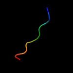
Transmembrane helices have been predicted in your sequence to adopt the topology shown below