Phyre Home Retrieve Phyre Job Id
Emailmdejesus@rockefeller.edu
DescriptionRVBD0550c_(-)_640641_640907
DateFri Jul 26 01:50:10 BST 2019
Unique Job IDd7e1e88b0afc47d4
SequenceLLSRRTKTIV ... Download FASTA
Job Typenormal
Job Expiry

Summary 

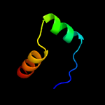
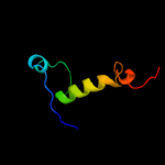
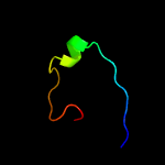
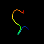
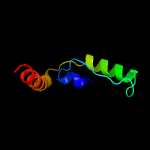
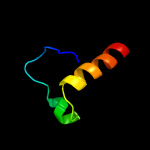
Top model
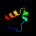
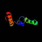
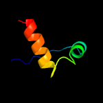
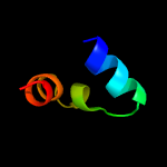
Image coloured by rainbow N → C terminus
Model dimensions (Å):X:20.487 Y:25.562 Z:24.710
Model (left) based on template c5yrzC_
Top template information
PDB header:antitoxin/hydrolase
Chain: C: PDB Molecule:hicb;
PDBTitle: toxin-antitoxin complex from streptococcus pneumoniae
Confidence and coverage
Confidence: 83.8% Coverage: 36%
32 residues ( 36% of your sequence) have been modelled with 83.8% confidence by the single highest scoring template.
You may wish to submit your sequence to Phyrealarm. This will automatically scan your sequence every week for new potential templates as they appear in the Phyre2 library.
Please note: You must be registered and logged in to use Phyrealarm.
3D viewing
Interactive 3D view in JSmol
For other options to view your downloaded structure offline see the FAQ

Sequence analysis 

Download FASTA version

Secondary structure and disorder prediction [Show] 

Domain analysis [Show] 

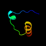
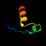
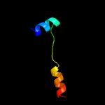
Detailed template information [Hide] 


#
Template Alignment Coverage3D Model Confidence
% i.d. Template Information
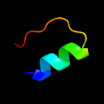
1c5yrzC_


83.8 25
PDB header:antitoxin/hydrolase
Chain: C: PDB Molecule:hicb;
PDBTitle: toxin-antitoxin complex from streptococcus pneumoniae
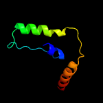
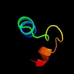
2c4p7dA_


69.5 12
PDB header:toxin
Chain: A: PDB Molecule:antitoxin hicb3;
PDBTitle: antitoxin hicb3 crystal structure
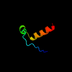
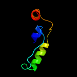
3c6g1nB_


62.4 27
PDB header:antitoxin
Chain: B: PDB Molecule:antitoxin hicb;
PDBTitle: crystal structure of the burkholderia pseudomallei antitoxin hicb
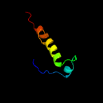
4c6a6xC_


37.9 62
PDB header:toxin
Chain: C: PDB Molecule:antitoxin maze7;
PDBTitle: the crystal structure of the mtb maze-mazf-mt9 complex
5c5x3tA_


26.8 43
PDB header:antitoxin/toxin
Chain: A: PDB Molecule:antitoxin vapb26;
PDBTitle: vapbc from mycobacterium tuberculosis
6c3qd5B_


24.4 23
PDB header:isomerase
Chain: B: PDB Molecule:putative ribose-5-phosphate isomerase;
PDBTitle: crystal structure of a putative ribose-5-phosphate isomerase from2 coccidioides immitis solved by combined iodide ion sad and mr
7c2k5jB_


23.1 27
PDB header:structural genomics, unknown function
Chain: B: PDB Molecule:uncharacterized protein yiif;
PDBTitle: solution structure of protein yiif from shigella flexneri2 serotype 5b (strain 8401) . northeast structural genomics3 consortium target sft1
8c2ca9B_


23.0 26
PDB header:transcription
Chain: B: PDB Molecule:putative nickel-responsive regulator;
PDBTitle: apo-nikr from helicobacter pylori in closed trans-2 conformation
9c2bj3D_


20.2 24
PDB header:transcription
Chain: D: PDB Molecule:nickel responsive regulator;
PDBTitle: nikr-apo
10d2vvpa1


19.4 19
Fold:Ribose/Galactose isomerase RpiB/AlsB
Superfamily:Ribose/Galactose isomerase RpiB/AlsB
Family:Ribose/Galactose isomerase RpiB/AlsB
11c1q5vB_


17.5 28
PDB header:transcription
Chain: B: PDB Molecule:nickel responsive regulator;
PDBTitle: apo-nikr
12c1b4aA_


12.6 50
PDB header:repressor
Chain: A: PDB Molecule:arginine repressor;
PDBTitle: structure of the arginine repressor from bacillus stearothermophilus
13c2k29A_


12.4 40
PDB header:transcription
Chain: A: PDB Molecule:antitoxin relb;
PDBTitle: structure of the dbd domain of e. coli antitoxin relb
14c3e38A_


11.0 32
PDB header:hydrolase
Chain: A: PDB Molecule:two-domain protein containing predicted php-like metal-
PDBTitle: crystal structure of a two-domain protein containing predicted php-2 like metal-dependent phosphoesterase (bvu_3505) from bacteroides3 vulgatus atcc 8482 at 2.20 a resolution
15d2p5ka1


10.8 39
Fold:DNA/RNA-binding 3-helical bundle
Superfamily:"Winged helix" DNA-binding domain
Family:Arginine repressor (ArgR), N-terminal DNA-binding domain
16c4lfnD_


10.0 22
PDB header:isomerase
Chain: D: PDB Molecule:galactose-6-phosphate isomerase subunit b;
PDBTitle: crystal structure of d-galactose-6-phosphate isomerase in complex with2 d-ribulose
17d1v0da_


9.9 60
Fold:His-Me finger endonucleases
Superfamily:His-Me finger endonucleases
Family:Caspase-activated DNase, CAD (DffB, DFF40)
18c1v0dA_


9.9 60
PDB header:hydrolase
Chain: A: PDB Molecule:dna fragmentation factor 40 kda subunit;
PDBTitle: crystal structure of caspase-activated dnase (cad)
19c6mu0A_


9.8 24
PDB header:isomerase
Chain: A: PDB Molecule:probable ribose-5-phosphate isomerase b;
PDBTitle: crystal structure of ribose-5-phosphate isomerase b from mycoplasma2 genitalium with bound ribulose-5-phosphate
20c2k9iB_


8.7 40
PDB header:dna binding protein
Chain: B: PDB Molecule:uncharacterized protein orf56;
PDBTitle: nmr structure of plasmid copy control protein orf56 from sulfolobus2 islandicus
21c6fxwA_


not modelled 8.6 13
PDB header:isomerase
Chain: A: PDB Molecule:putative ribose 5-phosphate isomerase;
PDBTitle: structure of leishmania infantum type b ribose 5-phosphate isomerase
22c6ae9B_


not modelled 8.1 20
PDB header:hydrolase
Chain: B: PDB Molecule:probable protein phosphatase 2c 1;
PDBTitle: x-ray structure of the photosystem ii phosphatase pbcp
23c3shgB_


not modelled 7.8 75
PDB header:transferase/protein binding
Chain: B: PDB Molecule:vbha;
PDBTitle: vbht fic protein from bartonella schoenbuchensis in complex with vbha2 antitoxin
24c3qgaD_


not modelled 6.3 24
PDB header:hydrolase
Chain: D: PDB Molecule:fusion of urease beta and gamma subunits;
PDBTitle: 3.0 a model of iron containing urease urea2b2 from helicobacter2 mustelae
25d1ugpa_


not modelled 6.3 50
Fold:Nitrile hydratase alpha chain
Superfamily:Nitrile hydratase alpha chain
Family:Nitrile hydratase alpha chain
26c3v4gA_


not modelled 6.2 29
PDB header:dna binding protein
Chain: A: PDB Molecule:arginine repressor;
PDBTitle: 1.60 angstrom resolution crystal structure of an arginine repressor2 from vibrio vulnificus cmcp6
27c5aj3g_


not modelled 6.2 30
PDB header:ribosome
Chain: G: PDB Molecule:mitoribosomal protein us7m, mrps7;
PDBTitle: structure of the small subunit of the mammalian mitoribosome
28c4yh5B_


not modelled 6.2 22
PDB header:transferase
Chain: B: PDB Molecule:levoglucosan kinase;
PDBTitle: lipomyces starkeyi levoglucosan kinase bound to adp and manganese
29c1e9zA_


not modelled 5.4 24
PDB header:hydrolase
Chain: A: PDB Molecule:urease subunit alpha;
PDBTitle: crystal structure of helicobacter pylori urease
30c3skqA_


not modelled 5.4 25
PDB header:metal transport
Chain: A: PDB Molecule:mitochondrial distribution and morphology protein 38;
PDBTitle: mdm38 is a 14-3-3-like receptor and associates with the protein2 synthesis machinery at the inner mitochondrial membrane
31c3ereD_


not modelled 5.2 46
PDB header:dna binding protein/dna
Chain: D: PDB Molecule:arginine repressor;
PDBTitle: crystal structure of the arginine repressor protein from mycobacterium2 tuberculosis in complex with the dna operator
32c3m1pA_


not modelled 5.1 22
PDB header:isomerase
Chain: A: PDB Molecule:ribose 5-phosphate isomerase;
PDBTitle: structure of ribose 5-phosphate isomerase type b from trypanosoma2 cruzi, soaked with allose-6-phosphate
33c3k7pA_


not modelled 5.1 22
PDB header:isomerase
Chain: A: PDB Molecule:ribose 5-phosphate isomerase;
PDBTitle: structure of mutant of ribose 5-phosphate isomerase type b from2 trypanosoma cruzi.

Binding site prediction 

Due to computational demand, binding site predictions are not run for batch jobs

If you want to predict binding sites, please manually submit your model of choice to 3DLigandSite