Phyre Home Retrieve Phyre Job Id
Emailmdejesus@rockefeller.edu
DescriptionRVBD0601c_(-)_698527_698997
DateFri Jul 26 01:50:16 BST 2019
Unique Job IDdef88cfa360277ab
SequenceMALVLAAAGA ... Download FASTA
Job Typenormal
Job Expiry

Summary 

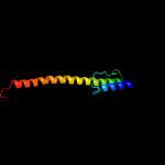
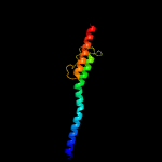
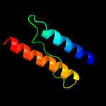
Top model
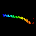
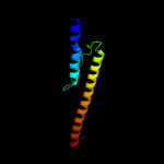
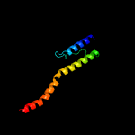
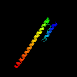
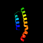
Image coloured by rainbow N → C terminus
Model dimensions (Å):X:52.348 Y:60.814 Z:53.201
Model (left) based on template c3zrwB_
Top template information
PDB header:signaling protein
Chain: B: PDB Molecule:af1503 protein, osmolarity sensor protein envz;
PDBTitle: the structure of the dimeric hamp-dhp fusion a291v mutant
Confidence and coverage
Confidence: 99.6% Coverage: 54%
84 residues ( 54% of your sequence) have been modelled with 99.6% confidence by the single highest scoring template.
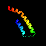
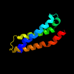
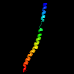
Additional confident templates have been detected (see Domain analysis) which cover other regions of your sequence.
124 residues ( 79%) could be modelled at >90% confidence using multiple-templates.
You may wish to try resubmitting your sequence in "intensive" mode to model more of your sequence.
3D viewing
Interactive 3D view in JSmol
For other options to view your downloaded structure offline see the FAQ

Sequence analysis 

Download FASTA version

Secondary structure and disorder prediction [Show] 

Domain analysis [Show] 

Detailed template information [Hide] 


#
Template Alignment Coverage3D Model Confidence
% i.d. Template Information
1c3zrwB_


99.6 23
PDB header:signaling protein
Chain: B: PDB Molecule:af1503 protein, osmolarity sensor protein envz;
PDBTitle: the structure of the dimeric hamp-dhp fusion a291v mutant
2c4biuB_


99.2 27
PDB header:transferase
Chain: B: PDB Molecule:sensor protein cpxa;
PDBTitle: crystal structure of cpxahdc (orthorhombic form 1)
3c4mt8A_


99.2 21
PDB header:transferase
Chain: A: PDB Molecule:ethylene response sensor 1;
PDBTitle: structure of the ers1 dimerization and histidine phosphotransfer2 domain from arabidopsis thaliana
4c5ukvA_


99.1 33
PDB header:transferase
Chain: A: PDB Molecule:atp-binding protein;
PDBTitle: dhp domain of phor of m. tuberculosis - semet
5c4gn0D_


99.0 17
PDB header:signaling protein
Chain: D: PDB Molecule:hamp domain of af1503;
PDBTitle: de novo phasing of a hamp-complex using an improved arcimboldo method
6d1joya_


98.9 24
Fold:ROP-like
Superfamily:Homodimeric domain of signal transducing histidine kinase
Family:Homodimeric domain of signal transducing histidine kinase
7d2c2aa1


98.9 31
Fold:ROP-like
Superfamily:Homodimeric domain of signal transducing histidine kinase
Family:Homodimeric domain of signal transducing histidine kinase
8c5jefA_


98.8 17
PDB header:transferase
Chain: A: PDB Molecule:nitrate/nitrite sensor protein narq;
PDBTitle: fragment of nitrate/nitrite sensor histidine kinase narq (wt) in2 asymmetric holo state
9c2c2aA_


98.6 31
PDB header:transferase
Chain: A: PDB Molecule:sensor histidine kinase;
PDBTitle: structure of the entire cytoplasmic portion of a sensor2 histidine kinase protein
10c3lnrA_


98.3 14
PDB header:signaling protein
Chain: A: PDB Molecule:aerotaxis transducer aer2;
PDBTitle: crystal structure of poly-hamp domains from the p. aeruginosa soluble2 receptor aer2
11c4ctiA_


98.1 25
PDB header:signaling protein
Chain: A: PDB Molecule:osmolarity sensor protein envz, af1503;
PDBTitle: escherichia coli envz histidine kinase catalytic part fused to2 archaeoglobus fulgidus af1503 hamp domain
12c3zx6A_


97.5 14
PDB header:signaling
Chain: A: PDB Molecule:hamp, methyl-accepting chemotaxis protein i;
PDBTitle: structure of hamp(af1503)-tsr fusion - hamp (a291v) mutant
13d2asxa1


97.5 23
Fold:HAMP domain-like
Superfamily:HAMP domain-like
Family:HAMP domain
14c2q8fA_


97.2 6
PDB header:transferase
Chain: A: PDB Molecule:[pyruvate dehydrogenase [lipoamide]] kinase isozyme 1;
PDBTitle: structure of pyruvate dehydrogenase kinase isoform 1
15c3d2rB_


96.9 11
PDB header:transferase
Chain: B: PDB Molecule:[pyruvate dehydrogenase [lipoamide]] kinase isozyme 4;
PDBTitle: crystal structure of pyruvate dehydrogenase kinase isoform 4 in2 complex with adp
16c3gieA_


95.7 19
PDB header:transferase
Chain: A: PDB Molecule:sensor histidine kinase desk;
PDBTitle: crystal structure of deskc_h188e in complex with amp-pcp
17c1y8oA_


95.6 6
PDB header:transferase
Chain: A: PDB Molecule:[pyruvate dehydrogenase [lipoamide]] kinase isozyme 3;
PDBTitle: crystal structure of the pdk3-l2 complex
18c3crlB_


95.2 8
PDB header:transferase
Chain: B: PDB Molecule:pyruvate dehydrogenase [lipoamide] kinase isozyme 2,
PDBTitle: crystal structure of the pdhk2-l2 complex.
19c4i5sA_


95.2 19
PDB header:transferase
Chain: A: PDB Molecule:putative histidine kinase covs; vick-like protein;
PDBTitle: structure and function of sensor histidine kinase
20c2rm8A_


92.5 21
PDB header:signaling protein
Chain: A: PDB Molecule:sensory rhodopsin ii transducer;
PDBTitle: the solution structure of phototactic transducer protein2 htrii linker region from natronomonas pharaonis
21c2bu8A_


not modelled 91.9 8
PDB header:transferase
Chain: A: PDB Molecule:pyruvate dehydrogenase kinase isoenzyme 2;
PDBTitle: crystal structures of human pyruvate dehydrogenase kinase 2 containing2 physiological and synthetic ligands
22c4gczB_


not modelled 91.3 14
PDB header:signaling protein, de novo protein
Chain: B: PDB Molecule:blue-light photoreceptor, sensor protein fixl;
PDBTitle: structure of a blue-light photoreceptor
23c3tz5A_


not modelled 87.4 16
PDB header:transferase/transferase inhibitor
Chain: A: PDB Molecule:[3-methyl-2-oxobutanoate dehydrogenase [lipoamide]] kinase,
PDBTitle: crystal structure of branched-chain alpha-ketoacid dehydrogenase2 kinase/phenylbutyrate complex with adp
24c1gjvA_


not modelled 86.1 16
PDB header:transferase
Chain: A: PDB Molecule:[3-methyl-2-oxobutanoate dehydrogenase [lipoamide]] kinase;
PDBTitle: branched-chain alpha-ketoacid dehydrogenase kinase (bck) complxed with2 atp-gamma-s
25c6e95A_


not modelled 77.9 10
PDB header:signaling protein
Chain: A: PDB Molecule:staphylococcus aureus agrc histidine kinase module fused to
PDBTitle: chimeric structure of saccharomyces cerevisiae gcn4 leucine zipper2 fused to staphylococcus aureus agrc cytoplasmic histidine kinase3 module (dataset isotropically truncated by hkl2000)
26c5c3lB_


not modelled 22.7 20
PDB header:transport protein
Chain: B: PDB Molecule:nucleoporin nup58;
PDBTitle: structure of the metazoan nup62.nup58.nup54 nucleoporin complex.
27c2oo4B_


not modelled 17.5 23
PDB header:cell cycle,signaling protein
Chain: B: PDB Molecule:neurogenic locus notch homolog protein 2;
PDBTitle: structure of lnr-hd (negative regulatory region) from human notch 2
28c2n28A_


not modelled 15.2 14
PDB header:viral protein
Chain: A: PDB Molecule:protein vpu;
PDBTitle: solid-state nmr structure of vpu
29d1li5a1


not modelled 13.8 30
Fold:Anticodon-binding domain of a subclass of class I aminoacyl-tRNA synthetases
Superfamily:Anticodon-binding domain of a subclass of class I aminoacyl-tRNA synthetases
Family:Anticodon-binding domain of a subclass of class I aminoacyl-tRNA synthetases
30c3i08C_


not modelled 11.3 18
PDB header:signaling protein
Chain: C: PDB Molecule:neurogenic locus notch homolog protein 1;
PDBTitle: crystal structure of the s1-cleaved notch1 negative2 regulatory region (nrr)
31c3etoB_


not modelled 10.9 18
PDB header:signaling protein
Chain: B: PDB Molecule:neurogenic locus notch homolog protein 1;
PDBTitle: 2 angstrom xray structure of the notch1 negative regulatory region2 (nrr)
32c3rtyA_


not modelled 10.8 8
PDB header:circadian clock protein
Chain: A: PDB Molecule:period circadian protein;
PDBTitle: structure of an enclosed dimer formed by the drosophila period protein
33c2momC_


not modelled 9.2 18
PDB header:membrane protein
Chain: C: PDB Molecule:lysosome-associated membrane glycoprotein 2;
PDBTitle: structural insights of tm domain of lamp-2a in dpc micelles
34c2momB_


not modelled 9.2 18
PDB header:membrane protein
Chain: B: PDB Molecule:lysosome-associated membrane glycoprotein 2;
PDBTitle: structural insights of tm domain of lamp-2a in dpc micelles
35c4o9pC_


not modelled 8.2 18
PDB header:membrane protein
Chain: C: PDB Molecule:nad(p) transhydrogenase subunit alpha 2;
PDBTitle: crystal structure of thermus thermophilis transhydrogeanse domain ii2 dimer semet derivative
36c5omdA_


not modelled 7.9 17
PDB header:protein binding
Chain: A: PDB Molecule:dna damage checkpoint protein lcd1;
PDBTitle: crystal structure of s. cerevisiae ddc2 n-terminal coiled-coil domain
37c2mi2A_


not modelled 7.7 16
PDB header:transport protein
Chain: A: PDB Molecule:sec-independent protein translocase protein tatb;
PDBTitle: solution structure of the e. coli tatb protein in dpc micelles
38c4hj6A_


not modelled 7.6 5
PDB header:signaling protein
Chain: A: PDB Molecule:lov protein;
PDBTitle: crystal structure of rhodobacter sphaeroides lov protein
39c1zzaA_


not modelled 7.4 23
PDB header:membrane protein
Chain: A: PDB Molecule:stannin;
PDBTitle: solution nmr structure of the membrane protein stannin
40c5v2sA_


not modelled 7.4 12
PDB header:viral protein
Chain: A: PDB Molecule:envelope glycoprotein b;
PDBTitle: crystal structure of glycoprotein b from herpes simplex virus type i
41c3qweA_


not modelled 6.7 20
PDB header:protein binding
Chain: A: PDB Molecule:gem-interacting protein;
PDBTitle: crystal structure of the n-terminal domain of the gem interacting2 protein
42c4zlpB_


not modelled 6.7 32
PDB header:transcription
Chain: B: PDB Molecule:neurogenic locus notch homolog protein 3;
PDBTitle: crystal structure of notch3 negative regulatory region
43c4o9rA_


not modelled 6.6 10
PDB header:signaling protein
Chain: A: PDB Molecule:smoothened homolog/soluble cytochrome b562 chimeric
PDBTitle: human smoothened receptor structure in complex with cyclopamine

Binding site prediction 

Due to computational demand, binding site predictions are not run for batch jobs

If you want to predict binding sites, please manually submit your model of choice to 3DLigandSite

Transmembrane helix prediction 

Transmembrane helices have been predicted in your sequence to adopt the topology shown below