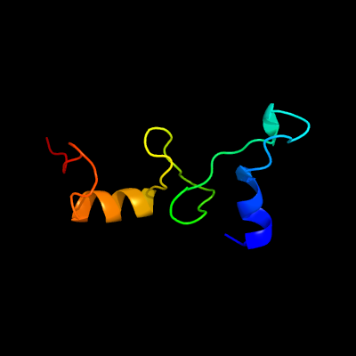
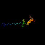
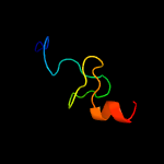
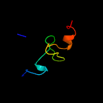
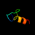
1 d2uubn1
100.0
70
Fold: Glucocorticoid receptor-like (DNA-binding domain)Superfamily: Glucocorticoid receptor-like (DNA-binding domain)Family: Ribosomal protein S14
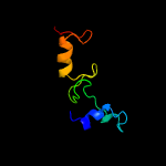
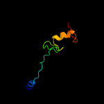
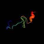
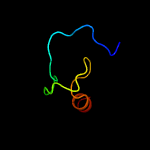
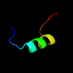
2 c6dzkN_
100.0
54
PDB header: ribosomeChain: N: PDB Molecule: 30s ribosomal protein s14;PDBTitle: cryo-em structure of mycobacterium smegmatis c(minus) 30s ribosomal2 subunit with mpy
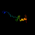
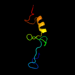
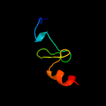
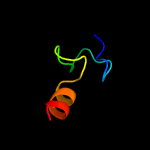
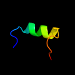
3 c3bbnN_
100.0
48
PDB header: ribosomeChain: N: PDB Molecule: ribosomal protein s14;PDBTitle: homology model for the spinach chloroplast 30s subunit fitted to 9.4a2 cryo-em map of the 70s chlororibosome.
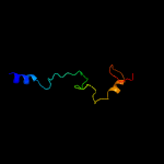
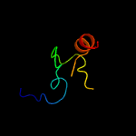
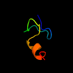
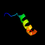
4 d2qaln1
100.0
46
Fold: Glucocorticoid receptor-like (DNA-binding domain)Superfamily: Glucocorticoid receptor-like (DNA-binding domain)Family: Ribosomal protein S14
5 c2gy9N_
100.0
49
PDB header: ribosomeChain: N: PDB Molecule: 30s ribosomal subunit protein s14;PDBTitle: structure of the 30s subunit of a pre-translocational e. coli ribosome2 obtained by fitting atomic models for rna and protein components into3 cryo-em map emd-1056
6 c3j6vN_
99.9
37
PDB header: ribosomeChain: N: PDB Molecule: 28s ribosomal protein s14, mitochondrial;PDBTitle: cryo-em structure of the small subunit of the mammalian mitochondrial2 ribosome
7 c5xyid_
96.2
38
PDB header: ribosomeChain: D: PDB Molecule: ribosomal protein s3, putative;PDBTitle: small subunit of trichomonas vaginalis ribosome
8 c3jyvN_
96.1
31
PDB header: ribosomeChain: N: PDB Molecule: 40s ribosomal protein s29(a);PDBTitle: structure of the 40s rrna and proteins and p/e trna for eukaryotic2 ribosome based on cryo-em map of thermomyces lanuginosus ribosome at3 8.9a resolution
9 c3j20P_
96.0
39
PDB header: ribosomeChain: P: PDB Molecule: 30s ribosomal protein s14p type z;PDBTitle: promiscuous behavior of proteins in archaeal ribosomes revealed by2 cryo-em: implications for evolution of eukaryotic ribosomes (30s3 ribosomal subunit)
10 c2xznN_
95.9
31
PDB header: ribosomeChain: N: PDB Molecule: rps29e;PDBTitle: crystal structure of the eukaryotic 40s ribosomal2 subunit in complex with initiation factor 1. this file3 contains the 40s subunit and initiation factor for4 molecule 2
11 c5xxud_
95.9
28
PDB header: ribosomeChain: D: PDB Molecule: ribosomal protein us3;PDBTitle: small subunit of toxoplasma gondii ribosome
12 c2zkqn_
95.8
29
PDB header: ribosomal protein/rnaChain: N: PDB Molecule: PDBTitle: structure of a mammalian ribosomal 40s subunit within an 80s complex2 obtained by docking homology models of the rna and proteins into an3 8.7 a cryo-em map
13 c3zey8_
95.3
43
PDB header: ribosomeChain: 8: PDB Molecule: ribosomal protein s29, putative;PDBTitle: high-resolution cryo-electron microscopy structure of the trypanosoma2 brucei ribosome
14 c6az1S_
95.1
39
PDB header: ribosome/antibioticChain: S: PDB Molecule: ribosomal protein s14;PDBTitle: cryo-em structure of the small subunit of leishmania ribosome bound to2 paromomycin
15 c1s1hN_
85.3
38
PDB header: ribosomeChain: N: PDB Molecule: 40s ribosomal protein s29-b;PDBTitle: structure of the ribosomal 80s-eef2-sordarin complex from yeast2 obtained by docking atomic models for rna and protein components into3 a 11.7 a cryo-em map. this file, 1s1h, contains 40s subunit. the 60s4 ribosomal subunit is in file 1s1i.
16 c4j2nB_
27.8
27
PDB header: viral proteinChain: B: PDB Molecule: gp37;PDBTitle: crystal structure of mycobacteriophage pukovnik xis
17 c4j2nA_
25.8
27
PDB header: viral proteinChain: A: PDB Molecule: gp37;PDBTitle: crystal structure of mycobacteriophage pukovnik xis
18 c6amaO_
23.7
19
PDB header: dna binding protein/dnaChain: O: PDB Molecule: putative dna-binding protein;PDBTitle: structure of s. coelicolor/s. venezuelae bldc-smea-ssfa complex to2 3.09 angstrom
19 d2bmfa1
18.0
30
Fold: P-loop containing nucleoside triphosphate hydrolasesSuperfamily: P-loop containing nucleoside triphosphate hydrolasesFamily: RNA helicase
20 c4lhfA_
11.5
30
PDB header: viral proteinChain: A: PDB Molecule: regulatory protein cox;PDBTitle: crystal structure of a dna binding protein from phage p2
21 c5ntbB_
not modelled
11.2
30
PDB header: protease inhibitorChain: B: PDB Molecule: papain inhibitor;PDBTitle: streptomyces papain inhibitor (spi)
22 c2zhhA_
not modelled
11.0
15
PDB header: transcriptionChain: A: PDB Molecule: redox-sensitive transcriptional activator soxr;PDBTitle: crystal structure of soxr
23 c1yuzB_
not modelled
7.9
29
PDB header: oxidoreductaseChain: B: PDB Molecule: nigerythrin;PDBTitle: partially reduced state of nigerythrin
24 c2xzm8_
not modelled
7.7
32
PDB header: ribosomeChain: 8: PDB Molecule: rps25e,;PDBTitle: crystal structure of the eukaryotic 40s ribosomal2 subunit in complex with initiation factor 1. this file3 contains the 40s subunit and initiation factor for4 molecule 1
25 d1n10a2
not modelled
7.4
25
Fold: Double psi beta-barrelSuperfamily: Barwin-like endoglucanasesFamily: Pollen allergen PHL P 1 N-terminal domain
26 c3zeyU_
not modelled
7.1
26
PDB header: ribosomeChain: U: PDB Molecule: 40s ribosomal protein s25, putative;PDBTitle: high-resolution cryo-electron microscopy structure of the trypanosoma2 brucei ribosome
27 d2ieaa3
not modelled
7.1
21
Fold: TK C-terminal domain-likeSuperfamily: TK C-terminal domain-likeFamily: Transketolase C-terminal domain-like
28 c2fugC_
not modelled
6.8
33
PDB header: oxidoreductaseChain: C: PDB Molecule: nadh-quinone oxidoreductase chain 3;PDBTitle: crystal structure of the hydrophilic domain of respiratory complex i2 from thermus thermophilus
29 d1r8da_
not modelled
6.5
12
Fold: Putative DNA-binding domainSuperfamily: Putative DNA-binding domainFamily: DNA-binding N-terminal domain of transcription activators
30 c1y6uA_
not modelled
6.2
27
PDB header: dna binding proteinChain: A: PDB Molecule: excisionase from transposon tn916;PDBTitle: the structure of the excisionase (xis) protein from2 conjugative transposon tn916 provides insights into the3 regulation of heterobivalent tyrosine recombinases
31 c3izbV_
not modelled
6.2
36
PDB header: ribosomeChain: V: PDB Molecule: 40s ribosomal protein rps25 (s25e);PDBTitle: localization of the small subunit ribosomal proteins into a 6.1 a2 cryo-em map of saccharomyces cerevisiae translating 80s ribosome
32 c4ivvA_
not modelled
5.9
22
PDB header: hydrolaseChain: A: PDB Molecule: autolysin;PDBTitle: catalytic amidase domain of the major autolysin lyta from2 streptococcus pneumaniae
33 c1z4hA_
not modelled
5.7
18
PDB header: protein binding, dna binding proteinChain: A: PDB Molecule: tor inhibition protein;PDBTitle: the response regulator tori belongs to a new family of2 atypical excisionase
34 c3iz6V_
not modelled
5.7
23
PDB header: ribosomeChain: V: PDB Molecule: 40s ribosomal protein s25 (s25e);PDBTitle: localization of the small subunit ribosomal proteins into a 5.5 a2 cryo-em map of triticum aestivum translating 80s ribosome
35 c5jyxB_
not modelled
5.5
19
PDB header: transferaseChain: B: PDB Molecule: archeaosine synthase quef-like;PDBTitle: crystal structure of the covalent thioimide intermediate of the2 archaeosine synthase quef-like
36 c3gpvA_
not modelled
5.3
19
PDB header: transcription regulatorChain: A: PDB Molecule: transcriptional regulator, merr family;PDBTitle: crystal structure of a transcriptional regulator, merr2 family from bacillus thuringiensis
37 c3j3aZ_
not modelled
5.3
32
PDB header: ribosomeChain: Z: PDB Molecule: 40s ribosomal protein s25;PDBTitle: structure of the human 40s ribosomal proteins