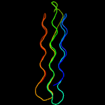
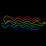
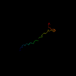
PDB header:structural protein/contractile protein Chain: A: PDB Molecule:collagen i alpha 1; PDBTitle: the structure of collagen type i. single type i collagen2 molecule: rigid refinment
Confidence and coverage
Confidence:
100.0%
Coverage:
99%
744 residues ( 99% of your sequence) have been modelled with 100.0% confidence by the single highest scoring template.
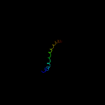
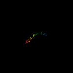
Warning: 100% of your sequence is predicted disordered. Disordered regions cannot be meaningfully predicted.
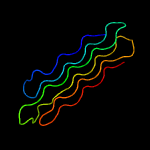
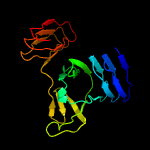
Region: 2 - 747 Aligned: 744 Modelled: 746 Confidence: 100.0% Identity: 29% PDB header:structural protein/contractile protein Chain: A: PDB Molecule:collagen i alpha 1; PDBTitle: the structure of collagen type i. single type i collagen2 molecule: rigid refinment
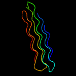
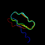
Region: 1 - 747 Aligned: 743 Modelled: 746 Confidence: 99.8% Identity: 29% PDB header:structural protein/contractile protein Chain: B: PDB Molecule:collagen i alpha 2; PDBTitle: the structure of collagen type i. single type i collagen2 molecule
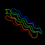
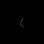
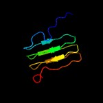
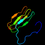
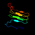
Region: 15 - 53 Aligned: 39 Modelled: 39 Confidence: 96.4% Identity: 28% PDB header:structural protein Chain: B: PDB Molecule:collagen alpha-2(i) chain,collagen alpha-2(ix) chain; PDBTitle: crystal structure of the type ix collagen nc2 hetero-trimerization2 domain with a guest fragment a2a1a1 of type i collagen
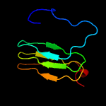
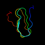
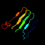
Region: 17 - 54 Aligned: 38 Modelled: 38 Confidence: 95.8% Identity: 37% PDB header:structural protein Chain: C: PDB Molecule:collagen alpha-1(i) chain,collagen alpha-3(ix) chain; PDBTitle: crystal structure of the type ix collagen nc2 hetero-trimerization2 domain with a guest fragment a2a1a1 of type i collagen (native form)
Region: 17 - 54 Aligned: 38 Modelled: 38 Confidence: 95.8% Identity: 37% PDB header:structural protein Chain: A: PDB Molecule:collagen alpha-1(i) chain,collagen alpha-1(ix) chain; PDBTitle: crystal structure of the type ix collagen nc2 hetero-trimerization2 domain with a guest fragment a2a1a1 of type i collagen