PDB 3sol chain A
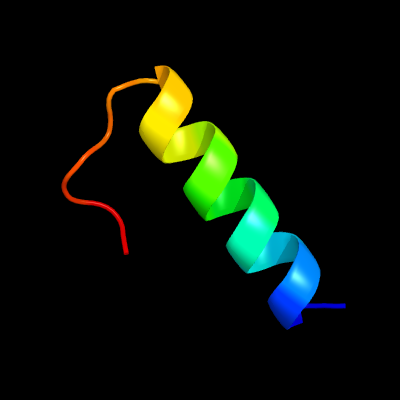
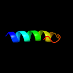
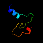
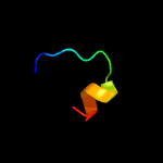
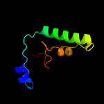
Region: 87 - 108
Aligned: 22
Modelled: 22
Confidence: 42.3%
Identity: 41%
PDB header:protein transport
Chain: A: PDB Molecule:type ii secretion pathway related protein;
PDBTitle: crystal structure of the type 2 secretion system pilotin gsps
Phyre2
PDB 3utk chain A
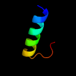
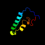
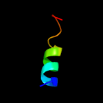
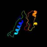
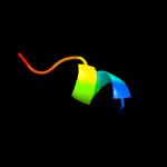
Region: 87 - 108
Aligned: 22
Modelled: 22
Confidence: 26.7%
Identity: 41%
PDB header:protein transport
Chain: A: PDB Molecule:lipoprotein outs;
PDBTitle: structure of the pilotin of the type ii secretion system
Phyre2
PDB 1ner chain A
Region: 109 - 120
Aligned: 12
Modelled: 12
Confidence: 22.9%
Identity: 50%
Fold: lambda repressor-like DNA-binding domains
Superfamily: lambda repressor-like DNA-binding domains
Family: Phage repressors
Phyre2
PDB 4a56 chain A
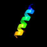
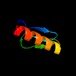
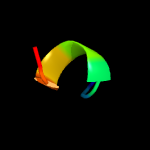
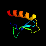
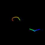
Region: 87 - 108
Aligned: 22
Modelled: 22
Confidence: 21.1%
Identity: 23%
PDB header:protein transport
Chain: A: PDB Molecule:pullulanase secretion protein puls;
PDBTitle: crystal structure of the type 2 secretion system pilotin2 from klebsiella oxytoca
Phyre2
PDB 5hxp chain A
Region: 22 - 80
Aligned: 53
Modelled: 59
Confidence: 16.0%
Identity: 15%
PDB header:transferase
Chain: A: PDB Molecule:(2z,6z)-farnesyl diphosphate synthase, chloroplastic;
PDBTitle: crystal structure of z,z-farnesyl diphosphate synthase (d71m, e75a and2 h103y mutants) complexed with ipp
Phyre2
PDB 1uxy chain A domain 2
Region: 4 - 48
Aligned: 45
Modelled: 45
Confidence: 13.5%
Identity: 20%
Fold: Uridine diphospho-N-Acetylenolpyruvylglucosamine reductase, MurB, C-terminal domain
Superfamily: Uridine diphospho-N-Acetylenolpyruvylglucosamine reductase, MurB, C-terminal domain
Family: Uridine diphospho-N-Acetylenolpyruvylglucosamine reductase, MurB, C-terminal domain
Phyre2
PDB 6jcn chain B
Region: 9 - 80
Aligned: 67
Modelled: 72
Confidence: 13.0%
Identity: 16%
PDB header:transferase
Chain: B: PDB Molecule:dehydrodolichyl diphosphate synthase complex subunit nus1;
PDBTitle: yeast dehydrodolichyl diphosphate synthase complex subunit nus1
Phyre2
PDB 2knp chain A
Region: 103 - 113
Aligned: 11
Modelled: 11
Confidence: 10.6%
Identity: 55%
PDB header:unknown function
Chain: A: PDB Molecule:mcocc-1;
PDBTitle: isolation and characterization of peptides from momordica2 cochinchinensis seeds.
Phyre2
PDB 3nvn chain A
Region: 83 - 137
Aligned: 42
Modelled: 42
Confidence: 10.5%
Identity: 26%
PDB header:viral protein/signaling protein
Chain: A: PDB Molecule:evm139;
PDBTitle: molecular mechanism of guidance cue recognition
Phyre2
PDB 1plp chain A
Region: 89 - 105
Aligned: 17
Modelled: 17
Confidence: 10.5%
Identity: 47%
PDB header:membrane protein
Chain: A: PDB Molecule:phospholamban;
PDBTitle: solution structure of the cytoplasmic domain of2 phospholamban
Phyre2
PDB 2bmt chain A
Region: 99 - 105
Aligned: 7
Modelled: 7
Confidence: 8.5%
Identity: 71%
Fold: Knottins (small inhibitors, toxins, lectins)
Superfamily: Scorpion toxin-like
Family: Short-chain scorpion toxins
Phyre2
PDB 4xal chain A
Region: 32 - 48
Aligned: 17
Modelled: 17
Confidence: 8.4%
Identity: 41%
PDB header:viral protein
Chain: A: PDB Molecule:tegument protein vp22;
PDBTitle: crystal structure of the conserved core domain of vp22 from hsv-1
Phyre2
PDB 2m0d chain A
Region: 125 - 138
Aligned: 14
Modelled: 14
Confidence: 7.4%
Identity: 57%
PDB header:transcription
Chain: A: PDB Molecule:zinc finger and btb domain-containing protein 17;
PDBTitle: solution structure of miz-1 zinc finger 5
Phyre2
PDB 2c8m chain A domain 1
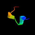
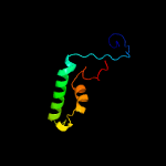
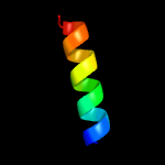
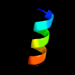
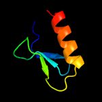
Region: 81 - 159
Aligned: 69
Modelled: 79
Confidence: 7.3%
Identity: 13%
Fold: Class II aaRS and biotin synthetases
Superfamily: Class II aaRS and biotin synthetases
Family: LplA-like
Phyre2
PDB 1hsk chain A domain 2
Region: 4 - 48
Aligned: 45
Modelled: 45
Confidence: 7.2%
Identity: 13%
Fold: Uridine diphospho-N-Acetylenolpyruvylglucosamine reductase, MurB, C-terminal domain
Superfamily: Uridine diphospho-N-Acetylenolpyruvylglucosamine reductase, MurB, C-terminal domain
Family: Uridine diphospho-N-Acetylenolpyruvylglucosamine reductase, MurB, C-terminal domain
Phyre2
PDB 3vh5 chain D
Region: 33 - 45
Aligned: 13
Modelled: 13
Confidence: 6.9%
Identity: 62%
PDB header:dna binding protein
Chain: D: PDB Molecule:cenp-x;
PDBTitle: crystal structure of the chicken cenp-t histone fold/cenp-w/cenp-2 s/cenp-x heterotetrameric complex, crystal form i
Phyre2
PDB 5hc7 chain A
Region: 9 - 80
Aligned: 69
Modelled: 72
Confidence: 6.3%
Identity: 32%
PDB header:transferase/transferase inhibitor
Chain: A: PDB Molecule:prenyltransference for protein;
PDBTitle: crystal structure of lavandulyl diphosphate synthase from lavandula x2 intermedia in complex with s-thiolo-isopentenyldiphosphate
Phyre2
PDB 5z1y chain A
Region: 139 - 147
Aligned: 9
Modelled: 9
Confidence: 6.1%
Identity: 67%
PDB header:antibiotic
Chain: A: PDB Molecule:mbjamp1 peptide;
PDBTitle: mbjamp1 structure
Phyre2
PDB 4v1a chain V
Region: 99 - 107
Aligned: 9
Modelled: 9
Confidence: 5.9%
Identity: 56%
PDB header:ribosome
Chain: V: PDB Molecule:
PDBTitle: structure of the large subunit of the mammalian mitoribosome, part 22 of 2
Phyre2
PDB 4pyt chain A
Region: 4 - 46
Aligned: 43
Modelled: 43
Confidence: 5.8%
Identity: 14%
PDB header:oxidoreductase
Chain: A: PDB Molecule:udp-n-acetylenolpyruvoylglucosamine reductase;
PDBTitle: crystal structure of a murb family ep-udp-n-acetylglucosamine2 reductase
Phyre2
1 |
|
2 |
|
3 |
|
4 |
|
5 |
|
6 |
|
7 |
|
8 |
|
9 |
|
10 |
|
11 |
|
12 |
|
13 |
|
14 |
|
15 |
|
16 |
|
17 |
|
18 |
|
19 |
|
20 |
|
21 |
|
22 |
|
23 |
|
24 |
|