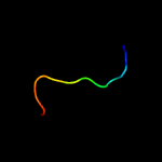
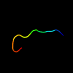
| 1 |
|
PDB 5mlc chain 8
Region: 52 - 58
Aligned: 7
Modelled: 7
Confidence: 13.1%
Identity: 57%
PDB header:ribosome
Chain: 8: PDB Molecule:psrp6, chloroplastic;
PDBTitle: cryo-em structure of the spinach chloroplast ribosome reveals the2 location of plastid-specific ribosomal proteins and extensions
Phyre2
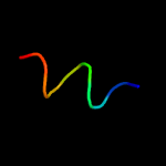
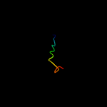
| 2 |
|
PDB 5mmi chain 7
Region: 52 - 58
Aligned: 7
Modelled: 7
Confidence: 13.0%
Identity: 57%
PDB header:ribosome
Chain: 7: PDB Molecule:50s ribosomal protein 6, chloroplastic;
PDBTitle: structure of the large subunit of the chloroplast ribosome
Phyre2
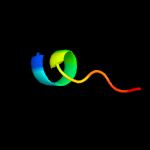
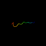
| 3 |
|
PDB 4j2u chain A
Region: 53 - 64
Aligned: 12
Modelled: 12
Confidence: 8.6%
Identity: 33%
PDB header:lyase
Chain: A: PDB Molecule:enoyl-coa hydratase;
PDBTitle: crystal structure of an enoyl-coa hydratase from rhodobacter2 sphaeroides 2.4.1
Phyre2
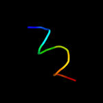
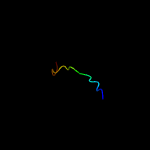
| 4 |
|
PDB 1dpk chain A
Region: 49 - 58
Aligned: 10
Modelled: 10
Confidence: 7.2%
Identity: 60%
PDB header:cell adhesion
Chain: A: PDB Molecule:integrin alpha-iib subunit;
PDBTitle: solution structure of the cytoplasmic domain of the2 integrin alpha-iib subunit
Phyre2
| 5 |
|
PDB 1s4w chain A
Region: 49 - 58
Aligned: 10
Modelled: 10
Confidence: 7.2%
Identity: 60%
PDB header:cell adhesion
Chain: A: PDB Molecule:integrin alpha-iib;
PDBTitle: nmr structure of the cytoplasmic domain of integrin aiib in2 dpc micelles
Phyre2
| 6 |
|
PDB 1m8o chain A
Region: 49 - 58
Aligned: 10
Modelled: 10
Confidence: 7.2%
Identity: 60%
PDB header:membrane protein
Chain: A: PDB Molecule:platelet integrin alfaiib subunit: cytoplasmic
PDBTitle: platelet integrin alfaiib-beta3 cytoplasmic domain
Phyre2
| 7 |
|
PDB 2ra9 chain A
Region: 17 - 26
Aligned: 10
Modelled: 10
Confidence: 7.1%
Identity: 50%
PDB header:unknown function
Chain: A: PDB Molecule:uncharacterized protein duf1285;
PDBTitle: crystal structure of a duf1285 family protein (sbal_2486) from2 shewanella baltica os155 at 1.40 a resolution
Phyre2
| 8 |
|
PDB 6a0a chain A
Region: 20 - 36
Aligned: 16
Modelled: 17
Confidence: 6.4%
Identity: 69%
PDB header:structural protein
Chain: A: PDB Molecule:collagen type iii peptide;
PDBTitle: structure of a triple-helix region of human collagen type iii
Phyre2
| 9 |
|
PDB 6a0a chain B
Region: 20 - 36
Aligned: 16
Modelled: 17
Confidence: 6.0%
Identity: 69%
PDB header:structural protein
Chain: B: PDB Molecule:collagen type iii peptide;
PDBTitle: structure of a triple-helix region of human collagen type iii
Phyre2
| 10 |
|
PDB 6a0a chain C
Region: 20 - 36
Aligned: 16
Modelled: 17
Confidence: 6.0%
Identity: 69%
PDB header:structural protein
Chain: C: PDB Molecule:collagen type iii peptide;
PDBTitle: structure of a triple-helix region of human collagen type iii
Phyre2