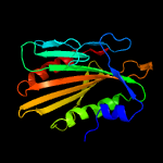
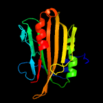
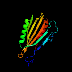
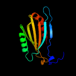
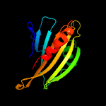
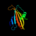
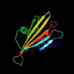
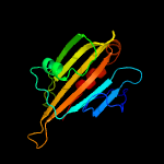
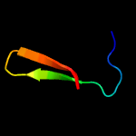
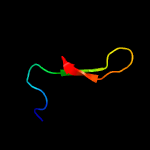
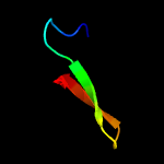
1 c6e5fA_
100.0
20
PDB header: lipid binding proteinChain: A: PDB Molecule: lipid binding protein lpqn;PDBTitle: crystal structure of lpqn involved in cell envelope biogenesis of2 mycobacterium tuberculosis
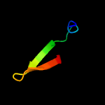
2 c4ol4A_
100.0
34
PDB header: lipid binding proteinChain: A: PDB Molecule: proline-rich 28 kda antigen;PDBTitle: crystal structure of secreted proline rich antigen mtc28 (rv0040c)2 from mycobacterium tuberculosis
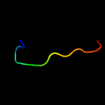
3 c3lydA_
100.0
16
PDB header: structural genomics, unknown functionChain: A: PDB Molecule: uncharacterized protein;PDBTitle: crystal structure of putative uncharacterized protein from jonesia2 denitrificans
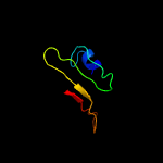
4 d1tu1a_
98.5
13
Fold: Mog1p/PsbP-likeSuperfamily: Mog1p/PsbP-likeFamily: PA0094-like
5 c2lnjA_
97.1
12
PDB header: photosynthesisChain: A: PDB Molecule: putative uncharacterized protein sll1418;PDBTitle: solution structure of cyanobacterial psbp (cyanop) from synechocystis2 sp. pcc 6803
6 c2xb3A_
96.0
10
PDB header: photosynthesisChain: A: PDB Molecule: psbp protein;PDBTitle: the structure of cyanobacterial psbp
7 d1v2ba_
89.8
16
Fold: Mog1p/PsbP-likeSuperfamily: Mog1p/PsbP-likeFamily: PsbP-like
8 c2vu4A_
80.1
17
PDB header: photosynthesisChain: A: PDB Molecule: oxygen-evolving enhancer protein 2;PDBTitle: structure of psbp protein from spinacia oleracea at 1.98 a2 resolution
9 c1ymzA_
61.4
25
PDB header: unknown functionChain: A: PDB Molecule: cc45;PDBTitle: cc45, an artificial ww domain designed using statistical2 coupling analysis
10 c6j69A_
59.1
25
PDB header: cell cycleChain: A: PDB Molecule: protein kibra;PDBTitle: structure of kibra and dendrin complex
11 c1e0mA_
56.8
33
PDB header: de novo proteinChain: A: PDB Molecule: wwprototype;PDBTitle: prototype ww domain
12 d2jmfa1
41.6
38
Fold: WW domain-likeSuperfamily: WW domainFamily: WW domain
13 c1yiuA_
38.9
46
PDB header: ligaseChain: A: PDB Molecule: itchy e3 ubiquitin protein ligase;PDBTitle: itch e3 ubiquitin ligase ww3 domain
14 c3n6zA_
33.0
23
PDB header: hydrolaseChain: A: PDB Molecule: putative immunoglobulin a1 protease;PDBTitle: crystal structure of a putative immunoglobulin a1 protease2 (bacova_03286) from bacteroides ovatus at 1.30 a resolution
15 c2kywA_
32.4
15
PDB header: cell adhesionChain: A: PDB Molecule: adhesion exoprotein;PDBTitle: solution nmr structure of a domain of adhesion exoprotein from2 pediococcus pentosaceus, northeast structural genomics consortium3 target ptr41o
16 c1wr7A_
31.9
27
PDB header: ligaseChain: A: PDB Molecule: nedd4-2;PDBTitle: solution structure of the third ww domain of nedd4-2
17 c2ysbA_
30.6
25
PDB header: protein bindingChain: A: PDB Molecule: salvador homolog 1 protein;PDBTitle: solution structure of the first ww domain from the mouse2 salvador homolog 1 protein (sav1)
18 c3l4hA_
30.3
14
PDB header: protein bindingChain: A: PDB Molecule: e3 ubiquitin-protein ligase hecw1;PDBTitle: helical box domain and second ww domain of the human e3 ubiquitin-2 protein ligase hecw1
19 c2lawA_
30.0
31
PDB header: signaling protein/transcriptionChain: A: PDB Molecule: yorkie homolog;PDBTitle: structure of the second ww domain from human yap in complex with a2 human smad1 derived peptide
20 d1q5fa_
29.5
9
Fold: Pili subunitsSuperfamily: Pili subunitsFamily: Pilin
21 c5ydxA_
not modelled
25.6
17
PDB header: signaling proteinChain: A: PDB Molecule: ww domain with ppxy motif;PDBTitle: nmr structure of yap1-2 ww1 domain with lats1 ppxy motif complex
22 d1k9ra_
not modelled
25.5
31
Fold: WW domain-likeSuperfamily: WW domainFamily: WW domain
23 c1wmvA_
not modelled
25.4
21
PDB header: oxidoreductase, apoptosisChain: A: PDB Molecule: ww domain containing oxidoreductase;PDBTitle: solution structure of the second ww domain of wwox
24 c1wr4A_
not modelled
25.2
31
PDB header: ligaseChain: A: PDB Molecule: ubiquitin-protein ligase nedd4-2;PDBTitle: solution structure of the second ww domain of nedd4-2
25 c2djyA_
not modelled
22.7
26
PDB header: ligase/signaling proteinChain: A: PDB Molecule: smad ubiquitination regulatory factor 2;PDBTitle: solution structure of smurf2 ww3 domain-smad7 py peptide2 complex
26 d1tk7a1
not modelled
22.7
29
Fold: WW domain-likeSuperfamily: WW domainFamily: WW domain
27 c2l4jA_
not modelled
21.8
30
PDB header: transcriptionChain: A: PDB Molecule: yes-associated protein 2 (yap2);PDBTitle: yap ww2
28 c2ysdA_
not modelled
20.6
21
PDB header: protein bindingChain: A: PDB Molecule: membrane-associated guanylate kinase, ww and pdzPDBTitle: solution structure of the first ww domain from the human2 membrane-associated guanylate kinase, ww and pdz domain-3 containing protein 1. magi-1
29 d1tk7a2
not modelled
20.4
38
Fold: WW domain-likeSuperfamily: WW domainFamily: WW domain
30 c2dmvA_
not modelled
20.1
31
PDB header: ligaseChain: A: PDB Molecule: itchy homolog e3 ubiquitin protein ligase;PDBTitle: solution structure of the second ww domain of itchy homolog2 e3 ubiquitin protein ligase (itch)
31 c2jmfA_
not modelled
20.1
26
PDB header: ligase/signaling proteinChain: A: PDB Molecule: e3 ubiquitin-protein ligase suppressor of deltex;PDBTitle: solution structure of the su(dx) ww4- notch py peptide2 complex
32 c4xz7A_
not modelled
19.7
19
PDB header: transferaseChain: A: PDB Molecule: putative uncharacterized protein;PDBTitle: crystal structure of a tgase
33 c2kykA_
not modelled
19.1
26
PDB header: ligaseChain: A: PDB Molecule: e3 ubiquitin-protein ligase itchy homolog;PDBTitle: the sandwich region between two lmp2a py motif regulates the2 interaction between aip4ww2domain and py motif
34 d4croa_
not modelled
18.7
29
Fold: lambda repressor-like DNA-binding domainsSuperfamily: lambda repressor-like DNA-binding domainsFamily: Phage repressors
35 d2f21a1
not modelled
18.6
31
Fold: WW domain-likeSuperfamily: WW domainFamily: WW domain
36 c2dwvB_
not modelled
18.6
17
PDB header: protein bindingChain: B: PDB Molecule: salvador homolog 1 protein;PDBTitle: solution structure of the second ww domain from mouse2 salvador homolog 1 protein (mww45)
37 c2pijB_
not modelled
18.3
54
PDB header: transcriptionChain: B: PDB Molecule: prophage pfl 6 cro;PDBTitle: structure of the cro protein from prophage pfl 6 in pseudomonas2 fluorescens pf-5
38 c2yshA_
not modelled
17.5
26
PDB header: protein bindingChain: A: PDB Molecule: growth-arrest-specific protein 7;PDBTitle: solution structure of the ww domain from the human growth-2 arrest-specific protein 7, gas-7
39 d1a4ia1
not modelled
17.3
18
Fold: NAD(P)-binding Rossmann-fold domainsSuperfamily: NAD(P)-binding Rossmann-fold domainsFamily: Aminoacid dehydrogenase-like, C-terminal domain
40 c6j1xB_
not modelled
16.6
26
PDB header: ligaseChain: B: PDB Molecule: nedd4-like e3 ubiquitin-protein ligase wwp1;PDBTitle: wwp1 close conformation
41 c3ud2C_
not modelled
16.5
36
PDB header: protein bindingChain: C: PDB Molecule: ankyrin-1;PDBTitle: crystal structure of selenomethionine zu5a-zu5b protein domains of2 human erythrocyte ankyrin
42 c2n8tA_
not modelled
15.4
21
PDB header: ligase/peptideChain: A: PDB Molecule: e3 ubiquitin-protein ligase nedd4;PDBTitle: solution structure of the rnedd4 ww2 domain-cx43ct peptide complex by2 nmr
43 c5ydyA_
not modelled
15.3
24
PDB header: signaling proteinChain: A: PDB Molecule: ww2 domain and ppxy motif complex;PDBTitle: nmr structure of yap1-2 ww2 domain with lats1 ppxy motif complex
44 d1i5hw_
not modelled
15.3
24
Fold: WW domain-likeSuperfamily: WW domainFamily: WW domain
45 c2vdaB_
not modelled
15.0
55
PDB header: protein transportChain: B: PDB Molecule: maltoporin;PDBTitle: solution structure of the seca-signal peptide complex
46 d1u2ca1
not modelled
14.9
16
Fold: Immunoglobulin-like beta-sandwichSuperfamily: Cadherin-likeFamily: Dystroglycan, N-terminal domain
47 c2mdwA_
not modelled
14.5
67
PDB header: de novo proteinChain: A: PDB Molecule: designed protein;PDBTitle: nmr structure of a strand-swapped dimer of the ww domain
48 d1t1ea2
not modelled
14.1
12
Fold: Ferredoxin-likeSuperfamily: Protease propeptides/inhibitorsFamily: Subtilase propeptides/inhibitors
49 c4rohA_
not modelled
14.1
46
PDB header: ligaseChain: A: PDB Molecule: e3 ubiquitin-protein ligase itchy homolog;PDBTitle: crystal structure of tandem ww domains of itch in complex with txnip2 peptide
50 d1d1la_
not modelled
14.1
29
Fold: lambda repressor-like DNA-binding domainsSuperfamily: lambda repressor-like DNA-binding domainsFamily: Phage repressors
51 c1ai4B_
not modelled
13.7
15
PDB header: antibiotic resistanceChain: B: PDB Molecule: penicillin amidohydrolase;PDBTitle: penicillin acylase complexed with 3,4-dihydroxyphenylacetic acid
52 c2ysgA_
not modelled
13.6
16
PDB header: protein bindingChain: A: PDB Molecule: syntaxin-binding protein 4;PDBTitle: solution structure of the ww domain from the human syntaxin-2 binding protein 4
53 c2ysfA_
not modelled
13.1
21
PDB header: protein bindingChain: A: PDB Molecule: e3 ubiquitin-protein ligase itchy homolog;PDBTitle: solution structure of the fourth ww domain from the human2 e3 ubiquitin-protein ligase itchy homolog, itch
54 d1w8oa1
not modelled
12.8
12
Fold: Immunoglobulin-like beta-sandwichSuperfamily: E set domainsFamily: E-set domains of sugar-utilizing enzymes
55 c4hsrB_
not modelled
12.7
12
PDB header: hydrolaseChain: B: PDB Molecule: glutaryl-7-aminocephalosporanic acid acylase beta chain;PDBTitle: crystal structure of a class iii engineered cephalosporin acylase
56 c2yscA_
not modelled
12.7
57
PDB header: protein bindingChain: A: PDB Molecule: amyloid beta a4 precursor protein-binding familyPDBTitle: solution structure of the ww domain from the human amyloid2 beta a4 precursor protein-binding family b member 3, apbb3
57 c2ez5W_
not modelled
12.7
21
PDB header: signalling protein,ligaseChain: W: PDB Molecule: e3 ubiquitin-protein ligase nedd4;PDBTitle: solution structure of the dnedd4 ww3* domain- comm lpsy2 peptide complex
58 c1t1eA_
not modelled
12.1
13
PDB header: hydrolaseChain: A: PDB Molecule: kumamolisin;PDBTitle: high resolution crystal structure of the intact pro-2 kumamolisin, a sedolisin type proteinase (previously3 called kumamolysin or kscp)
59 c6p0qA_
not modelled
12.0
38
PDB header: rna binding proteinChain: A: PDB Molecule: ribosome biogenesis protein wdr12;PDBTitle: crystal structure of ubiquitin-like domain of human wdr12
60 c2lb0A_
not modelled
12.0
15
PDB header: signaling protein/transcriptionChain: A: PDB Molecule: e3 ubiquitin-protein ligase smurf1;PDBTitle: structure of the first ww domain of human smurf1 in complex with a di-2 phosphorylated human smad1 derived peptide
61 c2kq0A_
not modelled
11.9
38
PDB header: ligaseChain: A: PDB Molecule: e3 ubiquitin-protein ligase nedd4;PDBTitle: human nedd4 3rd ww domain complex with ebola zaire virus matrix2 protein vp40 derived peptide ilptappeymea
62 c3edyA_
not modelled
11.8
17
PDB header: hydrolaseChain: A: PDB Molecule: tripeptidyl-peptidase 1;PDBTitle: crystal structure of the precursor form of human tripeptidyl-peptidase2 1
63 c2zajA_
not modelled
10.9
31
PDB header: protein bindingChain: A: PDB Molecule: membrane-associated guanylate kinase, ww and pdzPDBTitle: solution structure of the short-isoform of the second ww2 domain from the human membrane-associated guanylate kinase,3 ww and pdz domain-containing protein 1 (magi-1)
64 d1b0aa1
not modelled
10.9
25
Fold: NAD(P)-binding Rossmann-fold domainsSuperfamily: NAD(P)-binding Rossmann-fold domainsFamily: Aminoacid dehydrogenase-like, C-terminal domain
65 d1f8ab1
not modelled
10.7
31
Fold: WW domain-likeSuperfamily: WW domainFamily: WW domain
66 d1nmva1
not modelled
10.5
31
Fold: WW domain-likeSuperfamily: WW domainFamily: WW domain
67 d2ho2a1
not modelled
10.4
27
Fold: WW domain-likeSuperfamily: WW domainFamily: WW domain
68 c2lazA_
not modelled
10.3
15
PDB header: signaling protein/transcriptionChain: A: PDB Molecule: e3 ubiquitin-protein ligase smurf1;PDBTitle: structure of the first ww domain of human smurf1 in complex with a2 mono-phosphorylated human smad1 derived peptide
69 d2itka1
not modelled
9.9
33
Fold: WW domain-likeSuperfamily: WW domainFamily: WW domain
70 c3ee6A_
not modelled
9.9
17
PDB header: hydrolaseChain: A: PDB Molecule: tripeptidyl-peptidase 1;PDBTitle: crystal structure analysis of tripeptidyl peptidase -i
71 c3k3wB_
not modelled
9.7
12
PDB header: hydrolaseChain: B: PDB Molecule: penicillin g acylase;PDBTitle: thermostable penicillin g acylase from alcaligenes faecalis in2 orthorhombic form
72 d1m9sa4
not modelled
9.3
9
Fold: SH3-like barrelSuperfamily: Prokaryotic SH3-related domainFamily: GW domain
73 c5dtcA_
not modelled
9.2
31
PDB header: protein bindingChain: A: PDB Molecule: ribosome biogenesis protein ytm1;PDBTitle: ubl structure
74 c1cp9B_
not modelled
9.1
10
PDB header: hydrolaseChain: B: PDB Molecule: penicillin g amidase;PDBTitle: crystal structure of penicillin g acylase from the bro1 mutant strain2 of providencia rettgeri
75 c4fxwB_
not modelled
9.0
50
PDB header: protein bindingChain: B: PDB Molecule: splicing factor 1;PDBTitle: structure of phosphorylated sf1 complex with u2af65-uhm domain
76 c4yfaC_
not modelled
8.6
27
PDB header: hydrolaseChain: C: PDB Molecule: protein related to penicillin acylase;PDBTitle: structure of n-acylhomoserine lactone acylase macq in complex with2 decanoic acid
77 c1tk7A_
not modelled
8.6
31
PDB header: signaling proteinChain: A: PDB Molecule: cg4244-pb;PDBTitle: nmr structure of ww domains (ww3-4) from suppressor of2 deltex
78 c3nh8A_
not modelled
8.0
13
PDB header: hydrolaseChain: A: PDB Molecule: aspartoacylase-2;PDBTitle: crystal structure of murine aminoacylase 3 in complex with n-acetyl-s-2 1,2-dichlorovinyl-l-cysteine
79 d1nlta1
not modelled
7.8
9
Fold: HSP40/DnaJ peptide-binding domainSuperfamily: HSP40/DnaJ peptide-binding domainFamily: HSP40/DnaJ peptide-binding domain
80 d1jroa2
not modelled
7.8
26
Fold: beta-Grasp (ubiquitin-like)Superfamily: 2Fe-2S ferredoxin-likeFamily: 2Fe-2S ferredoxin domains from multidomain proteins
81 d1pina1
not modelled
7.7
33
Fold: WW domain-likeSuperfamily: WW domainFamily: WW domain
82 c2kxoA_
not modelled
7.7
19
PDB header: cell cycleChain: A: PDB Molecule: cell division topological specificity factor;PDBTitle: solution nmr structure of the cell division regulator mine protein2 from neisseria gonorrhoeae
83 d2gu2a1
not modelled
7.6
15
Fold: Phosphorylase/hydrolase-likeSuperfamily: Zn-dependent exopeptidasesFamily: AstE/AspA-like
84 d3orca_
not modelled
7.5
25
Fold: lambda repressor-like DNA-binding domainsSuperfamily: lambda repressor-like DNA-binding domainsFamily: Phage repressors
85 c4a5oB_
not modelled
7.4
20
PDB header: oxidoreductaseChain: B: PDB Molecule: bifunctional protein fold;PDBTitle: crystal structure of pseudomonas aeruginosa n5, n10-2 methylenetetrahydrofolate dehydrogenase-cyclohydrolase (fold)
86 d1c3ga1
not modelled
7.3
33
Fold: HSP40/DnaJ peptide-binding domainSuperfamily: HSP40/DnaJ peptide-binding domainFamily: HSP40/DnaJ peptide-binding domain
87 c5xmcA_
not modelled
7.3
36
PDB header: ligaseChain: A: PDB Molecule: e3 ubiquitin-protein ligase itchy;PDBTitle: crystal structure of the auto-inhibited nedd4 family e3 ligase itch
88 d1a9xa2
not modelled
7.1
13
Fold: Methylglyoxal synthase-likeSuperfamily: Methylglyoxal synthase-likeFamily: Carbamoyl phosphate synthetase, large subunit allosteric, C-terminal domain
89 c1yn9B_
not modelled
7.1
14
PDB header: hydrolaseChain: B: PDB Molecule: polynucleotide 5'-phosphatase;PDBTitle: crystal structure of baculovirus rna 5'-phosphatase2 complexed with phosphate
90 c1s1hO_
not modelled
7.0
24
PDB header: ribosomeChain: O: PDB Molecule: 40s ribosomal protein s13;PDBTitle: structure of the ribosomal 80s-eef2-sordarin complex from yeast2 obtained by docking atomic models for rna and protein components into3 a 11.7 a cryo-em map. this file, 1s1h, contains 40s subunit. the 60s4 ribosomal subunit is in file 1s1i.
91 c1a4iB_
not modelled
6.9
25
PDB header: oxidoreductaseChain: B: PDB Molecule: methylenetetrahydrofolate dehydrogenase /PDBTitle: human tetrahydrofolate dehydrogenase / cyclohydrolase
92 c4uzmA_
not modelled
6.7
28
PDB header: structural proteinChain: A: PDB Molecule: putative membrane protein igaa homolog;PDBTitle: shotgun proteolysis: a practical application
93 c6apeA_
not modelled
6.6
27
PDB header: oxidoreductase, hydrolaseChain: A: PDB Molecule: bifunctional protein fold;PDBTitle: crystal structure of bifunctional protein fold from helicobacter2 pylori
94 c2mdjA_
not modelled
6.6
24
PDB header: transferaseChain: A: PDB Molecule: histone-lysine n-methyltransferase setd2;PDBTitle: solution structure of ww domain with polyproline stretch (pp2ww) of2 hypb
95 c2kxqA_
not modelled
6.5
14
PDB header: protein bindingChain: A: PDB Molecule: e3 ubiquitin-protein ligase smurf2;PDBTitle: solution structure of smurf2 ww2 and ww3 bound to smad7 py motif2 containing peptide
96 c2muyA_
not modelled
6.5
25
PDB header: nucleotide binding proteinChain: A: PDB Molecule: atp-dependent zinc metalloprotease ftsh;PDBTitle: the solution structure of the ftsh periplasmic n-domain
97 c4v1al_
not modelled
6.4
33
PDB header: ribosomeChain: L: PDB Molecule: PDBTitle: structure of the large subunit of the mammalian mitoribosome, part 22 of 2
98 c4b4uB_
not modelled
6.4
8
PDB header: oxidoreductaseChain: B: PDB Molecule: bifunctional protein fold;PDBTitle: crystal structure of acinetobacter baumannii n5,2 n10-methylenetetrahydrofolate dehydrogenase-cyclohydrolase (fold)3 complexed with nadp cofactor
99 c5ag8A_
not modelled
6.3
22
PDB header: hydrolaseChain: A: PDB Molecule: gingipain r2;PDBTitle: crystal structure of a mutant (665i6h) of the c-terminal2 domain of rgpb


