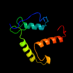
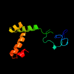
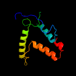
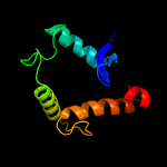
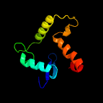
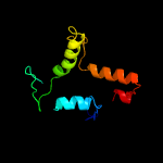
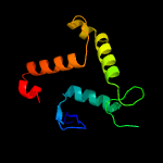
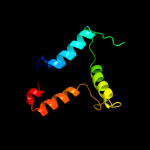
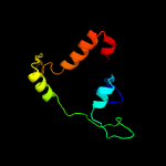
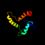
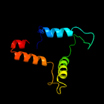
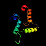
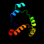
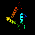
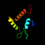
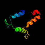
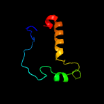
1 c3nkhB_
99.2
19
PDB header: recombinationChain: B: PDB Molecule: integrase;PDBTitle: crystal structure of integrase from mrsa strain staphylococcus aureus
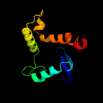
2 c1z1bA_
99.1
13
PDB header: dna binding protein/dnaChain: A: PDB Molecule: integrase;PDBTitle: crystal structure of a lambda integrase dimer bound to a2 coc' core site
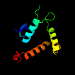
3 c5dcfA_
99.1
19
PDB header: recombinationChain: A: PDB Molecule: tyrosine recombinase xerd,dna translocase ftsk;PDBTitle: c-terminal domain of xerd recombinase in complex with gamma domain of2 ftsk
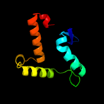
4 c5jjvA_
99.1
18
PDB header: recombinationChain: A: PDB Molecule: tyrosine recombinase xerh;PDBTitle: crystal structure of xerh site-specific recombinase bound to2 palindromic difh substrate: post-cleavage complex
5 c2a3vA_
98.8
15
PDB header: recombinationChain: A: PDB Molecule: site-specific recombinase inti4;PDBTitle: structural basis for broad dna-specificity in integron recombination
6 c6en2A_
98.6
17
PDB header: recombinationChain: A: PDB Molecule: int protein;PDBTitle: structure of the tn1549 transposon integrase (aa 82-397, r225k) in2 complex with a circular intermediate dna (ci6b-dna)
7 c5vfzA_
98.5
18
PDB header: dna binding proteinChain: A: PDB Molecule: gp33;PDBTitle: integrase from mycobacterium phage brujita
8 d1p7da_
98.4
13
Fold: DNA breaking-rejoining enzymesSuperfamily: DNA breaking-rejoining enzymesFamily: Lambda integrase-like, catalytic core
9 c1ma7A_
98.3
9
PDB header: hydrolase, ligase/dnaChain: A: PDB Molecule: cre recombinase;PDBTitle: crystal structure of cre site-specific recombinase2 complexed with a mutant dna substrate, loxp-a8/t27
10 c5hxyE_
98.2
19
PDB header: recombinationChain: E: PDB Molecule: tyrosine recombinase xera;PDBTitle: crystal structure of xera recombinase
11 c1crxA_
98.0
11
PDB header: replication/dnaChain: A: PDB Molecule: cre recombinase;PDBTitle: cre recombinase/dna complex reaction intermediate i
12 d1aiha_
98.0
19
Fold: DNA breaking-rejoining enzymesSuperfamily: DNA breaking-rejoining enzymesFamily: Lambda integrase-like, catalytic core
13 d1ae9a_
97.8
13
Fold: DNA breaking-rejoining enzymesSuperfamily: DNA breaking-rejoining enzymesFamily: Lambda integrase-like, catalytic core
14 d5crxb2
97.7
11
Fold: DNA breaking-rejoining enzymesSuperfamily: DNA breaking-rejoining enzymesFamily: Lambda integrase-like, catalytic core
15 c4a8eA_
97.6
19
PDB header: cell cycleChain: A: PDB Molecule: probable tyrosine recombinase xerc-like;PDBTitle: the structure of a dimeric xer recombinase from archaea
16 c5c6kB_
97.4
24
PDB header: hydrolaseChain: B: PDB Molecule: integrase;PDBTitle: bacteriophage p2 integrase catalytic domain
17 c1a0pA_
97.2
19
PDB header: dna recombinationChain: A: PDB Molecule: site-specific recombinase xerd;PDBTitle: site-specific recombinase, xerd
18 d1a0pa2
96.6
19
Fold: DNA breaking-rejoining enzymesSuperfamily: DNA breaking-rejoining enzymesFamily: Lambda integrase-like, catalytic core
19 d1f44a2
95.9
9
Fold: DNA breaking-rejoining enzymesSuperfamily: DNA breaking-rejoining enzymesFamily: Lambda integrase-like, catalytic core
20 c3uxuA_
95.4
13
PDB header: recombinationChain: A: PDB Molecule: probable integrase;PDBTitle: the structure of the catalytic domain of the sulfolobus spindle-shaped2 viral integrase reveals an evolutionarily conserved catalytic core3 and supports a mechanism of dna cleavage in trans
21 c5ks8F_
not modelled
58.4
4
PDB header: ligaseChain: F: PDB Molecule: pyruvate carboxylase subunit beta;PDBTitle: crystal structure of two-subunit pyruvate carboxylase from2 methylobacillus flagellatus
22 c5ks8D_
not modelled
47.5
4
PDB header: ligaseChain: D: PDB Molecule: pyruvate carboxylase subunit beta;PDBTitle: crystal structure of two-subunit pyruvate carboxylase from2 methylobacillus flagellatus
23 c3bleA_
not modelled
45.4
15
PDB header: transferaseChain: A: PDB Molecule: citramalate synthase from leptospira interrogans;PDBTitle: crystal structure of the catalytic domain of licms in complexed with2 malonate
24 c3ivuB_
not modelled
43.4
8
PDB header: transferaseChain: B: PDB Molecule: homocitrate synthase, mitochondrial;PDBTitle: homocitrate synthase lys4 bound to 2-og
25 c2nx9B_
not modelled
39.8
10
PDB header: lyaseChain: B: PDB Molecule: oxaloacetate decarboxylase 2, subunit alpha;PDBTitle: crystal structure of the carboxyltransferase domain of the2 oxaloacetate decarboxylase na+ pump from vibrio cholerae
26 c1ydnA_
not modelled
38.1
8
PDB header: lyaseChain: A: PDB Molecule: hydroxymethylglutaryl-coa lyase;PDBTitle: crystal structure of the hmg-coa lyase from brucella melitensis,2 northeast structural genomics target lr35.
27 d1rqba2
not modelled
36.5
10
Fold: TIM beta/alpha-barrelSuperfamily: AldolaseFamily: HMGL-like
28 c4lrtC_
not modelled
35.5
15
PDB header: lyase/oxidoreductaseChain: C: PDB Molecule: 4-hydroxy-2-oxovalerate aldolase;PDBTitle: crystal and solution structures of the bifunctional enzyme2 (aldolase/aldehyde dehydrogenase) from thermomonospora curvata,3 reveal a cofactor-binding domain motion during nad+ and coa4 accommodation whithin the shared cofactor-binding site
29 c4jn6C_
not modelled
35.4
13
PDB header: lyase/oxidoreductaseChain: C: PDB Molecule: 4-hydroxy-2-oxovalerate aldolase;PDBTitle: crystal structure of the aldolase-dehydrogenase complex from2 mycobacterium tuberculosis hrv37
30 c3dxiB_
not modelled
32.9
8
PDB header: structural genomics, unknown functionChain: B: PDB Molecule: putative aldolase;PDBTitle: crystal structure of the n-terminal domain of a putative2 aldolase (bvu_2661) from bacteroides vulgatus
31 c3ewbX_
not modelled
30.2
9
PDB header: transferaseChain: X: PDB Molecule: 2-isopropylmalate synthase;PDBTitle: crystal structure of n-terminal domain of putative 2-isopropylmalate2 synthase from listeria monocytogenes
32 c3rmjB_
not modelled
27.1
8
PDB header: transferaseChain: B: PDB Molecule: 2-isopropylmalate synthase;PDBTitle: crystal structure of truncated alpha-isopropylmalate synthase from2 neisseria meningitidis
33 c2qqdG_
not modelled
27.0
25
PDB header: lyaseChain: G: PDB Molecule: pyruvoyl-dependent arginine decarboxylase (ecPDBTitle: n47a mutant of pyruvoyl-dependent arginine decarboxylase2 from methanococcus jannashii
34 c6e1jB_
not modelled
26.2
9
PDB header: plant proteinChain: B: PDB Molecule: 2-isopropylmalate synthase, a genome specific 1;PDBTitle: crystal structure of methylthioalkylmalate synthase (bjumam1.1) from2 brassica juncea
35 c3eegB_
not modelled
25.2
13
PDB header: transferaseChain: B: PDB Molecule: 2-isopropylmalate synthase;PDBTitle: crystal structure of a 2-isopropylmalate synthase from2 cytophaga hutchinsonii
36 c2hjnA_
not modelled
24.8
14
PDB header: cell cycleChain: A: PDB Molecule: maintenance of ploidy protein mob1;PDBTitle: structural and functional analysis of saccharomyces2 cerevisiae mob1
37 c2xflB_
not modelled
24.8
19
PDB header: hydrolaseChain: B: PDB Molecule: dyne7;PDBTitle: induced-fit and allosteric effects upon polyene binding2 revealed by crystal structures of the dynemicin3 thioesterase
38 c2b9sA_
not modelled
23.6
18
PDB header: isomerase/dnaChain: A: PDB Molecule: topoisomerase i-like protein;PDBTitle: crystal structure of heterodimeric l. donovani topoisomerase i-2 vanadate-dna complex
39 c3hzpA_
not modelled
21.4
22
PDB header: structural genomics, unknown functionChain: A: PDB Molecule: ntf2-like protein of unknown function;PDBTitle: crystal structure of ntf2-like protein of unknown function mn2a_05052 from prochlorococcus marinus (yp_291699.1) from prochlorococcus sp.3 natl2a at 1.40 a resolution
40 c5eo4A_
not modelled
20.8
19
PDB header: hydrolaseChain: A: PDB Molecule: thioesterase;PDBTitle: structural and biochemical characterization of the hypothetical2 protein sav2348 from staphylococcus aureus.
41 c1nvmG_
not modelled
18.3
8
PDB header: lyase/oxidoreductaseChain: G: PDB Molecule: 4-hydroxy-2-oxovalerate aldolase;PDBTitle: crystal structure of a bifunctional aldolase-dehydrogenase :2 sequestering a reactive and volatile intermediate
42 c1ydoC_
not modelled
17.8
12
PDB header: lyaseChain: C: PDB Molecule: hmg-coa lyase;PDBTitle: crystal structure of the bacillis subtilis hmg-coa lyase, northeast2 structural genomics target sr181.
43 c2w3xE_
not modelled
17.7
20
PDB header: hydrolaseChain: E: PDB Molecule: cale7;PDBTitle: crystal structure of a bifunctional hotdog fold2 thioesterase in enediyne biosynthesis, cale7
44 c4ov9A_
not modelled
17.1
8
PDB header: transferaseChain: A: PDB Molecule: isopropylmalate synthase;PDBTitle: structure of isopropylmalate synthase binding with alpha-2 isopropylmalate
45 d1k4ta2
not modelled
17.0
16
Fold: DNA breaking-rejoining enzymesSuperfamily: DNA breaking-rejoining enzymesFamily: Eukaryotic DNA topoisomerase I, catalytic core
46 d1rr8c1
not modelled
16.4
15
Fold: DNA breaking-rejoining enzymesSuperfamily: DNA breaking-rejoining enzymesFamily: Eukaryotic DNA topoisomerase I, catalytic core
47 c3m0zD_
not modelled
14.9
13
PDB header: lyaseChain: D: PDB Molecule: putative aldolase;PDBTitle: crystal structure of putative aldolase from klebsiella pneumoniae.
48 c1rr2A_
not modelled
13.5
10
PDB header: transferaseChain: A: PDB Molecule: transcarboxylase 5s subunit;PDBTitle: propionibacterium shermanii transcarboxylase 5s subunit bound to 2-2 ketobutyric acid
49 c4lqqB_
not modelled
13.0
16
PDB header: transferase/transferase activatorChain: B: PDB Molecule: cbk1 kinase activator protein mob2;PDBTitle: crystal structure of the cbk1(t743e)-mob2 kinase-coactivator complex2 in crystal form b
50 c2bcwC_
not modelled
12.4
14
PDB header: ribosomeChain: C: PDB Molecule: elongation factor g;PDBTitle: coordinates of the n-terminal domain of ribosomal protein l11,c-2 terminal domain of ribosomal protein l7/l12 and a portion of the g'3 domain of elongation factor g, as fitted into cryo-em map of an4 escherichia coli 70s*ef-g*gdp*fusidic acid complex
51 c1a31A_
not modelled
11.6
19
PDB header: isomerase/dnaChain: A: PDB Molecule: protein (topoisomerase i);PDBTitle: human reconstituted dna topoisomerase i in covalent complex2 with a 22 base pair dna duplex
52 d2hlja1
not modelled
10.5
18
Fold: Thioesterase/thiol ester dehydrase-isomeraseSuperfamily: Thioesterase/thiol ester dehydrase-isomeraseFamily: 4HBT-like
53 d2oiwa1
not modelled
10.3
18
Fold: Thioesterase/thiol ester dehydrase-isomeraseSuperfamily: Thioesterase/thiol ester dehydrase-isomeraseFamily: 4HBT-like
54 d2oafa1
not modelled
10.2
14
Fold: Thioesterase/thiol ester dehydrase-isomeraseSuperfamily: Thioesterase/thiol ester dehydrase-isomeraseFamily: 4HBT-like
55 c2cw6B_
not modelled
10.0
11
PDB header: lyaseChain: B: PDB Molecule: hydroxymethylglutaryl-coa lyase, mitochondrial;PDBTitle: crystal structure of human hmg-coa lyase: insights into2 catalysis and the molecular basis for3 hydroxymethylglutaric aciduria
56 c2ftpA_
not modelled
9.8
12
PDB header: lyaseChain: A: PDB Molecule: hydroxymethylglutaryl-coa lyase;PDBTitle: crystal structure of hydroxymethylglutaryl-coa lyase from pseudomonas2 aeruginosa
57 d1x9ya2
not modelled
9.7
16
Fold: Cystatin-likeSuperfamily: Cystatin/monellinFamily: Staphopain B, prodomain
58 c3ck1B_
not modelled
9.3
18
PDB header: hydrolaseChain: B: PDB Molecule: putative thioesterase;PDBTitle: crystal structure of a putative thioesterase (reut_a2179) from2 ralstonia eutropha jmp134 at 1.74 a resolution
59 c1p6gE_
not modelled
9.3
13
PDB header: ribosomeChain: E: PDB Molecule: 30s ribosomal protein s5;PDBTitle: real space refined coordinates of the 30s subunit fitted into the low2 resolution cryo-em map of the ef-g.gtp state of e. coli 70s ribosome
60 d1hjra_
not modelled
8.9
15
Fold: Ribonuclease H-like motifSuperfamily: Ribonuclease H-likeFamily: RuvC resolvase
61 d2uube1
not modelled
8.6
11
Fold: Ribosomal protein S5 domain 2-likeSuperfamily: Ribosomal protein S5 domain 2-likeFamily: Translational machinery components
62 c6a4cA_
not modelled
8.5
22
PDB header: unknown functionChain: A: PDB Molecule: uncharacterized protein mxan_0049;PDBTitle: solution structure of mxan_0049
63 d1w26a2
not modelled
8.4
19
Fold: Ribosome binding domain-likeSuperfamily: Trigger factor ribosome-binding domainFamily: Trigger factor ribosome-binding domain
64 d1ka8a_
not modelled
7.8
13
Fold: DNA/RNA-binding 3-helical bundleSuperfamily: "Winged helix" DNA-binding domainFamily: P4 origin-binding domain-like
65 d2qale1
not modelled
7.6
11
Fold: Ribosomal protein S5 domain 2-likeSuperfamily: Ribosomal protein S5 domain 2-likeFamily: Translational machinery components
66 d1h3oa_
not modelled
7.5
19
Fold: Histone-foldSuperfamily: Histone-foldFamily: TBP-associated factors, TAFs
67 c1h3oA_
not modelled
7.5
19
PDB header: transcription/tbp-associated factorsChain: A: PDB Molecule: transcription initiation factor tfiid 135 kda subunit;PDBTitle: crystal structure of the human taf4-taf12 (tafii135-tafii20) complex
68 c4gj1A_
not modelled
7.5
11
PDB header: isomeraseChain: A: PDB Molecule: 1-(5-phosphoribosyl)-5-[(5-phosphoribosylamino)PDBTitle: crystal structure of 1-(5-phosphoribosyl)-5-[(5-phosphoribosylamino)2 methylideneamino] imidazole-4-carboxamide isomerase (hisa).
69 d1nvma2
not modelled
7.2
8
Fold: TIM beta/alpha-barrelSuperfamily: AldolaseFamily: HMGL-like
70 c4y9iA_
not modelled
7.1
18
PDB header: oxidoreductaseChain: A: PDB Molecule: mycobacterium tuberculosis paralogous family 11;PDBTitle: structure of f420-h2 dependent reductase (fdr-a) msmeg_2027
71 d1p9ya_
not modelled
7.1
19
Fold: Ribosome binding domain-likeSuperfamily: Trigger factor ribosome-binding domainFamily: Trigger factor ribosome-binding domain
72 c3bg3B_
not modelled
6.9
8
PDB header: ligaseChain: B: PDB Molecule: pyruvate carboxylase, mitochondrial;PDBTitle: crystal structure of human pyruvate carboxylase (missing the biotin2 carboxylase domain at the n-terminus)
73 d1t11a2
not modelled
6.8
10
Fold: Ribosome binding domain-likeSuperfamily: Trigger factor ribosome-binding domainFamily: Trigger factor ribosome-binding domain
74 d1hlva2
not modelled
6.5
11
Fold: DNA/RNA-binding 3-helical bundleSuperfamily: Homeodomain-likeFamily: Centromere-binding
75 d1p4wa_
not modelled
6.4
14
Fold: DNA/RNA-binding 3-helical bundleSuperfamily: C-terminal effector domain of the bipartite response regulatorsFamily: GerE-like (LuxR/UhpA family of transcriptional regulators)
76 c2d3o1_
not modelled
6.4
14
PDB header: ribosomeChain: 1: PDB Molecule: trigger factor;PDBTitle: structure of ribosome binding domain of the trigger factor on the 50s2 ribosomal subunit from d. radiodurans
77 c3a9iA_
not modelled
6.2
6
PDB header: transferase/transferase inhibitorChain: A: PDB Molecule: homocitrate synthase;PDBTitle: crystal structure of homocitrate synthase from thermus thermophilus2 complexed with lys
78 d2ia9a1
not modelled
5.8
12
Fold: SpoVG-likeSuperfamily: SpoVG-likeFamily: SpoVG-like
79 c2zyfA_
not modelled
5.6
8
PDB header: transferaseChain: A: PDB Molecule: homocitrate synthase;PDBTitle: crystal structure of homocitrate synthase from thermus thermophilus2 complexed with magnesuim ion and alpha-ketoglutarate
80 c4lfuA_
not modelled
5.6
5
PDB header: dna binding proteinChain: A: PDB Molecule: regulatory protein sdia;PDBTitle: crystal structure of escherichia coli sdia in the space group c2
81 d1hywa_
not modelled
5.5
23
Fold: gpW/XkdW-likeSuperfamily: Head-to-tail joining protein W, gpWFamily: Head-to-tail joining protein W, gpW
82 c2jwlB_
not modelled
5.5
22
PDB header: membrane proteinChain: B: PDB Molecule: protein tolr;PDBTitle: solution structure of periplasmic domain of tolr from h.2 influenzae with saxs data
83 c4exqA_
not modelled
5.3
22
PDB header: biosynthetic proteinChain: A: PDB Molecule: uroporphyrinogen decarboxylase;PDBTitle: crystal structure of uroporphyrinogen decarboxylase (upd) from2 burkholderia thailandensis e264
84 c2nscA_
not modelled
5.3
19
PDB header: chaperoneChain: A: PDB Molecule: trigger factor;PDBTitle: structures of and interactions between domains of trigger factor from2 themotoga maritima
85 c3r5wO_
not modelled
5.3
23
PDB header: oxidoreductaseChain: O: PDB Molecule: deazaflavin-dependent nitroreductase;PDBTitle: structure of ddn, the deazaflavin-dependent nitroreductase from2 mycobacterium tuberculosis involved in bioreductive activation of pa-3 824, with co-factor f420
86 d1r3ba_
not modelled
5.2
9
Fold: Bromodomain-likeSuperfamily: Mob1/phoceinFamily: Mob1/phocein
87 c3r5yC_
not modelled
5.2
25
PDB header: unknown functionChain: C: PDB Molecule: putative uncharacterized protein;PDBTitle: structure of a deazaflavin-dependent nitroreductase from nocardia2 farcinica, with co-factor f420
88 c2i9zB_
not modelled
5.1
15
PDB header: structural genomics, unknown functionChain: B: PDB Molecule: putative septation protein spovg;PDBTitle: structural genomics, the crystal structure of full-length spovg from2 staphylococcus epidermidis atcc 12228
89 d2i9xa1
not modelled
5.1
15
Fold: SpoVG-likeSuperfamily: SpoVG-likeFamily: SpoVG-like
90 c2ra4B_
not modelled
5.1
17
PDB header: cytokineChain: B: PDB Molecule: small-inducible cytokine a13;PDBTitle: crystal structure of human monocyte chemoattractant protein 4 (mcp-2 4/ccl13)


