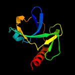
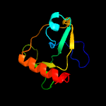
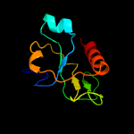
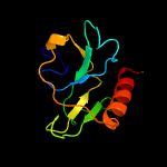
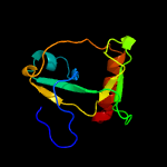
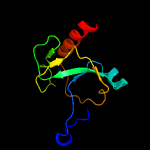
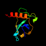
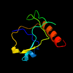
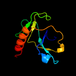
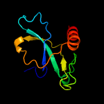
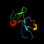
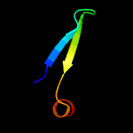
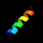
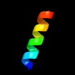
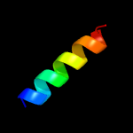
1 c5ccaA_
100.0
98
PDB header: hydrolaseChain: A: PDB Molecule: endoribonuclease mazf3;PDBTitle: crystal structure of mtb toxin
2 c4mzpC_
99.9
21
PDB header: hydrolaseChain: C: PDB Molecule: mazf mrna interferase;PDBTitle: mazf from s. aureus crystal form iii, c2221, 2.7 a
3 d1m1fa_
99.9
22
Fold: SH3-like barrelSuperfamily: Cell growth inhibitor/plasmid maintenance toxic componentFamily: Kid/PemK
4 c5hjzA_
99.9
22
PDB header: hydrolase/rnaChain: A: PDB Molecule: endoribonuclease mazf9;PDBTitle: structure of m. tuberculosis mazf-mt1 (rv2801c) in complex with rna
5 d1ne8a_
99.9
24
Fold: SH3-like barrelSuperfamily: Cell growth inhibitor/plasmid maintenance toxic componentFamily: Kid/PemK
6 d1ub4a_
99.9
18
Fold: SH3-like barrelSuperfamily: Cell growth inhibitor/plasmid maintenance toxic componentFamily: Kid/PemK
7 c5wygC_
99.9
20
PDB header: hydrolaseChain: C: PDB Molecule: probable endoribonuclease mazf7;PDBTitle: the crystal structure of the apo form of mtb mazf
8 c5xe3B_
99.9
24
PDB header: hydrolase/antitoxinChain: B: PDB Molecule: endoribonuclease mazf4;PDBTitle: endoribonuclease in complex with its cognate antitoxin from2 mycobacterial species
9 c5hk3B_
99.9
26
PDB header: hydrolase/dnaChain: B: PDB Molecule: endoribonuclease mazf6;PDBTitle: crystal structure of m. tuberculosis mazf-mt3 t52d-f62d mutant in2 complex with dna
10 c3jrzA_
97.1
12
PDB header: toxinChain: A: PDB Molecule: ccdb;PDBTitle: ccdbvfi-formii-ph5.6
11 d3vuba_
96.6
18
Fold: SH3-like barrelSuperfamily: Cell growth inhibitor/plasmid maintenance toxic componentFamily: CcdB
12 c3d55A_
11.0
14
PDB header: toxin inhibitorChain: A: PDB Molecule: uncharacterized protein rv3357/mt3465;PDBTitle: crystal structure of m. tuberculosis yefm antitoxin
13 c6bwqB_
8.8
45
PDB header: metal binding proteinChain: B: PDB Molecule: pyridinium-3,5-bisthiocarboxylic acid mononucleotide nickelPDBTitle: larc2, the c-terminal domain of a cyclometallase involved in the2 synthesis of the npn cofactor of lactate racemase, in complex with3 mnctp
14 c3c19A_
8.8
36
PDB header: structural genomics, unknown functionChain: A: PDB Molecule: uncharacterized protein mk0293;PDBTitle: crystal structure of protein mk0293 from methanopyrus kandleri av19
15 d1rz4a1
8.3
26
Fold: DNA/RNA-binding 3-helical bundleSuperfamily: "Winged helix" DNA-binding domainFamily: Eukaryotic translation initiation factor 3 subunit 12, eIF3k, C-terminal domain
16 c6n1bA_
7.8
13
PDB header: hydrolaseChain: A: PDB Molecule: carbohydrate-binding protein;PDBTitle: crystal structure of an n-acetylgalactosamine deacetylase from f.2 plautii in complex with blood group b trisaccharide
17 c4bxtD_
6.9
12
PDB header: viral proteinChain: D: PDB Molecule: phosphoprotein p;PDBTitle: crystal structure of the human metapneumovirus2 phosphoprotein tetramerization domain
18 c5oiyG_
6.9
12
PDB header: viral proteinChain: G: PDB Molecule: phosphoprotein;PDBTitle: structure of the hmpv p oligomerization domain at 2.2 a
19 c5oixA_
6.9
12
PDB header: viral proteinChain: A: PDB Molecule: phosphoprotein;PDBTitle: structure of the hmpv p oligomerization domain at 1.6 a
20 c5oiyE_
6.9
12
PDB header: viral proteinChain: E: PDB Molecule: phosphoprotein;PDBTitle: structure of the hmpv p oligomerization domain at 2.2 a
21 c5oixE_
not modelled
6.9
12
PDB header: viral proteinChain: E: PDB Molecule: phosphoprotein;PDBTitle: structure of the hmpv p oligomerization domain at 1.6 a
22 c5oiyC_
not modelled
6.9
12
PDB header: viral proteinChain: C: PDB Molecule: phosphoprotein;PDBTitle: structure of the hmpv p oligomerization domain at 2.2 a
23 c5oixF_
not modelled
6.9
12
PDB header: viral proteinChain: F: PDB Molecule: phosphoprotein;PDBTitle: structure of the hmpv p oligomerization domain at 1.6 a
24 c5oiyA_
not modelled
6.7
12
PDB header: viral proteinChain: A: PDB Molecule: phosphoprotein;PDBTitle: structure of the hmpv p oligomerization domain at 2.2 a
25 c5oiyB_
not modelled
6.7
12
PDB header: viral proteinChain: B: PDB Molecule: phosphoprotein;PDBTitle: structure of the hmpv p oligomerization domain at 2.2 a
26 c4bxtG_
not modelled
6.7
12
PDB header: viral proteinChain: G: PDB Molecule: phosphoprotein p;PDBTitle: crystal structure of the human metapneumovirus2 phosphoprotein tetramerization domain
27 c4bxtF_
not modelled
6.6
12
PDB header: viral proteinChain: F: PDB Molecule: phosphoprotein p;PDBTitle: crystal structure of the human metapneumovirus2 phosphoprotein tetramerization domain
28 c5oiyH_
not modelled
6.6
12
PDB header: viral proteinChain: H: PDB Molecule: phosphoprotein;PDBTitle: structure of the hmpv p oligomerization domain at 2.2 a
29 c4bxtB_
not modelled
6.6
12
PDB header: viral proteinChain: B: PDB Molecule: phosphoprotein p;PDBTitle: crystal structure of the human metapneumovirus2 phosphoprotein tetramerization domain
30 c5oixB_
not modelled
6.6
12
PDB header: viral proteinChain: B: PDB Molecule: phosphoprotein;PDBTitle: structure of the hmpv p oligomerization domain at 1.6 a
31 c4bxtE_
not modelled
6.6
12
PDB header: viral proteinChain: E: PDB Molecule: phosphoprotein p;PDBTitle: crystal structure of the human metapneumovirus2 phosphoprotein tetramerization domain
32 c5oixC_
not modelled
6.6
12
PDB header: viral proteinChain: C: PDB Molecule: phosphoprotein;PDBTitle: structure of the hmpv p oligomerization domain at 1.6 a
33 c3oeiB_
not modelled
6.5
6
PDB header: toxin, protein bindingChain: B: PDB Molecule: relj (antitoxin rv3357);PDBTitle: crystal structure of mycobacterium tuberculosis reljk (rv3357-rv3358-2 relbe3)
34 c5oixG_
not modelled
6.5
12
PDB header: viral proteinChain: G: PDB Molecule: phosphoprotein;PDBTitle: structure of the hmpv p oligomerization domain at 1.6 a
35 c4jhlA_
not modelled
6.1
22
PDB header: hydrolaseChain: A: PDB Molecule: acetyl xylan esterase;PDBTitle: crystal structure of of axe2, an acetylxylan esterase from geobacillus2 stearothermophilus
36 c5fmzB_
not modelled
6.0
9
PDB header: transcriptionChain: B: PDB Molecule: rna-directed rna polymerase catalytic subunit;PDBTitle: crystal structure of influenza b polymerase with bound 5' vrna
37 c4wsaB_
not modelled
5.8
9
PDB header: transferase/rnaChain: B: PDB Molecule: rna-directed rna polymerase catalytic subunit;PDBTitle: crystal structure of influenza b polymerase bound to the vrna promoter2 (flub1 form)
38 c4q3kB_
not modelled
5.3
11
PDB header: hydrolaseChain: B: PDB Molecule: mgs-m1;PDBTitle: crystal structure of mgs-m1, an alpha/beta hydrolase enzyme from a2 medee basin deep-sea metagenome library
39 c4bxtH_
not modelled
5.2
12
PDB header: viral proteinChain: H: PDB Molecule: phosphoprotein p;PDBTitle: crystal structure of the human metapneumovirus2 phosphoprotein tetramerization domain
40 c5oiyF_
not modelled
5.2
12
PDB header: viral proteinChain: F: PDB Molecule: phosphoprotein;PDBTitle: structure of the hmpv p oligomerization domain at 2.2 a
41 c2of3A_
not modelled
5.2
12
PDB header: structural protein, cell cycleChain: A: PDB Molecule: zyg-9;PDBTitle: tog domain structure from c.elegans zyg9
42 d2uubm1
not modelled
5.1
20
Fold: S13-like H2TH domainSuperfamily: S13-like H2TH domainFamily: Ribosomal protein S13
43 c5oixD_
not modelled
5.0
12
PDB header: viral proteinChain: D: PDB Molecule: phosphoprotein;PDBTitle: structure of the hmpv p oligomerization domain at 1.6 a
44 c5oixH_
not modelled
5.0
12
PDB header: viral proteinChain: H: PDB Molecule: phosphoprotein;PDBTitle: structure of the hmpv p oligomerization domain at 1.6 a
45 c5oiyD_
not modelled
5.0
12
PDB header: viral proteinChain: D: PDB Molecule: phosphoprotein;PDBTitle: structure of the hmpv p oligomerization domain at 2.2 a
46 c4bxtA_
not modelled
5.0
12
PDB header: viral proteinChain: A: PDB Molecule: phosphoprotein p;PDBTitle: crystal structure of the human metapneumovirus2 phosphoprotein tetramerization domain


