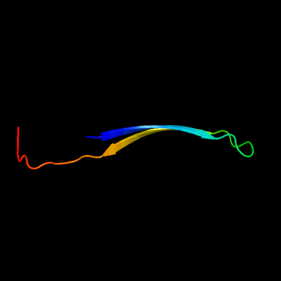
| 1 |
|
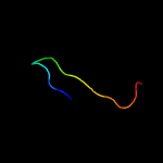
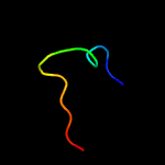
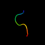
PDB 5mtw chain D
Region: 3 - 36
Aligned: 34
Modelled: 34
Confidence: 41.2%
Identity: 26%
PDB header:chaperone
Chain: D: PDB Molecule:secb-like chaperone rv1957;
PDBTitle: mycobacterium tuberculosis rv1957 secb-like chaperone in complex with2 a chad peptide from rv1956 higa1 antitoxin
Phyre2
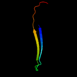
| 2 |
|
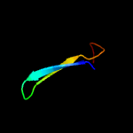
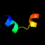
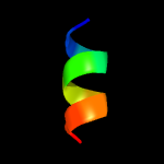
PDB 3ctr chain A
Region: 6 - 19
Aligned: 14
Modelled: 14
Confidence: 29.1%
Identity: 50%
PDB header:hydrolase
Chain: A: PDB Molecule:poly(a)-specific ribonuclease parn;
PDBTitle: crystal structure of the rrm-domain of the poly(a)-specific2 ribonuclease parn bound to m7gtp
Phyre2
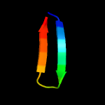
| 3 |
|
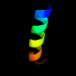
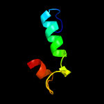
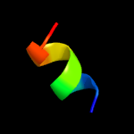
PDB 1whv chain A
Region: 8 - 24
Aligned: 17
Modelled: 17
Confidence: 17.7%
Identity: 47%
Fold: Ferredoxin-like
Superfamily: RNA-binding domain, RBD
Family: Canonical RBD
Phyre2
| 4 |
|
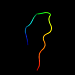
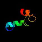
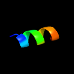
PDB 3d45 chain B
Region: 6 - 25
Aligned: 20
Modelled: 20
Confidence: 17.4%
Identity: 40%
PDB header:hydrolase
Chain: B: PDB Molecule:poly(a)-specific ribonuclease parn;
PDBTitle: crystal structure of mouse parn in complex with m7gpppg
Phyre2
| 5 |
|
PDB 3k7c chain C
Region: 14 - 30
Aligned: 17
Modelled: 17
Confidence: 12.9%
Identity: 35%
PDB header:protein binding
Chain: C: PDB Molecule:putative ntf2-like transpeptidase;
PDBTitle: crystal structure of putative ntf2-like transpeptidase (np_281412.1)2 from campylobacter jejuni at 2.00 a resolution
Phyre2
| 6 |
|
PDB 4f8l chain A
Region: 10 - 23
Aligned: 14
Modelled: 14
Confidence: 9.8%
Identity: 36%
PDB header:cell adhesion/inhibitor
Chain: A: PDB Molecule:ph 6 antigen;
PDBTitle: x-ray structure of psaa from yersinia pestis, in complex with2 galactose and aebsf
Phyre2
| 7 |
|
PDB 5lo7 chain B
Region: 10 - 23
Aligned: 13
Modelled: 14
Confidence: 8.6%
Identity: 54%
PDB header:cell adhesion
Chain: B: PDB Molecule:fimbrial protein myfa,fimbrial protein myfa;
PDBTitle: crystal structure of self-complemented myfa, the major subunit of myf2 fimbriae from yersinia enterocolitica
Phyre2
| 8 |
|
PDB 4x8k chain B
Region: 28 - 44
Aligned: 17
Modelled: 17
Confidence: 8.1%
Identity: 53%
PDB header:transcription activator
Chain: B: PDB Molecule:rna polymerase-binding protein rbpa;
PDBTitle: mycobacterium tuberculosis rbpa-sid in complex with sigmaa domain 2
Phyre2
| 9 |
|
PDB 2hl7 chain A
Region: 11 - 44
Aligned: 32
Modelled: 34
Confidence: 7.5%
Identity: 28%
PDB header:oxidoreductase
Chain: A: PDB Molecule:cytochrome c-type biogenesis protein ccmh;
PDBTitle: crystal structure of the periplasmic domain of ccmh from pseudomonas2 aeruginosa
Phyre2
| 10 |
|
PDB 2kw0 chain A
Region: 11 - 44
Aligned: 32
Modelled: 34
Confidence: 6.6%
Identity: 22%
PDB header:oxidoreductase
Chain: A: PDB Molecule:ccmh protein;
PDBTitle: solution structure of n-terminal domain of ccmh from escherichia.coli
Phyre2
| 11 |
|
PDB 2pjh chain A
Region: 26 - 32
Aligned: 7
Modelled: 7
Confidence: 6.0%
Identity: 57%
PDB header:transport protein
Chain: A: PDB Molecule:nuclear protein localization protein 4 homolog;
PDBTitle: strctural model of the p97 n domain- npl4 ubd complex
Phyre2
| 12 |
|
PDB 2v4h chain A
Region: 16 - 25
Aligned: 10
Modelled: 10
Confidence: 6.0%
Identity: 70%
PDB header:transcription
Chain: A: PDB Molecule:nf-kappa-b essential modulator;
PDBTitle: nemo cc2-lz domain - 1d5 darpin complex
Phyre2
| 13 |
|
PDB 4heo chain A
Region: 12 - 20
Aligned: 9
Modelled: 9
Confidence: 5.8%
Identity: 56%
PDB header:viral protein
Chain: A: PDB Molecule:phosphoprotein;
PDBTitle: hendra virus phosphoprotein c terminal domain
Phyre2
| 14 |
|
PDB 3vp8 chain B
Region: 17 - 28
Aligned: 12
Modelled: 12
Confidence: 5.2%
Identity: 25%
PDB header:transcription
Chain: B: PDB Molecule:general transcriptional corepressor tup1;
PDBTitle: crystal structure of the n-terminal domain of the yeast general2 corepressor tup1p
Phyre2