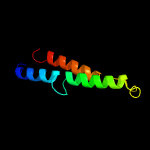
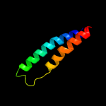
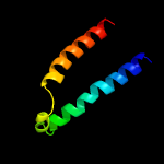
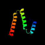
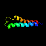
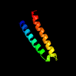
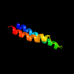
1 c4p79A_
88.7
9
PDB header: cell adhesionChain: A: PDB Molecule: claudin-15;PDBTitle: crystal structure of mouse claudin-15
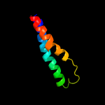
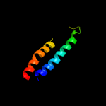
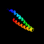
2 c5wivA_
85.6
11
PDB header: signaling protein/antagonistChain: A: PDB Molecule: d(4) dopamine receptor, soluble cytochrome b562 chimera;PDBTitle: structure of the sodium-bound human d4 dopamine receptor in complex2 with nemonapride
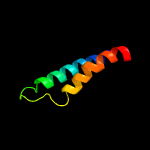
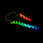
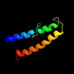
3 c4x5mB_
82.9
12
PDB header: transport proteinChain: B: PDB Molecule: uncharacterized protein;PDBTitle: crystal structure of semisweet in the inward-open conformation
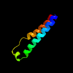
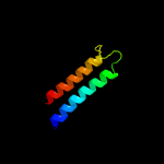
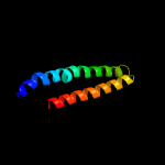
4 c2m67A_
82.3
20
PDB header: transport proteinChain: A: PDB Molecule: merf;PDBTitle: full-length mercury transporter protein merf in lipid bilayer2 membranes
5 c3v2yA_
82.2
27
PDB header: hydrolaseChain: A: PDB Molecule: sphingosine 1-phosphate receptor 1, lysozyme chimeraPDBTitle: crystal structure of a lipid g protein-coupled receptor at 2.80a
6 c6me5A_
80.9
22
PDB header: membrane proteinChain: A: PDB Molecule: chimera protein of melatonin receptor type 1a and glgaPDBTitle: xfel crystal structure of human melatonin receptor mt1 in complex with2 agomelatine
7 c4grvA_
80.5
20
PDB header: signaling protein/agonistChain: A: PDB Molecule: neurotensin receptor type 1, lysozyme chimera;PDBTitle: the crystal structure of the neurotensin receptor nts1 in complex with2 neurotensin (8-13)
8 c5ztyA_
78.8
20
PDB header: membrane proteinChain: A: PDB Molecule: g protein coupled receptor,t4 lysozyme,g protein coupledPDBTitle: crystal structure of human g protein coupled receptor
9 c5gliA_
77.6
17
PDB header: signaling proteinChain: A: PDB Molecule: endothelin receptor subtype-b;PDBTitle: human endothelin receptor type-b in the ligand-free form
10 c6m9tA_
77.2
16
PDB header: membrane proteinChain: A: PDB Molecule: prostaglandin e2 receptor ep3 subtype, endolysin chimera;PDBTitle: crystal structure of ep3 receptor bound to misoprostol-fa
11 c2yevB_
75.7
8
PDB header: electron transportChain: B: PDB Molecule: cytochrome c oxidase subunit 2;PDBTitle: structure of caa3-type cytochrome oxidase
12 c5zlgA_
75.4
11
PDB header: oxidoreductaseChain: A: PDB Molecule: cytochrome b reductase 1;PDBTitle: human duodenal cytochrome b (dcytb) in zinc ion and ascorbate bound2 form
13 c4rngE_
74.7
11
PDB header: sugar binding proteinChain: E: PDB Molecule: mtn3/saliva family;PDBTitle: crystal structure of a bacterial homologue of sweet transporters
14 c6g7oA_
74.5
17
PDB header: membrane proteinChain: A: PDB Molecule: alkaline ceramidase 3,soluble cytochrome b562;PDBTitle: crystal structure of human alkaline ceramidase 3 (acer3) at 2.72 angstrom resolution
15 c3rzeA_
74.5
15
PDB header: hydrolaseChain: A: PDB Molecule: histamine h1 receptor, lysozyme chimera;PDBTitle: structure of the human histamine h1 receptor in complex with doxepin
16 c4k5yA_
74.5
10
PDB header: membrane protein, receptorChain: A: PDB Molecule: corticotropin-releasing factor receptor 1, t4-lysozymePDBTitle: crystal structure of human corticotropin-releasing factor receptor 12 (crf1r) in complex with the antagonist cp-376395
17 c6e59A_
73.8
15
PDB header: signaling proteinChain: A: PDB Molecule: substance-p receptor, glga glycogen synthase, substance-pPDBTitle: crystal structure of the human nk1 tachykinin receptor
18 c4jkvA_
73.2
18
PDB header: membrane proteinChain: A: PDB Molecule: soluble cytochrome b562, smoothened homolog;PDBTitle: structure of the human smoothened 7tm receptor in complex with an2 antitumor agent
19 c5gasN_
73.1
18
PDB header: hydrolaseChain: N: PDB Molecule: archaeal/vacuolar-type h+-atpase subunit i;PDBTitle: thermus thermophilus v/a-atpase, conformation 2
20 c6o3cA_
71.7
18
PDB header: signaling proteinChain: A: PDB Molecule: smoothened homolog;PDBTitle: crystal structure of active smoothened bound to sag21k, cholesterol,2 and nbsmo8
21 c5zbhA_
not modelled
71.7
13
PDB header: signaling proteinChain: A: PDB Molecule: neuropeptide y receptor type 1,t4 lysozyme,neuropeptide yPDBTitle: the crystal structure of human neuropeptide y y1 receptor with bms-2 193885
22 c4rnbA_
not modelled
71.6
17
PDB header: signaling proteinChain: A: PDB Molecule: orexin receptor type 2, glga glycogen synthase;PDBTitle: crystal structure of the human ox2 orexin receptor bound to the2 insomnia drug suvorexant
23 c2lj2A_
not modelled
71.4
11
PDB header: membrane proteinChain: A: PDB Molecule: merf;PDBTitle: integral membrane core domain of the mercury transporter merf in lipid2 bilayer membranes
24 c5xpdA_
not modelled
71.2
15
PDB header: transport proteinChain: A: PDB Molecule: sugar transporter;PDBTitle: sugar transporter of atsweet13 in inward-facing state with a substrate2 analog
25 c5b2gG_
not modelled
70.4
23
PDB header: membrane proteinChain: G: PDB Molecule: endolysin,claudin-4;PDBTitle: crystal structure of human claudin-4 in complex with c-terminal2 fragment of clostridium perfringens enterotoxin
26 c4rfsS_
not modelled
69.3
16
PDB header: hydrolase, transport proteinChain: S: PDB Molecule: substrate binding pritein s;PDBTitle: structure of a pantothenate energy coupling factor transporter
27 c5z62M_
not modelled
69.0
14
PDB header: electron transportChain: M: PDB Molecule: cytochrome c oxidase subunit 8a, mitochondrial;PDBTitle: structure of human cytochrome c oxidase
28 c2jwaA_
not modelled
68.9
20
PDB header: transferaseChain: A: PDB Molecule: receptor tyrosine-protein kinase erbb-2;PDBTitle: erbb2 transmembrane segment dimer spatial structure
29 c2ks1A_
not modelled
68.9
20
PDB header: transferaseChain: A: PDB Molecule: receptor tyrosine-protein kinase erbb-2;PDBTitle: heterodimeric association of transmembrane domains of erbb1 and erbb22 receptors enabling kinase activation
30 c4zwjA_
not modelled
68.3
11
PDB header: signaling proteinChain: A: PDB Molecule: chimera protein of human rhodopsin, mouse s-arrestin, andPDBTitle: crystal structure of rhodopsin bound to arrestin by femtosecond x-ray2 laser
31 c3kp9A_
not modelled
66.2
18
PDB header: blood coagulation,oxidoreductaseChain: A: PDB Molecule: vkorc1/thioredoxin domain protein;PDBTitle: structure of a bacterial homolog of vitamin k epoxide reductase
32 c4zwjC_
not modelled
66.1
11
PDB header: signaling proteinChain: C: PDB Molecule: chimera protein of human rhodopsin, mouse s-arrestin, andPDBTitle: crystal structure of rhodopsin bound to arrestin by femtosecond x-ray2 laser
33 c5x33A_
not modelled
66.0
15
PDB header: membrane proteinChain: A: PDB Molecule: ltb4 receptor,lysozyme,ltb4 receptor;PDBTitle: leukotriene b4 receptor blt1 in complex with biil260
34 c4u16A_
not modelled
65.1
22
PDB header: membrane protein/inhibitorChain: A: PDB Molecule: muscarinic acetylcholine receptor m3,lysozyme,muscarinicPDBTitle: m3-mt4l receptor bound to nms
35 c3jbrE_
not modelled
64.5
17
PDB header: membrane proteinChain: E: PDB Molecule: voltage-dependent calcium channel gamma-1 subunit;PDBTitle: cryo-em structure of the rabbit voltage-gated calcium channel cav1.12 complex at 4.2 angstrom
36 c2rlfA_
not modelled
64.5
35
PDB header: proton transportChain: A: PDB Molecule: matrix protein 2;PDBTitle: proton channel m2 from influenza a in complex with2 inhibitor rimantadine
37 c3uonA_
not modelled
64.4
20
PDB header: signaling protein/antagonistChain: A: PDB Molecule: human m2 muscarinic acetylcholine, receptor t4 lysozymePDBTitle: structure of the human m2 muscarinic acetylcholine receptor bound to2 an antagonist
38 c5tjvA_
not modelled
62.4
15
PDB header: membrane proteinChain: A: PDB Molecule: cannabinoid receptor 1,glga glycogen synthase,cannabinoidPDBTitle: high-resolution crystal structure of the human cb1 cannabinoid2 receptor
39 c2l35B_
not modelled
62.2
32
PDB header: protein bindingChain: B: PDB Molecule: tyro protein tyrosine kinase-binding protein;PDBTitle: structure of the dap12-nkg2c transmembrane heterotrimer
40 c6bkkH_
not modelled
62.2
39
PDB header: membrane proteinChain: H: PDB Molecule: matrix protein 2;PDBTitle: influenza a m2 transmembrane domain bound to amantadine
41 c6bmzN_
not modelled
62.2
39
PDB header: membrane proteinChain: N: PDB Molecule: matrix protein 2;PDBTitle: influenza a m2 transmembrane domain bound to a spiroadamantane2 inhibitor
42 c1nyjD_
not modelled
62.2
39
PDB header: viral proteinChain: D: PDB Molecule: matrix protein m2;PDBTitle: the closed state structure of m2 protein h+ channel by2 solid state nmr spectroscopy
43 c6bmzG_
not modelled
62.2
39
PDB header: membrane proteinChain: G: PDB Molecule: matrix protein 2;PDBTitle: influenza a m2 transmembrane domain bound to a spiroadamantane2 inhibitor
44 c6bkkG_
not modelled
62.2
39
PDB header: membrane proteinChain: G: PDB Molecule: matrix protein 2;PDBTitle: influenza a m2 transmembrane domain bound to amantadine
45 c5jooA_
not modelled
62.2
39
PDB header: viral proteinChain: A: PDB Molecule: matrix protein 2;PDBTitle: xfel structure of influenza a m2 wild type tm domain at low ph in the2 lipidic cubic phase at room temperature
46 c5um1A_
not modelled
62.2
39
PDB header: proton transportChain: A: PDB Molecule: matrix protein 2;PDBTitle: xfel structure of influenza a m2 wild type tm domain at intermediate2 ph in the lipidic cubic phase at room temperature
47 c2kqtB_
not modelled
62.2
39
PDB header: transport proteinChain: B: PDB Molecule: m2 protein;PDBTitle: solid-state nmr structure of the m2 transmembrane peptide of the2 influenza a virus in dmpc lipid bilayers bound to deuterated3 amantadine
48 c1nyjB_
not modelled
62.2
39
PDB header: viral proteinChain: B: PDB Molecule: matrix protein m2;PDBTitle: the closed state structure of m2 protein h+ channel by2 solid state nmr spectroscopy
49 c4qkcA_
not modelled
62.2
39
PDB header: viral proteinChain: A: PDB Molecule: influenza m2 monomer;PDBTitle: influenza a m2 wild type tm domain at low ph in the lipidic cubic2 phase under cryo diffraction conditions
50 c5ttcA_
not modelled
62.2
39
PDB header: membrane proteinChain: A: PDB Molecule: matrix protein 2;PDBTitle: xfel structure of influenza a m2 wild type tm domain at high ph in the2 lipidic cubic phase at room temperature
51 c2kqtD_
not modelled
62.2
39
PDB header: transport proteinChain: D: PDB Molecule: m2 protein;PDBTitle: solid-state nmr structure of the m2 transmembrane peptide of the2 influenza a virus in dmpc lipid bilayers bound to deuterated3 amantadine
52 c6bkkF_
not modelled
62.2
39
PDB header: membrane proteinChain: F: PDB Molecule: matrix protein 2;PDBTitle: influenza a m2 transmembrane domain bound to amantadine
53 c1nyjC_
not modelled
62.2
39
PDB header: viral proteinChain: C: PDB Molecule: matrix protein m2;PDBTitle: the closed state structure of m2 protein h+ channel by2 solid state nmr spectroscopy
54 c6bkkE_
not modelled
62.2
39
PDB header: membrane proteinChain: E: PDB Molecule: matrix protein 2;PDBTitle: influenza a m2 transmembrane domain bound to amantadine
55 c2kqtC_
not modelled
62.2
39
PDB header: transport proteinChain: C: PDB Molecule: m2 protein;PDBTitle: solid-state nmr structure of the m2 transmembrane peptide of the2 influenza a virus in dmpc lipid bilayers bound to deuterated3 amantadine
56 c6bkkC_
not modelled
62.2
39
PDB header: membrane proteinChain: C: PDB Molecule: matrix protein 2;PDBTitle: influenza a m2 transmembrane domain bound to amantadine
57 c4qkmA_
not modelled
62.2
39
PDB header: viral proteinChain: A: PDB Molecule: influenza m2 monomer;PDBTitle: influenza a m2 wild type tm domain at low ph in the lipidic cubic2 phase under room temperature diffraction conditions
58 c6bkkB_
not modelled
62.2
39
PDB header: membrane proteinChain: B: PDB Molecule: matrix protein 2;PDBTitle: influenza a m2 transmembrane domain bound to amantadine
59 c6bocA_
not modelled
62.2
39
PDB header: membrane proteinChain: A: PDB Molecule: matrix protein 2;PDBTitle: influenza a m2 transmembrane domain bound to rimantadine in the2 inward(open) conformation
60 c6bocD_
not modelled
62.2
39
PDB header: membrane proteinChain: D: PDB Molecule: matrix protein 2;PDBTitle: influenza a m2 transmembrane domain bound to rimantadine in the2 inward(open) conformation
61 c6bocC_
not modelled
62.2
39
PDB header: membrane proteinChain: C: PDB Molecule: matrix protein 2;PDBTitle: influenza a m2 transmembrane domain bound to rimantadine in the2 inward(open) conformation
62 c6bkkA_
not modelled
62.2
39
PDB header: membrane proteinChain: A: PDB Molecule: matrix protein 2;PDBTitle: influenza a m2 transmembrane domain bound to amantadine
63 c2kqtA_
not modelled
62.2
39
PDB header: transport proteinChain: A: PDB Molecule: m2 protein;PDBTitle: solid-state nmr structure of the m2 transmembrane peptide of the2 influenza a virus in dmpc lipid bilayers bound to deuterated3 amantadine
64 c4qk7A_
not modelled
62.2
39
PDB header: viral proteinChain: A: PDB Molecule: influenza m2 monomer;PDBTitle: influenza a m2 wild type tm domain at high ph in the lipidic cubic2 phase under cryo diffraction conditions
65 c4qklA_
not modelled
62.2
39
PDB header: viral proteinChain: A: PDB Molecule: influenza m2 monomer, tm domain (22-46);PDBTitle: influenza a m2 wild type tm domain at high ph in the lipidic cubic2 phase under room temperature diffraction conditions
66 c1mp6A_
not modelled
62.2
39
PDB header: membrane proteinChain: A: PDB Molecule: matrix protein m2;PDBTitle: structure of the transmembrane region of the m2 protein h+2 channel by solid state nmr spectroscopy
67 c1nyjA_
not modelled
62.2
39
PDB header: viral proteinChain: A: PDB Molecule: matrix protein m2;PDBTitle: the closed state structure of m2 protein h+ channel by2 solid state nmr spectroscopy
68 c6bkkD_
not modelled
62.2
39
PDB header: membrane proteinChain: D: PDB Molecule: matrix protein 2;PDBTitle: influenza a m2 transmembrane domain bound to amantadine
69 c6bocB_
not modelled
62.2
39
PDB header: membrane proteinChain: B: PDB Molecule: matrix protein 2;PDBTitle: influenza a m2 transmembrane domain bound to rimantadine in the2 inward(open) conformation
70 c2y69Z_
not modelled
62.2
11
PDB header: electron transportChain: Z: PDB Molecule: cytochrome c oxidase polypeptide 8h;PDBTitle: bovine heart cytochrome c oxidase re-refined with molecular oxygen
71 c2bl2F_
not modelled
61.9
16
PDB header: hydrolaseChain: F: PDB Molecule: v-type sodium atp synthase subunit k;PDBTitle: the membrane rotor of the v-type atpase from enterococcus2 hirae
72 c6bmzM_
not modelled
61.8
39
PDB header: membrane proteinChain: M: PDB Molecule: matrix protein 2;PDBTitle: influenza a m2 transmembrane domain bound to a spiroadamantane2 inhibitor
73 c6bmzE_
not modelled
61.8
39
PDB header: membrane proteinChain: E: PDB Molecule: matrix protein 2;PDBTitle: influenza a m2 transmembrane domain bound to a spiroadamantane2 inhibitor
74 c6bmzL_
not modelled
61.8
39
PDB header: membrane proteinChain: L: PDB Molecule: matrix protein 2;PDBTitle: influenza a m2 transmembrane domain bound to a spiroadamantane2 inhibitor
75 c6bmzH_
not modelled
61.8
39
PDB header: membrane proteinChain: H: PDB Molecule: matrix protein 2;PDBTitle: influenza a m2 transmembrane domain bound to a spiroadamantane2 inhibitor
76 c6bklE_
not modelled
61.8
39
PDB header: membrane proteinChain: E: PDB Molecule: matrix protein 2;PDBTitle: influenza a m2 transmembrane domain bound to rimantadine
77 c6bmzA_
not modelled
61.8
39
PDB header: membrane proteinChain: A: PDB Molecule: matrix protein 2;PDBTitle: influenza a m2 transmembrane domain bound to a spiroadamantane2 inhibitor
78 c6bklA_
not modelled
61.8
39
PDB header: membrane proteinChain: A: PDB Molecule: matrix protein 2;PDBTitle: influenza a m2 transmembrane domain bound to rimantadine
79 c6bmzO_
not modelled
61.8
39
PDB header: membrane proteinChain: O: PDB Molecule: matrix protein 2;PDBTitle: influenza a m2 transmembrane domain bound to a spiroadamantane2 inhibitor
80 c6bmzP_
not modelled
61.8
39
PDB header: membrane proteinChain: P: PDB Molecule: matrix protein 2;PDBTitle: influenza a m2 transmembrane domain bound to a spiroadamantane2 inhibitor
81 c6bmzC_
not modelled
61.8
39
PDB header: membrane proteinChain: C: PDB Molecule: matrix protein 2;PDBTitle: influenza a m2 transmembrane domain bound to a spiroadamantane2 inhibitor
82 c6bmzK_
not modelled
61.8
39
PDB header: membrane proteinChain: K: PDB Molecule: matrix protein 2;PDBTitle: influenza a m2 transmembrane domain bound to a spiroadamantane2 inhibitor
83 c6bmzI_
not modelled
61.8
39
PDB header: membrane proteinChain: I: PDB Molecule: matrix protein 2;PDBTitle: influenza a m2 transmembrane domain bound to a spiroadamantane2 inhibitor
84 c6bklG_
not modelled
61.4
39
PDB header: membrane proteinChain: G: PDB Molecule: matrix protein 2;PDBTitle: influenza a m2 transmembrane domain bound to rimantadine
85 c6bmzJ_
not modelled
61.4
39
PDB header: membrane proteinChain: J: PDB Molecule: matrix protein 2;PDBTitle: influenza a m2 transmembrane domain bound to a spiroadamantane2 inhibitor
86 c6bklD_
not modelled
61.4
39
PDB header: membrane proteinChain: D: PDB Molecule: matrix protein 2;PDBTitle: influenza a m2 transmembrane domain bound to rimantadine
87 c6bmzD_
not modelled
61.4
39
PDB header: membrane proteinChain: D: PDB Molecule: matrix protein 2;PDBTitle: influenza a m2 transmembrane domain bound to a spiroadamantane2 inhibitor
88 c6bklB_
not modelled
61.4
39
PDB header: membrane proteinChain: B: PDB Molecule: matrix protein 2;PDBTitle: influenza a m2 transmembrane domain bound to rimantadine
89 c6bklF_
not modelled
61.4
39
PDB header: membrane proteinChain: F: PDB Molecule: matrix protein 2;PDBTitle: influenza a m2 transmembrane domain bound to rimantadine
90 c6bmzF_
not modelled
61.4
39
PDB header: membrane proteinChain: F: PDB Molecule: matrix protein 2;PDBTitle: influenza a m2 transmembrane domain bound to a spiroadamantane2 inhibitor
91 c6bmzB_
not modelled
61.4
39
PDB header: membrane proteinChain: B: PDB Molecule: matrix protein 2;PDBTitle: influenza a m2 transmembrane domain bound to a spiroadamantane2 inhibitor
92 c6bklC_
not modelled
61.4
39
PDB header: membrane proteinChain: C: PDB Molecule: matrix protein 2;PDBTitle: influenza a m2 transmembrane domain bound to rimantadine
93 c6bklH_
not modelled
61.4
39
PDB header: membrane proteinChain: H: PDB Molecule: matrix protein 2;PDBTitle: influenza a m2 transmembrane domain bound to rimantadine
94 c4wolA_
not modelled
61.4
32
PDB header: signaling proteinChain: A: PDB Molecule: tyro protein tyrosine kinase-binding protein;PDBTitle: crystal structure of the dap12 transmembrane domain in lipidic cubic2 phase
95 c2l34B_
not modelled
61.4
32
PDB header: protein bindingChain: B: PDB Molecule: tyro protein tyrosine kinase-binding protein;PDBTitle: structure of the dap12 transmembrane homodimer
96 c2l34A_
not modelled
61.4
32
PDB header: protein bindingChain: A: PDB Molecule: tyro protein tyrosine kinase-binding protein;PDBTitle: structure of the dap12 transmembrane homodimer
97 c4wo1A_
not modelled
61.3
32
PDB header: signaling proteinChain: A: PDB Molecule: tyro protein tyrosine kinase-binding protein;PDBTitle: crystal structure of the dap12 transmembrane domain in lipid cubic2 phase
98 c4wo1B_
not modelled
61.3
32
PDB header: signaling proteinChain: B: PDB Molecule: tyro protein tyrosine kinase-binding protein;PDBTitle: crystal structure of the dap12 transmembrane domain in lipid cubic2 phase
99 c4wo1D_
not modelled
61.3
32
PDB header: signaling proteinChain: D: PDB Molecule: tyro protein tyrosine kinase-binding protein;PDBTitle: crystal structure of the dap12 transmembrane domain in lipid cubic2 phase
100 c4wo1C_
not modelled
61.3
32
PDB header: signaling proteinChain: C: PDB Molecule: tyro protein tyrosine kinase-binding protein;PDBTitle: crystal structure of the dap12 transmembrane domain in lipid cubic2 phase
101 c5sv0C_
not modelled
60.0
22
PDB header: transport proteinChain: C: PDB Molecule: biopolymer transport protein exbb;PDBTitle: structure of the exbb/exbd complex from e. coli at ph 7.0
102 c5ws4A_
not modelled
59.8
16
PDB header: membrane proteinChain: A: PDB Molecule: macrolide export atp-binding/permease protein macb;PDBTitle: crystal structure of tripartite-type abc transporter macb from2 acinetobacter baumannii
103 c4wolC_
not modelled
59.6
32
PDB header: signaling proteinChain: C: PDB Molecule: tyro protein tyrosine kinase-binding protein;PDBTitle: crystal structure of the dap12 transmembrane domain in lipidic cubic2 phase
104 c4wolB_
not modelled
59.6
32
PDB header: signaling proteinChain: B: PDB Molecule: tyro protein tyrosine kinase-binding protein;PDBTitle: crystal structure of the dap12 transmembrane domain in lipidic cubic2 phase
105 c5xjjA_
not modelled
59.5
14
PDB header: transport proteinChain: A: PDB Molecule: multi drug efflux transporter;PDBTitle: crystal structure of a mate family protein
106 c4or2A_
not modelled
59.0
16
PDB header: signaling proteinChain: A: PDB Molecule: soluble cytochrome b562, metabotropic glutamate receptor 1;PDBTitle: human class c g protein-coupled metabotropic glutamate receptor 1 in2 complex with a negative allosteric modulator
107 c6mjhE_
not modelled
58.6
39
PDB header: membrane proteinChain: E: PDB Molecule: matrix protein 2;PDBTitle: the s31n mutant of the influenza a m2 proton channel in two distinct2 conformational states
108 c6mjhC_
not modelled
58.6
39
PDB header: membrane proteinChain: C: PDB Molecule: matrix protein 2;PDBTitle: the s31n mutant of the influenza a m2 proton channel in two distinct2 conformational states
109 c5c02A_
not modelled
58.6
39
PDB header: membrane proteinChain: A: PDB Molecule: matrix protein 2;PDBTitle: influenza a m2 transmembrane domain drug-resistant s31n mutant at ph2 8.0
110 c6mjhD_
not modelled
58.6
39
PDB header: membrane proteinChain: D: PDB Molecule: matrix protein 2;PDBTitle: the s31n mutant of the influenza a m2 proton channel in two distinct2 conformational states
111 c6mjhG_
not modelled
58.6
39
PDB header: membrane proteinChain: G: PDB Molecule: matrix protein 2;PDBTitle: the s31n mutant of the influenza a m2 proton channel in two distinct2 conformational states
112 c6mjhF_
not modelled
58.6
39
PDB header: membrane proteinChain: F: PDB Molecule: matrix protein 2;PDBTitle: the s31n mutant of the influenza a m2 proton channel in two distinct2 conformational states
113 c6mjhB_
not modelled
58.6
39
PDB header: membrane proteinChain: B: PDB Molecule: matrix protein 2;PDBTitle: the s31n mutant of the influenza a m2 proton channel in two distinct2 conformational states
114 c6mjhA_
not modelled
58.6
39
PDB header: membrane proteinChain: A: PDB Molecule: matrix protein 2;PDBTitle: the s31n mutant of the influenza a m2 proton channel in two distinct2 conformational states
115 c6mjhH_
not modelled
58.6
39
PDB header: membrane proteinChain: H: PDB Molecule: matrix protein 2;PDBTitle: the s31n mutant of the influenza a m2 proton channel in two distinct2 conformational states
116 c5xu1M_
not modelled
58.6
13
PDB header: transport proteinChain: M: PDB Molecule: abc transporter permeae;PDBTitle: structure of a non-canonical abc transporter from streptococcus2 pneumoniae r6
117 c6akfC_
not modelled
58.4
21
PDB header: membrane protein/toxinChain: C: PDB Molecule: claudin-3;PDBTitle: crystal structure of mouse claudin-3 p134a mutant in complex with c-2 terminal fragment of clostridium perfringens enterotoxin
118 c4nl6C_
not modelled
57.5
21
PDB header: splicingChain: C: PDB Molecule: survival motor neuron protein;PDBTitle: structure of the full-length form of the protein smn found in healthy2 patients
119 c6c14A_
not modelled
57.2
13
PDB header: membrane protein, metal transportChain: A: PDB Molecule: protocadherin-15;PDBTitle: cryoem structure of mouse pcdh15-1ec-lhfpl5 complex
120 c5o9hA_
not modelled
56.8
12
PDB header: membrane proteinChain: A: PDB Molecule: c5a anaphylatoxin chemotactic receptor 1;PDBTitle: crystal structure of thermostabilised human c5a anaphylatoxin2 chemotactic receptor 1 (c5ar) in complex with ndt9513727


