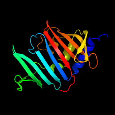
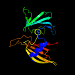
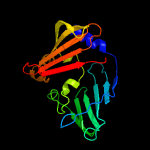
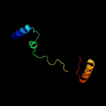
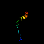
1 c3mhaB_
100.0
100
PDB header: lipid binding proteinChain: B: PDB Molecule: lipoprotein lprg;PDBTitle: crystal structure of lprg from mycobacterium tuberculosis bound to pim
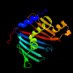
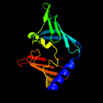
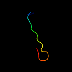
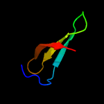
2 d2byoa1
100.0
30
Fold: LolA-like prokaryotic lipoproteins and lipoprotein localization factorsSuperfamily: Prokaryotic lipoproteins and lipoprotein localization factorsFamily: LppX-like
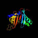
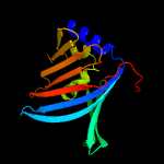
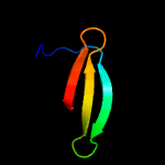
3 c4qa8A_
100.0
30
PDB header: lipid transportChain: A: PDB Molecule: putative lipoprotein lprf;PDBTitle: crystal structure of lprf from mycobacterium bovis
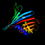
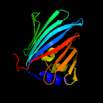
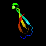
4 c3buuB_
98.6
12
PDB header: structural genomics, unknown functionChain: B: PDB Molecule: uncharacterized lola superfamily protein ne2245;PDBTitle: crystal structure of lola superfamily protein ne2245 from2 nitrosomonas europaea
5 c4mxtA_
98.0
11
PDB header: protein transportChain: A: PDB Molecule: uncharacterized protein;PDBTitle: crystal structure of an outer-membrane lipoprotein carrier protein2 (bacuni_04723) from bacteroides uniformis atcc 8492 at 1.40 a3 resolution
6 c6in8A_
97.3
13
PDB header: membrane proteinChain: A: PDB Molecule: sigma factor algu regulatory protein mucb;PDBTitle: crystal structure of mucb
7 c2v43A_
94.9
15
PDB header: regulatorChain: A: PDB Molecule: sigma-e factor regulatory protein rseb;PDBTitle: crystal structure of rseb: a sensor for periplasmic stress2 response in e. coli
8 c3bk5A_
92.6
10
PDB header: structural genomics, unknown functionChain: A: PDB Molecule: putative outer membrane lipoprotein-sorting protein;PDBTitle: crystal structure of putative outer membrane lipoprotein-sorting2 protein domain from vibrio parahaemolyticus
9 c4z48B_
88.5
13
PDB header: structural biology, unknown functionChain: B: PDB Molecule: uncharacterized protein;PDBTitle: crystal structure of a duf1329 family protein (despig_00262) from2 desulfovibrio piger atcc 29098 at 1.75 a resolution
10 c2w7qB_
86.1
9
PDB header: protein transportChain: B: PDB Molecule: outer-membrane lipoprotein carrier protein;PDBTitle: structure of pseudomonas aeruginosa lola
11 d1iwla_
66.4
14
Fold: LolA-like prokaryotic lipoproteins and lipoprotein localization factorsSuperfamily: Prokaryotic lipoproteins and lipoprotein localization factorsFamily: Outer-membrane lipoproteins carrier protein LolA
12 c3woaA_
54.0
6
PDB header: dna binding protein, sugar binding proteChain: A: PDB Molecule: repressor protein ci, maltose-binding periplasmic protein;PDBTitle: crystal structure of lambda repressor (1-45) fused with maltose-2 binding protein
13 c4mjsQ_
45.5
38
PDB header: transferase/protein bindingChain: Q: PDB Molecule: protein kinase c zeta type;PDBTitle: crystal structure of a pb1 complex
14 c4a4fA_
19.6
15
PDB header: rna binding proteinChain: A: PDB Molecule: survival of motor neuron-related-splicing factor 30;PDBTitle: solution structure of spf30 tudor domain in complex with symmetrically2 dimethylated arginine
15 d1mhna_
19.1
11
Fold: SH3-like barrelSuperfamily: Tudor/PWWP/MBTFamily: Tudor domain
16 c3ls1A_
18.3
12
PDB header: photosynthesisChain: A: PDB Molecule: sll1638 protein;PDBTitle: crystal structure of cyanobacterial psbq from synechocystis2 sp. pcc 6803 complexed with zn2+
17 d2d9ta1
16.7
17
Fold: SH3-like barrelSuperfamily: Tudor/PWWP/MBTFamily: Tudor domain
18 c3pnwX_
16.4
18
PDB header: protein binding/immune systemChain: X: PDB Molecule: tudor domain-containing protein 3;PDBTitle: crystal structure of the tudor domain of human tdrd3 in complex with2 an anti-tdrd3 fab
19 c1g5vA_
16.2
11
PDB header: translationChain: A: PDB Molecule: survival motor neuron protein 1;PDBTitle: solution structure of the tudor domain of the human smn2 protein
20 c2d9tA_
16.2
17
PDB header: structural genomics, unknown functionChain: A: PDB Molecule: tudor domain-containing protein 3;PDBTitle: solution structure of the tudor domain of tudor domain2 containing protein 3 from mouse
21 d1ueba2
not modelled
14.5
24
Fold: OB-foldSuperfamily: Nucleic acid-binding proteinsFamily: Cold shock DNA-binding domain-like
22 c6gwjD_
not modelled
13.3
10
PDB header: rna binding proteinChain: D: PDB Molecule: ekc/keops complex subunit gon7;PDBTitle: protein complex
23 d1v0aa1
not modelled
12.9
9
Fold: Galactose-binding domain-likeSuperfamily: Galactose-binding domain-likeFamily: CBM11
24 d1jrma_
not modelled
12.1
24
Fold: YggU-likeSuperfamily: YggU-likeFamily: YggU-like
25 c6e5fA_
not modelled
9.1
23
PDB header: lipid binding proteinChain: A: PDB Molecule: lipid binding protein lpqn;PDBTitle: crystal structure of lpqn involved in cell envelope biogenesis of2 mycobacterium tuberculosis
26 d2gysa2
not modelled
7.4
20
Fold: Immunoglobulin-like beta-sandwichSuperfamily: Fibronectin type IIIFamily: Fibronectin type III
27 c2m1hA_
not modelled
7.1
11
PDB header: transcriptionChain: A: PDB Molecule: transcription elongation factor s-ii;PDBTitle: solution structure of a pwwp domain from trypanosoma brucei
28 c4f98A_
not modelled
7.1
20
PDB header: structural genomics, unknown functionChain: A: PDB Molecule: hypothetical protein;PDBTitle: crystal structure of a duf2790 family protein (pa3229) from2 pseudomonas aeruginosa pao1 at 1.26 a resolution
29 c2lojA_
not modelled
6.9
20
PDB header: structural genomics, unknown functionChain: A: PDB Molecule: putative cytoplasmic protein;PDBTitle: solution nmr structure of tstm1273 from salmonella typhimurium lt2,2 nesg target stt322, csgid target idp01027 and ocsp target tstm1273
30 c2jraB_
not modelled
6.2
10
PDB header: structural genomics, unknown functionChain: B: PDB Molecule: protein rpa2121;PDBTitle: a novel domain-swapped solution nmr structure of protein rpa2121 from2 rhodopseudomonas palustris. northeast structural genomics target rpt6
31 c5o60Y_
not modelled
6.0
21
PDB header: ribosomeChain: Y: PDB Molecule: 50s ribosomal protein l28;PDBTitle: structure of the 50s large ribosomal subunit from mycobacterium2 smegmatis
32 c6gvwJ_
not modelled
5.9
33
PDB header: signaling proteinChain: J: PDB Molecule: brca1-a complex subunit rap80;PDBTitle: crystal structure of the brca1-a complex
33 c5e7tI_
not modelled
5.5
21
PDB header: viral proteinChain: I: PDB Molecule: major structural protein 1;PDBTitle: structure of the tripod (bppuct-a-l) from the baseplate of2 bacteriophage tuc2009
34 c3p8dB_
not modelled
5.5
16
PDB header: protein bindingChain: B: PDB Molecule: medulloblastoma antigen mu-mb-50.72;PDBTitle: crystal structure of the second tudor domain of human phf20 (homodimer2 form)
35 c2x34A_
not modelled
5.4
18
PDB header: carbohydrate-binding proteinChain: A: PDB Molecule: cellulose-binding protein, x158;PDBTitle: structure of a polyisoprenoid binding domain from saccharophagus2 degradans implicated in plant cell wall breakdown
36 c5x7hA_
not modelled
5.2
13
PDB header: transferaseChain: A: PDB Molecule: cycloisomaltooligosaccharide glucanotransferase;PDBTitle: crystal structure of paenibacillus sp. 598k2 cycloisomaltooligosaccharide glucanotransferase complexed with3 cycloisomaltoheptaose
37 c2hqxB_
not modelled
5.2
9
PDB header: transcriptionChain: B: PDB Molecule: p100 co-activator tudor domain;PDBTitle: crystal structure of human p100 tudor domain conserved2 region