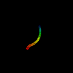
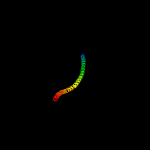
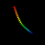
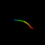
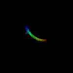
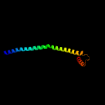
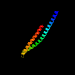
1 c6gajA_
98.9
12
PDB header: viral proteinChain: A: PDB Molecule: outer capsid protein sigma-1;PDBTitle: crystal structure of the t1l reovirus sigma1 coiled coil tail (iodide)
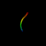
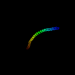
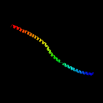
2 c6gapB_
98.1
13
PDB header: viral proteinChain: B: PDB Molecule: outer capsid protein sigma-1;PDBTitle: crystal structure of the t3d reovirus sigma1 coiled coil tail and body
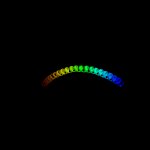
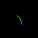
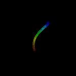
3 c4fmyE_
97.8
17
PDB header: viral proteinChain: E: PDB Molecule: dna stabilization protein;PDBTitle: hk620 tail needle crystal form i
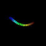
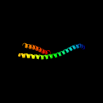
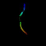
4 c5bu8A_
96.5
14
PDB header: viral proteinChain: A: PDB Molecule: dna stabilization protein;PDBTitle: hk620 tail needle crystallized at ph 7.5 and derivatized with xenon
5 c6gaoC_
96.3
11
PDB header: viral proteinChain: C: PDB Molecule: outer capsid protein sigma-1;PDBTitle: crystal structure of the t1l reovirus sigma1 coiled coil tail and body
6 c3u1aC_
96.2
12
PDB header: contractile proteinChain: C: PDB Molecule: smooth muscle tropomyosin alpha;PDBTitle: n-terminal 81-aa fragment of smooth muscle tropomyosin alpha
7 c2d3eD_
96.1
11
PDB header: contractile proteinChain: D: PDB Molecule: general control protein gcn4 and tropomyosin 1 alpha chain;PDBTitle: crystal structure of the c-terminal fragment of rabbit skeletal alpha-2 tropomyosin
8 c2fxmB_
96.1
13
PDB header: contractile proteinChain: B: PDB Molecule: myosin heavy chain, cardiac muscle beta isoform;PDBTitle: structure of the human beta-myosin s2 fragment
9 c4abxB_
95.6
11
PDB header: dna binding proteinChain: B: PDB Molecule: dna repair protein recn;PDBTitle: crystal structure of deinococcus radiodurans recn coiled-2 coil domain
10 c3ghgI_
95.5
9
PDB header: blood clottingChain: I: PDB Molecule: fibrinogen gamma chain;PDBTitle: crystal structure of human fibrinogen
11 c5wjbA_
95.4
11
PDB header: actin/dna binding proteinChain: A: PDB Molecule: capsid assembly scaffolding protein,myosin-7;PDBTitle: crystal structure of amino acids 1733-1797 of human beta cardiac2 myosin fused to gp7
12 c3u59C_
94.9
11
PDB header: contractile proteinChain: C: PDB Molecule: tropomyosin beta chain;PDBTitle: n-terminal 98-aa fragment of smooth muscle tropomyosin beta
13 c5cwsE_
94.8
12
PDB header: protein transportChain: E: PDB Molecule: nucleoporin nup57;PDBTitle: crystal structure of the intact chaetomium thermophilum nsp1-nup49-2 nup57 channel nucleoporin heterotrimer bound to its nic96 nuclear3 pore complex attachment site
14 c2pohA_
94.5
8
PDB header: viral proteinChain: A: PDB Molecule: head completion protein;PDBTitle: structure of phage p22 tail needle gp26
15 c2b9cA_
94.4
16
PDB header: contractile proteinChain: A: PDB Molecule: striated-muscle alpha tropomyosin;PDBTitle: structure of tropomyosin's mid-region: bending and binding sites for2 actin
16 c5nmoA_
94.4
14
PDB header: cell cycleChain: A: PDB Molecule: chromosome partition protein smc,chromosome partitionPDBTitle: structure of the bacillus subtilis smc joint domain
17 c2gl2B_
94.2
14
PDB header: cell adhesionChain: B: PDB Molecule: adhesion a;PDBTitle: crystal structure of the tetra muntant (t66g,r67g,f68g,y69g) of2 bacterial adhesin fada
18 c1ei3C_
94.1
7
PDB header: blood clottingChain: C: PDB Molecule: fibrinogen;PDBTitle: crystal structure of native chicken fibrinogen
19 c2efrB_
93.8
11
PDB header: contractile proteinChain: B: PDB Molecule: general control protein gcn4 and tropomyosin 1 alpha chain;PDBTitle: crystal structure of the c-terminal tropomyosin fragment with n- and2 c-terminal extensions of the leucine zipper at 1.8 angstroms3 resolution
20 c1deqF_
93.5
10
PDB header: blood clottingChain: F: PDB Molecule: fibrinogen (gamma chain);PDBTitle: the crystal structure of modified bovine fibrinogen (at ~42 angstrom resolution)
21 c5c3lC_
not modelled
93.4
17
PDB header: transport proteinChain: C: PDB Molecule: nucleoporin nup62;PDBTitle: structure of the metazoan nup62.nup58.nup54 nucleoporin complex.
22 c3ojaB_
not modelled
92.5
6
PDB header: protein bindingChain: B: PDB Molecule: anopheles plasmodium-responsive leucine-rich repeat proteinPDBTitle: crystal structure of lrim1/apl1c complex
23 c4rh7A_
not modelled
92.4
10
PDB header: motor proteinChain: A: PDB Molecule: green fluorescent protein/cytoplasmic dynein 2 heavy chainPDBTitle: crystal structure of human cytoplasmic dynein 2 motor domain in2 complex with adp.vi
24 c6ewyA_
not modelled
92.3
12
PDB header: structural proteinChain: A: PDB Molecule: peptidoglycan endopeptidase ripa;PDBTitle: ripa peptidoglycan hydrolase (rv1477, mycobacterium tuberculosis) n-2 terminal domain
25 c4a7fB_
not modelled
92.3
12
PDB header: structural protein/hydrolaseChain: B: PDB Molecule: tropomyosin 1 alpha;PDBTitle: structure of the actin-tropomyosin-myosin complex (rigor atm 3)
26 c1c1gA_
not modelled
92.1
10
PDB header: contractile proteinChain: A: PDB Molecule: tropomyosin;PDBTitle: crystal structure of tropomyosin at 7 angstroms resolution in the2 spermine-induced crystal form
27 c5ijnF_
not modelled
91.6
11
PDB header: transport proteinChain: F: PDB Molecule: nuclear pore complex protein nup54;PDBTitle: composite structure of the inner ring of the human nuclear pore2 complex (32 copies of nup205)
28 c3cwgA_
not modelled
91.5
9
PDB header: transcriptionChain: A: PDB Molecule: signal transducer and activator of transcriptionPDBTitle: unphosphorylated mouse stat3 core fragment
29 c3j99M_
not modelled
91.4
11
PDB header: hydrolaseChain: M: PDB Molecule: synaptosomal-associated protein 25;PDBTitle: structure of 20s supercomplex determined by single particle2 cryoelectron microscopy (state iiib)
30 c5nugB_
not modelled
90.8
10
PDB header: motor proteinChain: B: PDB Molecule: cytoplasmic dynein 1 heavy chain 1;PDBTitle: motor domains from human cytoplasmic dynein-1 in the phi-particle2 conformation
31 c1deqO_
not modelled
90.7
13
PDB header: blood clottingChain: O: PDB Molecule: fibrinogen (beta chain);PDBTitle: the crystal structure of modified bovine fibrinogen (at ~42 angstrom resolution)
32 c5gnaB_
not modelled
90.5
19
PDB header: gene regulationChain: B: PDB Molecule: flagellar hook-associated protein 2;PDBTitle: crystal structure of flagellin assembly related protein
33 c2ba2A_
not modelled
90.5
17
PDB header: structural genomics, unknown functionChain: A: PDB Molecule: hypothetical upf0134 protein mpn010;PDBTitle: crystal structure of the duf16 domain of mpn010 from2 mycoplasma pneumoniae
34 c5ijnT_
not modelled
90.1
14
PDB header: transport proteinChain: T: PDB Molecule: nuclear pore glycoprotein p62;PDBTitle: composite structure of the inner ring of the human nuclear pore2 complex (32 copies of nup205)
35 c5xauB_
not modelled
89.2
12
PDB header: cell adhesionChain: B: PDB Molecule: laminin subunit beta-1;PDBTitle: crystal structure of integrin binding fragment of laminin-511
36 c3q8tB_
not modelled
88.3
12
PDB header: apoptosisChain: B: PDB Molecule: beclin-1;PDBTitle: crystal structure of the coiled coil domain of beclin 1, an essential2 autophagy protein
37 c1ei3E_
not modelled
87.8
12
PDB header: blood clottingChain: E: PDB Molecule: fibrinogen;PDBTitle: crystal structure of native chicken fibrinogen
38 c6o7xa_
not modelled
87.7
9
PDB header: membrane proteinChain: A: PDB Molecule: vacuolar atp synthase catalytic subunit a;PDBTitle: saccharomyces cerevisiae v-atpase stv1-v1vo state 3
39 c2ykqC_
not modelled
87.4
5
PDB header: rna binding proteinChain: C: PDB Molecule: line-1 orf1p;PDBTitle: structure of the human line-1 orf1p trimer
40 c3swfA_
not modelled
87.1
16
PDB header: transport proteinChain: A: PDB Molecule: cgmp-gated cation channel alpha-1;PDBTitle: cnga1 621-690 containing clz domain
41 c3dtpA_
not modelled
87.1
11
PDB header: contractile proteinChain: A: PDB Molecule: myosin 2 heavy chain chimera of smooth and cardiac muscle;PDBTitle: tarantula heavy meromyosin obtained by flexible docking to tarantula2 muscle thick filament cryo-em 3d-map
42 c1ic2B_
not modelled
87.0
16
PDB header: contractile proteinChain: B: PDB Molecule: tropomyosin alpha chain, skeletal muscle;PDBTitle: deciphering the design of the tropomyosin molecule
43 c6ec0A_
not modelled
87.0
9
PDB header: protein fibrilChain: A: PDB Molecule: keratin 1;PDBTitle: crystal structure of the wild-type heterocomplex between coil 1b2 domains of human intermediate filament proteins keratin 1 (krt1) and3 keratin 10 (krt10)
44 c5j1gA_
not modelled
86.8
16
PDB header: structural proteinChain: A: PDB Molecule: plectin;PDBTitle: structure of the spectrin repeats 7 and 8 of the plakin domain of2 plectin
45 c5tvbB_
not modelled
85.7
10
PDB header: transferaseChain: B: PDB Molecule: nucleoprotein tpr;PDBTitle: structure of the tpr oligomerization domain
46 c5mg8B_
not modelled
85.5
10
PDB header: recombinationChain: B: PDB Molecule: structural maintenance of chromosomes protein 6;PDBTitle: crystal structure of the s.pombe smc5/6 hinge domain
47 c5lm2B_
not modelled
85.5
8
PDB header: hydrolaseChain: B: PDB Molecule: tyrosine-protein phosphatase non-receptor type 23;PDBTitle: crystal structure of hd-ptp phosphatase
48 c5gasN_
not modelled
85.1
15
PDB header: hydrolaseChain: N: PDB Molecule: archaeal/vacuolar-type h+-atpase subunit i;PDBTitle: thermus thermophilus v/a-atpase, conformation 2
49 c6o7ua_
not modelled
84.6
20
PDB header: membrane proteinChain: A: PDB Molecule: PDBTitle: saccharomyces cerevisiae v-atpase stv1-vo
50 c5yfpE_
not modelled
84.0
10
PDB header: exocytosisChain: E: PDB Molecule: exocyst complex component sec10;PDBTitle: cryo-em structure of the exocyst complex
51 c4xa3A_
not modelled
83.9
12
PDB header: motor proteinChain: A: PDB Molecule: gp7-myh7(1361-1425)-eb1 chimera protein;PDBTitle: crystal structure of the coiled-coil surrounding skip 2 of myh7
52 c3g67A_
not modelled
82.5
9
PDB header: signaling proteinChain: A: PDB Molecule: methyl-accepting chemotaxis protein;PDBTitle: crystal structure of a soluble chemoreceptor from thermotoga2 maritima
53 c1l4aD_
not modelled
82.3
8
PDB header: endocytosis/exocytosisChain: D: PDB Molecule: s-snap25 fusion protein;PDBTitle: x-ray structure of the neuronal complexin/snare complex2 from the squid loligo pealei
54 c1wyyB_
not modelled
82.1
16
PDB header: viral proteinChain: B: PDB Molecule: e2 glycoprotein;PDBTitle: post-fusion hairpin conformation of the sars coronavirus spike2 glycoprotein
55 c5xg2A_
not modelled
81.9
12
PDB header: dna binding proteinChain: A: PDB Molecule: chromosome partition protein smc;PDBTitle: crystal structure of a coiled-coil segment (residues 345-468 and 694-2 814) of pyrococcus yayanosii smc
56 c1sfcD_
not modelled
81.7
14
PDB header: transport proteinChain: D: PDB Molecule: protein (snap-25b);PDBTitle: neuronal synaptic fusion complex
57 c1qu7A_
not modelled
81.7
13
PDB header: signaling proteinChain: A: PDB Molecule: methyl-accepting chemotaxis protein i;PDBTitle: four helical-bundle structure of the cytoplasmic domain of a serine2 chemotaxis receptor
58 c3ghgK_
not modelled
81.4
11
PDB header: blood clottingChain: K: PDB Molecule: fibrinogen beta chain;PDBTitle: crystal structure of human fibrinogen
59 c3b5nL_
not modelled
80.8
19
PDB header: membrane proteinChain: L: PDB Molecule: protein transport protein sec9;PDBTitle: structure of the yeast plasma membrane snare complex
60 c5c3lA_
not modelled
80.4
11
PDB header: transport proteinChain: A: PDB Molecule: nup54;PDBTitle: structure of the metazoan nup62.nup58.nup54 nucleoporin complex.
61 c6a9pD_
not modelled
79.1
8
PDB header: structural proteinChain: D: PDB Molecule: glial fibrillary acidic protein;PDBTitle: crystal structure of the human glial fibrillary acidic protein 1b2 domain
62 c3vkgA_
not modelled
78.6
9
PDB header: motor proteinChain: A: PDB Molecule: dynein heavy chain, cytoplasmic;PDBTitle: x-ray structure of an mtbd truncation mutant of dynein motor domain
63 c2wpqA_
not modelled
78.4
8
PDB header: membrane proteinChain: A: PDB Molecule: trimeric autotransporter adhesin fragment;PDBTitle: salmonella enterica sada 479-519 fused to gcn4 adaptors (sadak3, in-2 register fusion)
64 c3ol1A_
not modelled
78.1
7
PDB header: structural proteinChain: A: PDB Molecule: vimentin;PDBTitle: crystal structure of vimentin (fragment 144-251) from homo sapiens,2 northeast structural genomics consortium target hr4796b
65 c5yixA_
not modelled
76.7
13
PDB header: dna binding proteinChain: A: PDB Molecule: rna polymerase sigma factor rpod;PDBTitle: caulobacter crescentus gcra sigma-interacting domain (sid) in complex2 with domain 2 of sigma 70
66 c6e2jB_
not modelled
75.5
11
PDB header: protein fibrilChain: B: PDB Molecule: keratin, type i cytoskeletal 10;PDBTitle: crystal structure of the heterocomplex between human keratin 1 coil 1b2 containing s233l mutation and wild-type human keratin 10 coil 1b
67 c2ch7A_
not modelled
75.0
9
PDB header: chemotaxisChain: A: PDB Molecule: methyl-accepting chemotaxis protein;PDBTitle: crystal structure of the cytoplasmic domain of a bacterial2 chemoreceptor from thermotoga maritima
68 c1deqD_
not modelled
74.9
8
PDB header: blood clottingChain: D: PDB Molecule: fibrinogen (alpha chain);PDBTitle: the crystal structure of modified bovine fibrinogen (at ~42 angstrom resolution)
69 c3o0zD_
not modelled
74.5
9
PDB header: transferaseChain: D: PDB Molecule: rho-associated protein kinase 1;PDBTitle: crystal structure of a coiled-coil domain from human rock i
70 d1i6za_
not modelled
74.2
16
Fold: Spectrin repeat-likeSuperfamily: BAG domainFamily: BAG domain
71 c4cgbA_
not modelled
73.5
12
PDB header: cell cycleChain: A: PDB Molecule: echinoderm microtubule-associated protein-like 2;PDBTitle: crystal structure of the trimerization domain of eml2
72 c4ll8E_
not modelled
71.9
7
PDB header: motor protein/transport proteinChain: E: PDB Molecule: swi5-dependent ho expression protein 3;PDBTitle: complex of carboxy terminal domain of myo4p and she3p middle fragment
73 c2npsD_
not modelled
71.6
25
PDB header: transport proteinChain: D: PDB Molecule: syntaxin-6;PDBTitle: crystal structure of the early endosomal snare complex
74 c3pltB_
not modelled
70.9
19
PDB header: structural proteinChain: B: PDB Molecule: sphingolipid long chain base-responsive protein lsp1;PDBTitle: crystal structure of lsp1 from saccharomyces cerevisiae
75 c2y3aB_
not modelled
70.8
7
PDB header: transferaseChain: B: PDB Molecule: phosphatidylinositol 3-kinase regulatory subunit beta;PDBTitle: crystal structure of p110beta in complex with icsh2 of p85beta and the2 drug gdc-0941
76 c4rsiB_
not modelled
70.6
5
PDB header: cell cycleChain: B: PDB Molecule: structural maintenance of chromosomes protein 4;PDBTitle: yeast smc2-smc4 hinge domain with extended coiled coils
77 c3wuqA_
not modelled
70.5
9
PDB header: motor proteinChain: A: PDB Molecule: cytoplasmic dynein 1 heavy chain 1;PDBTitle: structure of the entire stalk region of the dynein motor domain
78 c4gkwB_
not modelled
70.4
8
PDB header: structural proteinChain: B: PDB Molecule: spindle assembly abnormal protein 6;PDBTitle: crystal structure of the coiled-coil domain of c. elegans sas-6
79 c5u0pU_
not modelled
70.0
9
PDB header: transcriptionChain: U: PDB Molecule: mediator complex subunit 21;PDBTitle: cryo-em structure of the transcriptional mediator
80 c4cgbE_
not modelled
69.8
13
PDB header: cell cycleChain: E: PDB Molecule: echinoderm microtubule-associated protein-like 2;PDBTitle: crystal structure of the trimerization domain of eml2
81 c3hnwB_
not modelled
69.7
13
PDB header: structural genomics, unknown functionChain: B: PDB Molecule: uncharacterized protein;PDBTitle: crystal structure of a basic coiled-coil protein of unknown function2 from eubacterium eligens atcc 27750
82 c6f1tX_
not modelled
69.5
10
PDB header: motor proteinChain: X: PDB Molecule: bicd family-like cargo adapter 1,bicd family-like cargoPDBTitle: cryo-em structure of two dynein tail domains bound to dynactin and2 bicdr1
83 c6bwfB_
not modelled
68.4
16
PDB header: membrane proteinChain: B: PDB Molecule: trpm7;PDBTitle: 4.1 angstrom mg2+-unbound structure of mouse trpm7
84 c6bwfD_
not modelled
68.4
16
PDB header: membrane proteinChain: D: PDB Molecule: trpm7;PDBTitle: 4.1 angstrom mg2+-unbound structure of mouse trpm7
85 c6bwfC_
not modelled
68.4
16
PDB header: membrane proteinChain: C: PDB Molecule: trpm7;PDBTitle: 4.1 angstrom mg2+-unbound structure of mouse trpm7
86 c6bwfA_
not modelled
68.4
16
PDB header: membrane proteinChain: A: PDB Molecule: trpm7;PDBTitle: 4.1 angstrom mg2+-unbound structure of mouse trpm7
87 c6f1tx_
not modelled
68.1
10
PDB header: motor proteinChain: X: PDB Molecule: bicd family-like cargo adapter 1,bicd family-like cargoPDBTitle: cryo-em structure of two dynein tail domains bound to dynactin and2 bicdr1
88 c6nzkB_
not modelled
66.6
16
PDB header: viral proteinChain: B: PDB Molecule: spike surface glycoprotein;PDBTitle: structural basis for human coronavirus attachment to sialic acid2 receptors
89 c4qkvB_
not modelled
65.8
5
PDB header: transcriptionChain: B: PDB Molecule: polymerase i and transcript release factor;PDBTitle: crystal structure of the mouse cavin1 hr1 domain
90 c4iloA_
not modelled
65.6
10
PDB header: unknown functionChain: A: PDB Molecule: ct398;PDBTitle: 2.12a resolution structure of ct398 from chlamydia trachomatis
91 c3ci9B_
not modelled
65.1
7
PDB header: transcriptionChain: B: PDB Molecule: heat shock factor-binding protein 1;PDBTitle: crystal structure of the human hsbp1
92 c4qkwB_
not modelled
64.5
11
PDB header: signaling proteinChain: B: PDB Molecule: muscle-related coiled-coil protein;PDBTitle: crystal structure of the zebrafish cavin4a hr1 domain
93 c4rfxA_
not modelled
64.1
11
PDB header: protein transportChain: A: PDB Molecule: dynactin subunit 1;PDBTitle: crystal structure of the dynactin dctn1 fragment involved in dynein2 interaction
94 c4wy4D_
not modelled
63.9
20
PDB header: membrane proteinChain: D: PDB Molecule: synaptosomal-associated protein 29;PDBTitle: crystal structure of autophagic snare complex
95 c6b3oB_
not modelled
62.6
18
PDB header: viral proteinChain: B: PDB Molecule: spike glycoprotein;PDBTitle: tectonic conformational changes of a coronavirus spike glycoprotein2 promote membrane fusion
96 c5cwsJ_
not modelled
62.6
13
PDB header: protein transportChain: J: PDB Molecule: nucleoporin nup49;PDBTitle: crystal structure of the intact chaetomium thermophilum nsp1-nup49-2 nup57 channel nucleoporin heterotrimer bound to its nic96 nuclear3 pore complex attachment site
97 c3jbhA_
not modelled
62.1
12
PDB header: contractile proteinChain: A: PDB Molecule: myosin 2 heavy chain striated muscle;PDBTitle: two heavy meromyosin interacting-heads motifs flexible docked into2 tarantula thick filament 3d-map allows in depth study of intra- and3 intermolecular interactions
98 c6fiaB_
not modelled
61.9
11
PDB header: rna binding proteinChain: B: PDB Molecule: line-1 retrotransposable element orf1 protein;PDBTitle: structure of the human line-1 orf1p coiled coil domain
99 c3oa7A_
not modelled
61.0
13
PDB header: structural proteinChain: A: PDB Molecule: head morphogenesis protein, chaotic nuclear migrationPDBTitle: structure of the c-terminal domain of cnm67, a core component of the2 spindle pole body of saccharomyces cerevisiae
100 c5yfpG_
not modelled
60.4
15
PDB header: exocytosisChain: G: PDB Molecule: exocyst complex component exo70;PDBTitle: cryo-em structure of the exocyst complex
101 c5y06A_
not modelled
60.3
16
PDB header: unknown functionChain: A: PDB Molecule: msmeg_4306;PDBTitle: structural characterization of msmeg_4306 from mycobacterium smegmatis
102 c5j1iA_
not modelled
59.7
11
PDB header: structural proteinChain: A: PDB Molecule: plectin;PDBTitle: structure of the spectrin repeats 7, 8, and 9 of the plakin domain of2 plectin
103 c3vkhD_
not modelled
58.6
12
PDB header: motor proteinChain: D: PDB Molecule: PDBTitle: x-ray structure of a functional full-length dynein motor domain
104 c5voxb_
not modelled
58.4
9
PDB header: hydrolaseChain: B: PDB Molecule: v-type proton atpase subunit b;PDBTitle: yeast v-atpase in complex with legionella pneumophila effector sidk2 (rotational state 1)
105 c5frgA_
not modelled
58.3
16
PDB header: protein bindingChain: A: PDB Molecule: formin-binding protein 1-like;PDBTitle: the nmr structure of the cdc42-interacting region of toca1
106 d1eq1a_
not modelled
58.0
14
Fold: Apolipophorin-IIISuperfamily: Apolipophorin-IIIFamily: Apolipophorin-III
107 c5oenB_
not modelled
57.4
15
PDB header: transcriptionChain: B: PDB Molecule: signal transducer and activator of transcription;PDBTitle: crystal structure of stat2 in complex with irf9
108 c1gl2D_
not modelled
57.1
19
PDB header: membrane proteinChain: D: PDB Molecule: syntaxin 8;PDBTitle: crystal structure of an endosomal snare core complex
109 c6djlE_
not modelled
56.8
7
PDB header: signaling protein/protein transportChain: E: PDB Molecule: sh3 domain-binding protein 5;PDBTitle: crystal structure of the rab11 gef sh3bp5 bound to nucleotide free2 rab11a
110 c5lobG_
not modelled
56.4
18
PDB header: exocytosisChain: G: PDB Molecule: synaptosomal-associated protein 25;PDBTitle: structure of the ca2+-bound rabphilin3a c2b- snap25 complex (c2 space2 group)
111 d1t7sa_
not modelled
56.3
12
Fold: Spectrin repeat-likeSuperfamily: BAG domainFamily: BAG domain
112 c3zx6A_
not modelled
56.2
13
PDB header: signalingChain: A: PDB Molecule: hamp, methyl-accepting chemotaxis protein i;PDBTitle: structure of hamp(af1503)-tsr fusion - hamp (a291v) mutant
113 c3vkhA_
not modelled
55.7
9
PDB header: motor proteinChain: A: PDB Molecule: dynein heavy chain, cytoplasmic;PDBTitle: x-ray structure of a functional full-length dynein motor domain
114 c4ll7C_
not modelled
55.6
13
PDB header: transport proteinChain: C: PDB Molecule: swi5-dependent ho expression protein 3;PDBTitle: structure of she3p amino terminus.
115 c5tbyA_
not modelled
55.5
14
PDB header: contractile proteinChain: A: PDB Molecule: myosin-7;PDBTitle: human beta cardiac heavy meromyosin interacting-heads motif obtained2 by homology modeling (using swiss-model) of human sequence from3 aphonopelma homology model (pdb-3jbh), rigidly fitted to human beta-4 cardiac negatively stained thick filament 3d-reconstruction (emd-5 2240)
116 c3uulB_
not modelled
55.4
14
PDB header: structural proteinChain: B: PDB Molecule: utrophin;PDBTitle: crystal structure of first n-terminal utrophin spectrin repeat
117 c3na7A_
not modelled
54.5
6
PDB header: gene regulation, chaperoneChain: A: PDB Molecule: hp0958;PDBTitle: 2.2 angstrom structure of the hp0958 protein from helicobacter pylori2 ccug 17874
118 c2ke4A_
not modelled
53.9
15
PDB header: membrane proteinChain: A: PDB Molecule: cdc42-interacting protein 4;PDBTitle: the nmr structure of the tc10 and cdc42 interacting domain2 of cip4
119 c5cwsC_
not modelled
53.2
9
PDB header: protein transportChain: C: PDB Molecule: nucleoporin nsp1;PDBTitle: crystal structure of the intact chaetomium thermophilum nsp1-nup49-2 nup57 channel nucleoporin heterotrimer bound to its nic96 nuclear3 pore complex attachment site
120 c2ieqC_
not modelled
53.1
17
PDB header: viral proteinChain: C: PDB Molecule: spike glycoprotein;PDBTitle: core structure of s2 from the human coronavirus nl63 spike2 glycoprotein


