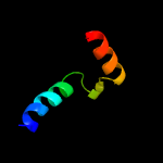
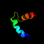
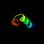
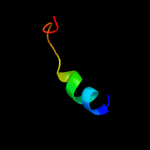
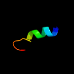
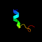
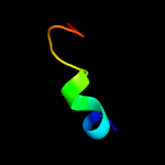
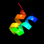
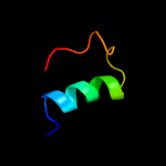
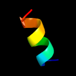
1 c5o6vC_
37.6
33
PDB header: virusChain: C: PDB Molecule: envelope protein;PDBTitle: the cryo-em structure of tick-borne encephalitis virus complexed with2 fab fragment of neutralizing antibody 19/1786
2 c4b03A_
28.3
15
PDB header: virusChain: A: PDB Molecule: dengue virus 1 e protein;PDBTitle: 6a electron cryomicroscopy structure of immature dengue virus serotype2 1
3 c5n1qD_
27.8
52
PDB header: transferaseChain: D: PDB Molecule: methyl-coenzyme m reductase iii from methanothermococcusPDBTitle: methyl-coenzyme m reductase iii from methanothermococcus2 thermolithotrophicus at 1.9 a resolution
4 d1v4sa2
26.1
60
Fold: Ribonuclease H-like motifSuperfamily: Actin-like ATPase domainFamily: Hexokinase
5 d1r3ba_
25.9
18
Fold: Bromodomain-likeSuperfamily: Mob1/phoceinFamily: Mob1/phocein
6 d1bg3a4
24.3
64
Fold: Ribonuclease H-like motifSuperfamily: Actin-like ATPase domainFamily: Hexokinase
7 c3hm8D_
22.9
64
PDB header: transferaseChain: D: PDB Molecule: hexokinase-3;PDBTitle: crystal structure of the c-terminal hexokinase domain of human hk3
8 d1czan4
22.9
70
Fold: Ribonuclease H-like motifSuperfamily: Actin-like ATPase domainFamily: Hexokinase
9 c5wvxA_
22.3
29
PDB header: hydrolase inhibitorChain: A: PDB Molecule: trypsin/chymotrypsin inhibitor;PDBTitle: crystal structure of bifunctional kunitz type trypsin /amylase2 inhibitor (amtin) from the tubers of alocasia macrorrhiza
10 c5yf4A_
22.2
29
PDB header: protein bindingChain: A: PDB Molecule: mob-like protein phocein;PDBTitle: a kinase complex mst4-mob4
11 d1pi1a_
22.0
24
Fold: Bromodomain-likeSuperfamily: Mob1/phoceinFamily: Mob1/phocein
12 c2hjnA_
21.7
29
PDB header: cell cycleChain: A: PDB Molecule: maintenance of ploidy protein mob1;PDBTitle: structural and functional analysis of saccharomyces2 cerevisiae mob1
13 c2kxaA_
21.6
42
PDB header: viral protein, immune systemChain: A: PDB Molecule: haemagglutinin ha2 chain peptide;PDBTitle: the hemagglutinin fusion peptide (h1 subtype) at ph 7.4
14 d1bg3a2
21.6
70
Fold: Ribonuclease H-like motifSuperfamily: Actin-like ATPase domainFamily: Hexokinase
15 d1czan2
21.3
47
Fold: Ribonuclease H-like motifSuperfamily: Actin-like ATPase domainFamily: Hexokinase
16 c6el1F_
20.7
21
PDB header: membrane proteinChain: F: PDB Molecule: yaxa;PDBTitle: yaxab pore complex
17 c1v4sA_
20.0
55
PDB header: transferaseChain: A: PDB Molecule: glucokinase isoform 2;PDBTitle: crystal structure of human glucokinase
18 d1ig8a2
19.1
45
Fold: Ribonuclease H-like motifSuperfamily: Actin-like ATPase domainFamily: Hexokinase
19 c2k27A_
17.0
36
PDB header: transcription regulatorChain: A: PDB Molecule: paired box protein pax-8;PDBTitle: solution structure of human pax8 paired box domain
20 c5zqtA_
16.9
55
PDB header: transferaseChain: A: PDB Molecule: hexokinase-6;PDBTitle: crystal structure of oryza sativa hexokinase 6
21 c2lbgA_
not modelled
16.7
45
PDB header: membrane proteinChain: A: PDB Molecule: major prion protein;PDBTitle: structure of the chr of the prion protein in dpc micelles
22 d1k78a2
not modelled
15.3
35
Fold: DNA/RNA-binding 3-helical bundleSuperfamily: Homeodomain-likeFamily: Paired domain
23 c1ciiA_
not modelled
15.0
10
PDB header: transmembrane proteinChain: A: PDB Molecule: colicin ia;PDBTitle: colicin ia
24 c5wsnC_
not modelled
15.0
22
PDB header: virusChain: C: PDB Molecule: e protein;PDBTitle: structure of japanese encephalitis virus
25 c1ig8A_
not modelled
14.5
45
PDB header: transferaseChain: A: PDB Molecule: hexokinase pii;PDBTitle: crystal structure of yeast hexokinase pii with the correct2 amino acid sequence
26 d1i8ya_
not modelled
14.3
44
Fold: Knottins (small inhibitors, toxins, lectins)Superfamily: Granulin repeatFamily: Granulin repeat
27 c1i8yA_
not modelled
14.3
44
PDB header: cytokineChain: A: PDB Molecule: granulin-1;PDBTitle: semi-automatic structure determination of the cg1 3-302 peptide based on aria
28 d1bdga2
not modelled
14.3
56
Fold: Ribonuclease H-like motifSuperfamily: Actin-like ATPase domainFamily: Hexokinase
29 d6paxa2
not modelled
14.3
27
Fold: DNA/RNA-binding 3-helical bundleSuperfamily: Homeodomain-likeFamily: Paired domain
30 c4ii0A_
not modelled
14.3
38
PDB header: hydrolase inhibitorChain: A: PDB Molecule: cratabl;PDBTitle: crystal structure of cratabl, a trypsin inhibitor from crataeva tapia
31 c2w8mB_
not modelled
14.0
63
PDB header: hydrolaseChain: B: PDB Molecule: orf d212;PDBTitle: structure of d212, a nuclease from a fusselovirus.
32 c4qs9A_
not modelled
13.8
55
PDB header: transferaseChain: A: PDB Molecule: hexokinase-1;PDBTitle: arabidopsis hexokinase 1 (athxk1) mutant s177a structure in glucose-2 bound form
33 d3bx1c1
not modelled
13.7
17
Fold: beta-TrefoilSuperfamily: STI-likeFamily: Kunitz (STI) inhibitors
34 c6ijjK_
not modelled
13.3
14
PDB header: membrane proteinChain: K: PDB Molecule: psak;PDBTitle: photosystem i of chlamydomonas reinhardtii
35 c1hkgA_
not modelled
13.0
6
PDB header: transferaseChain: A: PDB Molecule: hexokinase a;PDBTitle: structural dynamics of yeast hexokinase during catalysis
36 c6c6lO_
not modelled
12.9
21
PDB header: membrane proteinChain: O: PDB Molecule: v-type proton atpase subunit f;PDBTitle: yeast vacuolar atpase vo in lipid nanodisc
37 c4nl6C_
not modelled
12.8
78
PDB header: splicingChain: C: PDB Molecule: survival motor neuron protein;PDBTitle: structure of the full-length form of the protein smn found in healthy2 patients
38 c5dvhA_
not modelled
12.6
18
PDB header: protease inhibitorChain: A: PDB Molecule: pcpi-3;PDBTitle: structure of the kunitz-type cysteine protease inhibitor pcpi-3 from2 potato
39 c6nk6B_
not modelled
12.4
38
PDB header: virus like particle/signaling proteinChain: B: PDB Molecule: e1 glycoprotein;PDBTitle: electron cryo-microscopy of chikungunya vlp in complex with mouse2 mxra8 receptor
40 c1r8oA_
not modelled
11.7
29
PDB header: hydrolase inhibitorChain: A: PDB Molecule: kunitz trypsin inhibitor;PDBTitle: crystal structure of an unusual kunitz-type trypsin inhibitor from2 copaifera langsdorffii seeds
41 d1tiea_
not modelled
11.6
30
Fold: beta-TrefoilSuperfamily: STI-likeFamily: Kunitz (STI) inhibitors
42 c5ireA_
not modelled
11.5
24
PDB header: virusChain: A: PDB Molecule: e protein;PDBTitle: the cryo-em structure of zika virus
43 d2i1sa1
not modelled
11.3
10
Fold: MM3350-likeSuperfamily: MM3350-likeFamily: MM3350-like
44 c5dssB_
not modelled
11.2
29
PDB header: plant proteinChain: B: PDB Molecule: mp-4;PDBTitle: mp-4 contributes to snake venom neutralization by mucuna pruriens2 seeds through stimulation of cross-reactive antibodies
45 c2xfcD_
not modelled
11.0
38
PDB header: virusChain: D: PDB Molecule: e1 envelope glycoprotein;PDBTitle: the chikungunya e1 e2 envelope glycoprotein complex fit into2 the semliki forest virus cryo-em map
46 d1avwb_
not modelled
10.9
15
Fold: beta-TrefoilSuperfamily: STI-likeFamily: Kunitz (STI) inhibitors
47 c2yewB_
not modelled
10.8
86
PDB header: virusChain: B: PDB Molecule: e1 envelope glycoprotein;PDBTitle: modeling barmah forest virus structural proteins
48 d1m3va1
not modelled
10.8
60
Fold: Glucocorticoid receptor-like (DNA-binding domain)Superfamily: Glucocorticoid receptor-like (DNA-binding domain)Family: LIM domain
49 c5hpzB_
not modelled
10.8
37
PDB header: chlorophyll binding proteinChain: B: PDB Molecule: water-soluble chlorophyll protein;PDBTitle: type ii water soluble chl binding proteins
50 c3izxE_
not modelled
10.7
29
PDB header: virusChain: E: PDB Molecule: viral structural protein 5;PDBTitle: 3.1 angstrom cryoem structure of cytoplasmic polyhedrosis virus
51 d1wbaa_
not modelled
10.7
14
Fold: beta-TrefoilSuperfamily: STI-likeFamily: Kunitz (STI) inhibitors
52 d1xqoa_
not modelled
10.6
13
Fold: DNA-glycosylaseSuperfamily: DNA-glycosylaseFamily: AgoG-like
53 c3n42F_
not modelled
10.6
38
PDB header: viral proteinChain: F: PDB Molecule: e1 envelope glycoprotein;PDBTitle: crystal structures of the mature envelope glycoprotein complex (furin2 cleavage) of chikungunya virus.
54 c5yh4A_
not modelled
10.6
38
PDB header: hydrolase inhibitorChain: A: PDB Molecule: mirauclin-like protein;PDBTitle: miraculin-like protein from vitis vinifera
55 c2xfbF_
not modelled
10.6
38
PDB header: virusChain: F: PDB Molecule: e1 envelope glycoprotein;PDBTitle: the chikungunya e1 e2 envelope glycoprotein complex fit into2 the sindbis virus cryo-em map
56 c1ld4O_
not modelled
10.6
42
PDB header: virusChain: O: PDB Molecule: spike glycoprotein e1;PDBTitle: placement of the structural proteins in sindbis virus
57 d1hbna1
not modelled
10.5
44
Fold: Methyl-coenzyme M reductase alpha and beta chain C-terminal domainSuperfamily: Methyl-coenzyme M reductase alpha and beta chain C-terminal domainFamily: Methyl-coenzyme M reductase alpha and beta chain C-terminal domain
58 c1z8yE_
not modelled
10.4
86
PDB header: virusChain: E: PDB Molecule: spike glycoprotein e1;PDBTitle: mapping the e2 glycoprotein of alphaviruses
59 c3tc2C_
not modelled
10.4
21
PDB header: hydrolase inhibitorChain: C: PDB Molecule: kunitz-type proteinase inhibitor p1h5;PDBTitle: crystal structure of potato serine protease inhibitor.
60 d2alaa2
not modelled
10.2
38
Fold: Viral glycoprotein, central and dimerisation domainsSuperfamily: Viral glycoprotein, central and dimerisation domainsFamily: Viral glycoprotein, central and dimerisation domains
61 c3lw5K_
not modelled
10.2
27
PDB header: photosynthesisChain: K: PDB Molecule: photosystem i reaction center subunit x psak;PDBTitle: improved model of plant photosystem i
62 c6fgnA_
not modelled
10.1
35
PDB header: antitumor proteinChain: A: PDB Molecule: histone acetyltransferase p300,tumor protein 63;PDBTitle: solution structure of p300taz2-p63ta
63 c1qhaA_
not modelled
9.5
70
PDB header: transferaseChain: A: PDB Molecule: protein (hexokinase);PDBTitle: human hexokinase type i complexed with atp analogue amp-pnp
64 c4j2yA_
not modelled
9.5
29
PDB header: hydrolase/hydrolase inhibitorChain: A: PDB Molecule: trypsin inhibitor;PDBTitle: crystal structure of a plant trypsin inhibitor ecti in complex with2 bovine trypsin.
65 c3j0fG_
not modelled
9.5
42
PDB header: virusChain: G: PDB Molecule: e1 envelope glycoprotein;PDBTitle: sindbis virion
66 c3iirA_
not modelled
9.5
37
PDB header: hydrolase inhibitorChain: A: PDB Molecule: trypsin inhibitor;PDBTitle: crystal structure of miraculin like protein from seeds of murraya2 koenigii
67 c2qn4B_
not modelled
9.5
21
PDB header: hydrolase inhibitorChain: B: PDB Molecule: alpha-amylase/subtilisin inhibitor;PDBTitle: structure and function study of rice bifunctional alpha-2 amylase/subtilisin inhibitor from oryza sativa
68 c2q2kA_
not modelled
9.5
23
PDB header: dna binding protein/dnaChain: A: PDB Molecule: hypothetical protein;PDBTitle: structure of nucleic-acid binding protein
69 c1bdgA_
not modelled
9.2
56
PDB header: hexokinaseChain: A: PDB Molecule: hexokinase;PDBTitle: hexokinase from schistosoma mansoni complexed with glucose
70 c2q2kB_
not modelled
9.1
23
PDB header: dna binding protein/dnaChain: B: PDB Molecule: hypothetical protein;PDBTitle: structure of nucleic-acid binding protein
71 c1cirA_
not modelled
9.0
60
PDB header: serine protease inhibitorChain: A: PDB Molecule: chymotrypsin inhibitor 2;PDBTitle: complex of two fragments of ci2 [(1-40)(dot)(41-64)]
72 d1eyla_
not modelled
8.9
31
Fold: beta-TrefoilSuperfamily: STI-likeFamily: Kunitz (STI) inhibitors
73 d1s7ba_
not modelled
8.9
23
Fold: Multidrug resistance efflux transporter EmrESuperfamily: Multidrug resistance efflux transporter EmrEFamily: Multidrug resistance efflux transporter EmrE
74 c1hbuD_
not modelled
8.9
41
PDB header: methanogenesisChain: D: PDB Molecule: methyl-coenzyme m reductase i alpha subunit;PDBTitle: methyl-coenzyme m reductase in the mcr-red1-silent state in complex2 with coenzyme m
75 d1r8na_
not modelled
8.7
29
Fold: beta-TrefoilSuperfamily: STI-likeFamily: Kunitz (STI) inhibitors
76 c3s8jB_
not modelled
8.7
29
PDB header: hydrolase inhibitorChain: B: PDB Molecule: latex serine proteinase inhibitor;PDBTitle: crystal structure of a papaya latex serine protease inhibitor (ppi) at2 2.6a resolution
77 c2alaA_
not modelled
8.6
38
PDB header: viral proteinChain: A: PDB Molecule: structural polyprotein (p130);PDBTitle: crystal structure of the semliki forest virus envelope protein e1 in2 its monomeric conformation.
78 d1p4ea2
not modelled
8.6
22
Fold: DNA breaking-rejoining enzymesSuperfamily: DNA breaking-rejoining enzymesFamily: Lambda integrase-like, catalytic core
79 c3d3sA_
not modelled
8.6
15
PDB header: transferaseChain: A: PDB Molecule: l-2,4-diaminobutyric acid acetyltransferase;PDBTitle: crystal structure of l-2,4-diaminobutyric acid acetyltransferase from2 bordetella parapertussis
80 c4xb6D_
not modelled
8.6
39
PDB header: transferaseChain: D: PDB Molecule: alpha-d-ribose 1-methylphosphonate 5-phosphate c-p lyase;PDBTitle: structure of the e. coli c-p lyase core complex
81 c1e6yA_
not modelled
8.5
40
PDB header: oxidoreductaseChain: A: PDB Molecule: methyl-coenzyme m reductase subunit alpha;PDBTitle: methyl-coenzyme m reductase from methanosarcina barkeri
82 c6gy6Q_
not modelled
8.4
21
PDB header: toxinChain: Q: PDB Molecule: xaxa;PDBTitle: xaxab pore complex from xenorhabdus nematophila
83 c6mx4J_
not modelled
8.3
38
PDB header: virusChain: J: PDB Molecule: e1;PDBTitle: cryoem structure of chimeric eastern equine encephalitis virus
84 d1j2oa1
not modelled
8.3
60
Fold: Glucocorticoid receptor-like (DNA-binding domain)Superfamily: Glucocorticoid receptor-like (DNA-binding domain)Family: LIM domain
85 c3muwE_
not modelled
8.3
42
PDB header: virusChain: E: PDB Molecule: structural polyprotein;PDBTitle: pseudo-atomic structure of the e2-e1 protein shell in sindbis virus
86 d1xg7a_
not modelled
8.0
21
Fold: DNA-glycosylaseSuperfamily: DNA-glycosylaseFamily: AgoG-like
87 c5hg1A_
not modelled
8.0
70
PDB header: transferase/transferase inhibitorChain: A: PDB Molecule: hexokinase-2;PDBTitle: crystal structure of human hexokinase 2 with cmpd 1, a c-2-substituted2 glucosamine
88 c6igzK_
not modelled
7.8
21
PDB header: plant proteinChain: K: PDB Molecule: psak;PDBTitle: structure of psi-lhci
89 c3j0cG_
not modelled
7.6
42
PDB header: virusChain: G: PDB Molecule: e1 envelope glycoprotein;PDBTitle: models of e1, e2 and cp of venezuelan equine encephalitis virus tc-832 strain restrained by a near atomic resolution cryo-em map
90 c1bctA_
not modelled
7.6
57
PDB header: photoreceptorChain: A: PDB Molecule: bacteriorhodopsin;PDBTitle: three-dimensional structure of proteolytic fragment 163-2312 of bacterioopsin determined from nuclear magnetic3 resonance data in solution
91 c3io2A_
not modelled
7.4
35
PDB header: transferaseChain: A: PDB Molecule: histone acetyltransferase p300;PDBTitle: crystal structure of the taz2 domain of p300
92 c2dreA_
not modelled
7.2
17
PDB header: plant proteinChain: A: PDB Molecule: water-soluble chlorophyll protein;PDBTitle: crystal structure of water-soluble chlorophyll protein from2 lepidium virginicum at 2.00 angstrom resolution
93 c2l3fA_
not modelled
7.2
28
PDB header: structural genomics, unknown functionChain: A: PDB Molecule: uncharacterized protein;PDBTitle: solution nmr structure of a putative uracil dna glycosylase from2 methanosarcina acetivorans, northeast structural genomics consortium3 target mvr76
94 c2go2A_
not modelled
7.1
32
PDB header: protein bindingChain: A: PDB Molecule: kunitz-type serine protease inhibitor bbki;PDBTitle: crystal structure of bbki, a kunitz-type kallikrein inhibitor
95 c2aj6A_
not modelled
7.0
24
PDB header: transferaseChain: A: PDB Molecule: hypothetical protein mw0638;PDBTitle: crystal structure of a putative gnat family acetyltransferase (mw0638)2 from staphylococcus aureus subsp. aureus at 1.63 a resolution
96 c1p58C_
not modelled
6.8
27
PDB header: virusChain: C: PDB Molecule: major envelope protein e;PDBTitle: complex organization of dengue virus membrane proteins as revealed by2 9.5 angstrom cryo-em reconstruction
97 d1rutx1
not modelled
6.8
60
Fold: Glucocorticoid receptor-like (DNA-binding domain)Superfamily: Glucocorticoid receptor-like (DNA-binding domain)Family: LIM domain
98 c4e72A_
not modelled
6.7
36
PDB header: structural genomics, unknown functionChain: A: PDB Molecule: uncharacterized protein;PDBTitle: crystal structure of a duf3298 family protein (pa4972) from2 pseudomonas aeruginosa pao1 at 2.15 a resolution
99 c3iz3D_
not modelled
6.6
32
PDB header: virusChain: D: PDB Molecule: viral structural protein 5;PDBTitle: cryoem structure of cytoplasmic polyhedrosis virus


