| 1 |
|
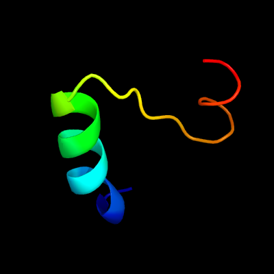
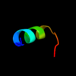
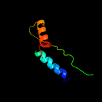
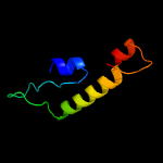
PDB 2csu chain A domain 3
Region: 42 - 68
Aligned: 27
Modelled: 27
Confidence: 27.4%
Identity: 19%
Fold: Flavodoxin-like
Superfamily: Succinyl-CoA synthetase domains
Family: Succinyl-CoA synthetase domains
Phyre2
| 2 |
|
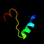
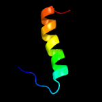
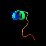
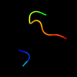
PDB 1mke chain A domain 1
Region: 56 - 78
Aligned: 23
Modelled: 23
Confidence: 19.6%
Identity: 4%
Fold: PH domain-like barrel
Superfamily: PH domain-like
Family: Enabled/VASP homology 1 domain (EVH1 domain)
Phyre2
| 3 |
|
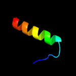
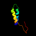
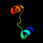
PDB 5uqd chain A
Region: 45 - 60
Aligned: 16
Modelled: 16
Confidence: 14.3%
Identity: 31%
PDB header:oxidoreductase
Chain: A: PDB Molecule:dumpy: shorter than wild-type;
PDBTitle: dpy-21 in complex with fe(ii) and alpha-ketoglutarate
Phyre2
| 4 |
|
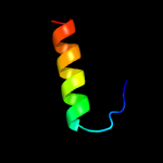
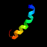
PDB 2ifs chain A
Region: 55 - 81
Aligned: 27
Modelled: 27
Confidence: 13.3%
Identity: 4%
PDB header:signaling protein
Chain: A: PDB Molecule:wiskott-aldrich syndrome protien interacting protein and
PDBTitle: structure of the n-wasp evh1 domain in complex with an extended wip2 peptide
Phyre2
| 5 |
|
PDB 6c3r chain B
Region: 73 - 124
Aligned: 51
Modelled: 52
Confidence: 11.3%
Identity: 10%
PDB header:viral protein
Chain: B: PDB Molecule:cricket paralysis virus 1a protein;
PDBTitle: cricket paralysis virus rnai suppressor protein crpv-1a
Phyre2
| 6 |
|
PDB 1xod chain A domain 1
Region: 56 - 77
Aligned: 22
Modelled: 22
Confidence: 8.4%
Identity: 23%
Fold: PH domain-like barrel
Superfamily: PH domain-like
Family: Enabled/VASP homology 1 domain (EVH1 domain)
Phyre2
| 7 |
|
PDB 1nye chain D
Region: 58 - 114
Aligned: 56
Modelled: 57
Confidence: 8.2%
Identity: 16%
Fold: OsmC-like
Superfamily: OsmC-like
Family: Ohr/OsmC resistance proteins
Phyre2
| 8 |
|
PDB 3ss4 chain C
Region: 47 - 62
Aligned: 16
Modelled: 16
Confidence: 6.7%
Identity: 38%
PDB header:hydrolase
Chain: C: PDB Molecule:glutaminase c;
PDBTitle: crystal structure of mouse glutaminase c, phosphate-bound form
Phyre2
| 9 |
|
PDB 1euc chain B domain 1
Region: 46 - 72
Aligned: 27
Modelled: 27
Confidence: 6.6%
Identity: 22%
Fold: Flavodoxin-like
Superfamily: Succinyl-CoA synthetase domains
Family: Succinyl-CoA synthetase domains
Phyre2
| 10 |
|
PDB 5dgg chain B
Region: 37 - 58
Aligned: 22
Modelled: 22
Confidence: 6.1%
Identity: 23%
PDB header:unknown function
Chain: B: PDB Molecule:uncharacterized protein;
PDBTitle: central domain of uncharacterized lpg1148 protein from legionella2 pneumophila
Phyre2
| 11 |
|
PDB 2lwe chain A
Region: 46 - 60
Aligned: 15
Modelled: 15
Confidence: 6.1%
Identity: 13%
PDB header:signaling protein
Chain: A: PDB Molecule:probable atp-dependent rna helicase ddx58;
PDBTitle: solution structure of mutant (t170e) second card of human rig-i
Phyre2
| 12 |
|
PDB 5h75 chain B
Region: 49 - 105
Aligned: 54
Modelled: 57
Confidence: 6.0%
Identity: 11%
PDB header:lyase
Chain: B: PDB Molecule:mersacidin decarboxylase,immunoglobulin g-binding protein
PDBTitle: crystal structure of the mrsd-protein a fusion protein
Phyre2
| 13 |
|
PDB 1vjq chain B
Region: 48 - 59
Aligned: 12
Modelled: 11
Confidence: 6.0%
Identity: 42%
PDB header:structural genomics, de novo protein
Chain: B: PDB Molecule:designed protein;
PDBTitle: designed protein based on backbone conformation of2 procarboxypeptidase-a (1aye) with sidechains chosen for maximal3 predicted stability.
Phyre2