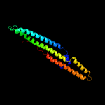
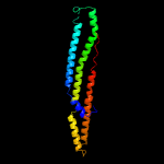
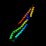
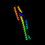
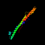
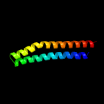
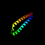
1 c5xfsB_
100.0
61
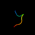
PDB header: protein transportChain: B: PDB Molecule: ppe family protein ppe15;PDBTitle: crystal structure of pe8-ppe15 in complex with espg5 from m.2 tuberculosis
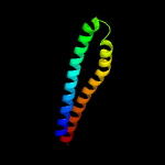
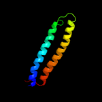
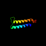
2 d2g38b1
100.0
32
Fold: Ferritin-likeSuperfamily: PE/PPE dimer-likeFamily: PPE
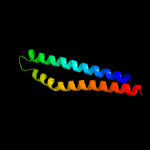
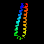
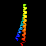
3 c2g38B_
100.0
32
PDB header: structural genomics, unknown functionChain: B: PDB Molecule: ppe family protein;PDBTitle: a pe/ppe protein complex from mycobacterium tuberculosis
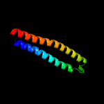
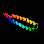
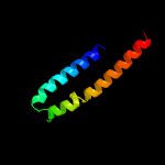
4 c4xy3A_
100.0
21
PDB header: protein transportChain: A: PDB Molecule: esx-1 secretion-associated protein espb;PDBTitle: structure of esx-1 secreted protein espb
5 c4wj2A_
98.5
15
PDB header: unknown functionChain: A: PDB Molecule: antigen mtb48;PDBTitle: mycobacterial protein
6 c2vs0B_
97.6
15
PDB header: cell invasionChain: B: PDB Molecule: virulence factor esxa;PDBTitle: structural analysis of homodimeric staphylococcal aureus2 virulence factor esxa
7 c3gvmA_
97.4
16
PDB header: viral proteinChain: A: PDB Molecule: putative uncharacterized protein sag1039;PDBTitle: structure of the homodimeric wxg-100 family protein from streptococcus2 agalactiae
8 c4iogD_
97.4
15
PDB header: unknown functionChain: D: PDB Molecule: secreted protein esxb;PDBTitle: the crystal structure of a secreted protein esxb (wild-type, in p212 space group) from bacillus anthracis str. sterne
9 c3zbhC_
97.2
14
PDB header: unknown functionChain: C: PDB Molecule: esxa;PDBTitle: geobacillus thermodenitrificans esxa crystal form i
10 d1wa8a1
96.5
17
Fold: Ferritin-likeSuperfamily: EsxAB dimer-likeFamily: ESAT-6 like
11 c4lwsB_
95.2
16
PDB header: unknown functionChain: B: PDB Molecule: uncharacterized protein;PDBTitle: esxa : esxb (semet) hetero-dimer from thermomonospora curvata
12 d1wa8b1
94.8
17
Fold: Ferritin-likeSuperfamily: EsxAB dimer-likeFamily: ESAT-6 like
13 c4lwsA_
94.3
18
PDB header: unknown functionChain: A: PDB Molecule: uncharacterized protein;PDBTitle: esxa : esxb (semet) hetero-dimer from thermomonospora curvata
14 c2kg7B_
86.4
15
PDB header: unknown functionChain: B: PDB Molecule: esat-6-like protein esxh;PDBTitle: structure and features of the complex formed by the tuberculosis2 virulence factors rv0287 and rv0288
15 c4i0xA_
85.7
18
PDB header: structural genomics, unknown functionChain: A: PDB Molecule: esat-6-like protein mab_3112;PDBTitle: crystal structure of the mycobacterum abscessus esxef (mab_3112-2 mab_3113) complex
16 c4i0xJ_
53.9
20
PDB header: structural genomics, unknown functionChain: J: PDB Molecule: esat-6-like protein mab_3113;PDBTitle: crystal structure of the mycobacterum abscessus esxef (mab_3112-2 mab_3113) complex
17 c5frgA_
21.0
63
PDB header: protein bindingChain: A: PDB Molecule: formin-binding protein 1-like;PDBTitle: the nmr structure of the cdc42-interacting region of toca1
18 c1bkvA_
18.0
50
PDB header: structural proteinChain: A: PDB Molecule: t3-785;PDBTitle: collagen
19 c1bkvC_
17.3
50
PDB header: structural proteinChain: C: PDB Molecule: t3-785;PDBTitle: collagen
20 c1bkvB_
17.3
50
PDB header: structural proteinChain: B: PDB Molecule: t3-785;PDBTitle: collagen
21 c4bz4D_
not modelled
16.7
58
PDB header: copper-binding proteinChain: D: PDB Molecule: copper-repressible polypeptide;PDBTitle: cora is a surface-associated copper-binding protein2 important in methylomicrobium album bg8 copper acquisition
22 c2ke4A_
not modelled
14.0
63
PDB header: membrane proteinChain: A: PDB Molecule: cdc42-interacting protein 4;PDBTitle: the nmr structure of the tc10 and cdc42 interacting domain2 of cip4
23 d1ui5a2
not modelled
11.8
19
Fold: Tetracyclin repressor-like, C-terminal domainSuperfamily: Tetracyclin repressor-like, C-terminal domainFamily: Tetracyclin repressor-like, C-terminal domain
24 c5h9xA_
not modelled
10.8
30
PDB header: hydrolaseChain: A: PDB Molecule: beta-1,3-glucanase;PDBTitle: crystal structure of gh family 64 laminaripentaose-producing beta-1,3-2 glucanase from paenibacillus barengoltzii
25 c5vzmB_
not modelled
10.4
63
PDB header: protein bindingChain: B: PDB Molecule: dna repair protein rev1;PDBTitle: solution nmr structure of human rev1 (932-1039) in complex with2 ubiquitin
26 c4lzxB_
not modelled
9.7
31
PDB header: metal binding proteinChain: B: PDB Molecule: iq domain-containing protein g;PDBTitle: complex of iqcg and ca2+-free cam
27 d2fcla1
not modelled
9.4
28
Fold: NucleotidyltransferaseSuperfamily: NucleotidyltransferaseFamily: TM1012-like
28 c4m1lB_
not modelled
8.2
36
PDB header: metal binding proteinChain: B: PDB Molecule: iq domain-containing protein g;PDBTitle: complex of iqcg and ca2+-bound cam
29 d2np5a2
not modelled
7.7
30
Fold: Tetracyclin repressor-like, C-terminal domainSuperfamily: Tetracyclin repressor-like, C-terminal domainFamily: Tetracyclin repressor-like, C-terminal domain
30 d1khba2
not modelled
7.7
20
Fold: PEP carboxykinase N-terminal domainSuperfamily: PEP carboxykinase N-terminal domainFamily: PEP carboxykinase N-terminal domain
31 c2iu1A_
not modelled
7.6
22
PDB header: transcriptionChain: A: PDB Molecule: eukaryotic translation initiation factor 5;PDBTitle: crystal structure of eif5 c-terminal domain
32 c4is4G_
not modelled
7.3
8
PDB header: ligaseChain: G: PDB Molecule: glutamine synthetase;PDBTitle: the glutamine synthetase from the dicotyledonous plant m. truncatula2 is a decamer
33 c1vytF_
not modelled
7.2
13
PDB header: transport proteinChain: F: PDB Molecule: voltage-dependent l-type calcium channelPDBTitle: beta3 subunit complexed with aid
34 c6rlxA_
not modelled
6.9
71
PDB header: hormone(muscle relaxant)Chain: A: PDB Molecule: relaxin, a-chain;PDBTitle: x-ray structure of human relaxin at 1.5 angstroms. comparison to2 insulin and implications for receptor binding determinants
35 c6rlxC_
not modelled
6.9
71
PDB header: hormone(muscle relaxant)Chain: C: PDB Molecule: relaxin, a-chain;PDBTitle: x-ray structure of human relaxin at 1.5 angstroms. comparison to2 insulin and implications for receptor binding determinants
36 c2mv1A_
not modelled
6.9
71
PDB header: signaling proteinChain: A: PDB Molecule: relaxin a chain;PDBTitle: solution nmr structure of human relaxin-2
37 c2v36D_
not modelled
6.8
35
PDB header: transferaseChain: D: PDB Molecule: gamma-glutamyltranspeptidase small chain;PDBTitle: crystal structure of gamma-glutamyl transferase from bacillus subtilis
38 c2fulE_
not modelled
6.8
39
PDB header: translationChain: E: PDB Molecule: eukaryotic translation initiation factor 5;PDBTitle: crystal structure of the c-terminal domain of s. cerevisiae eif5
39 c1bcvA_
not modelled
6.6
44
PDB header: synthetic peptideChain: A: PDB Molecule: peptide corresponding to the major immunogen site of fmdPDBTitle: synthetic peptide corresponding to the major immunogen site of fmd2 virus, nmr, 10 structures
40 c3ibzA_
not modelled
6.4
23
PDB header: structural genomics, unknown functionChain: A: PDB Molecule: putative tellurium resistant like protein terd;PDBTitle: crystal structure of putative tellurium resistant like protein (terd)2 from streptomyces coelicolor a3(2)
41 c2k6tA_
not modelled
6.3
71
PDB header: hormoneChain: A: PDB Molecule: insulin-like 3 a chain;PDBTitle: solution structure of the relaxin-like factor
42 c2h8bA_
not modelled
6.3
71
PDB header: hormone/growth factorChain: A: PDB Molecule: insulin-like 3;PDBTitle: solution structure of insl3
43 c4baxH_
not modelled
6.3
8
PDB header: ligaseChain: H: PDB Molecule: glutamine synthetase;PDBTitle: crystal structure of glutamine synthetase from streptomyces2 coelicolor
44 c2d3aJ_
not modelled
6.2
17
PDB header: ligaseChain: J: PDB Molecule: glutamine synthetase;PDBTitle: crystal structure of the maize glutamine synthetase2 complexed with adp and methionine sulfoximine phosphate
45 c4i6jB_
not modelled
6.1
33
PDB header: transcriptionChain: B: PDB Molecule: f-box/lrr-repeat protein 3;PDBTitle: a ubiquitin ligase-substrate complex
46 c2k6uA_
not modelled
6.1
71
PDB header: hormoneChain: A: PDB Molecule: insulin-like 3 a chain;PDBTitle: the solution structure of a conformationally restricted2 fully active derivative of the human relaxin-like factor3 (rlf)
47 c3fkyD_
not modelled
6.0
8
PDB header: ligaseChain: D: PDB Molecule: glutamine synthetase;PDBTitle: crystal structure of the glutamine synthetase gln1deltan182 from the yeast saccharomyces cerevisiae
48 d1kshb_
not modelled
5.9
18
Fold: Immunoglobulin-like beta-sandwichSuperfamily: E set domainsFamily: RhoGDI-like
49 c5uc0B_
not modelled
5.9
60
PDB header: hydrolaseChain: B: PDB Molecule: uncharacterized protein cog5400;PDBTitle: crystal structure of beta-barrel-like, uncharacterized protein of2 cog5400 from brucella abortus
50 d1k47a1
not modelled
5.7
13
Fold: Ribosomal protein S5 domain 2-likeSuperfamily: Ribosomal protein S5 domain 2-likeFamily: GHMP Kinase, N-terminal domain
51 c3r5zB_
not modelled
5.6
19
PDB header: unknown functionChain: B: PDB Molecule: putative uncharacterized protein;PDBTitle: structure of a deazaflavin-dependent reductase from nocardia2 farcinica, with co-factor f420
52 c1t0jC_
not modelled
5.5
14
PDB header: signaling proteinChain: C: PDB Molecule: voltage-dependent l-type calcium channel alpha-1c subunit;PDBTitle: crystal structure of a complex between voltage-gated calcium channel2 beta2a subunit and a peptide of the alpha1c subunit
53 c4deyB_
not modelled
5.5
6
PDB header: transport proteinChain: B: PDB Molecule: voltage-dependent l-type calcium channel subunit alpha-1c;PDBTitle: crystal structure of the voltage dependent calcium channel beta-22 subunit in complex with the cav1.2 i-ii linker.
54 c2lkqA_
not modelled
5.5
44
PDB header: immune systemChain: A: PDB Molecule: immunoglobulin lambda-like polypeptide 1;PDBTitle: nmr structure of the lambda 5 22-45 peptide
55 d2q09a1
not modelled
5.4
30
Fold: Composite domain of metallo-dependent hydrolasesSuperfamily: Composite domain of metallo-dependent hydrolasesFamily: Imidazolonepropionase-like
56 c4hppA_
not modelled
5.4
17
PDB header: ligaseChain: A: PDB Molecule: probable glutamine synthetase;PDBTitle: crystal structure of novel glutamine synthase homolog
57 c2le2B_
not modelled
5.2
60
PDB header: hydrolase inhibitorChain: B: PDB Molecule: p56;PDBTitle: novel dimeric structure of phage phi29-encoded protein p56: insights2 into uracil-dna glycosylase inhibition
58 c3ng0A_
not modelled
5.2
33
PDB header: ligaseChain: A: PDB Molecule: glutamine synthetase;PDBTitle: crystal structure of glutamine synthetase from synechocystis sp. pcc2 6803
59 c2y5tG_
not modelled
5.2
83
PDB header: immune systemChain: G: PDB Molecule: c1;PDBTitle: crystal structure of the pathogenic autoantibody ciic1 in complex with2 the triple-helical c1 peptide
60 c4ndvB_
not modelled
5.2
63
PDB header: sugar binding proteinChain: B: PDB Molecule: alpha-galactosyl-binding lectin;PDBTitle: crystal structure of l. decastes alpha-galactosyl-binding lectin in2 complex with globotriose
61 c6cgvW_
not modelled
5.2
31
PDB header: virusChain: W: PDB Molecule: pre-protein vi;PDBTitle: revised crystal structure of human adenovirus
62 c3j21i_
not modelled
5.2
31
PDB header: ribosomeChain: I: PDB Molecule: 50s ribosomal protein l13p;PDBTitle: promiscuous behavior of proteins in archaeal ribosomes revealed by2 cryo-em: implications for evolution of eukaryotic ribosomes (50s3 ribosomal proteins)
63 c3izrm_
not modelled
5.1
31
PDB header: ribosomeChain: M: PDB Molecule: 60s ribosomal protein l23 (l14p);PDBTitle: localization of the large subunit ribosomal proteins into a 5.5 a2 cryo-em map of triticum aestivum translating 80s ribosome
64 c1htoB_
not modelled
5.1
25
PDB header: ligaseChain: B: PDB Molecule: glutamine synthetase;PDBTitle: crystallographic structure of a relaxed glutamine synthetase from2 mycobacterium tuberculosis
65 c2qsrA_
not modelled
5.1
38
PDB header: transcriptionChain: A: PDB Molecule: transcription-repair coupling factor;PDBTitle: crystal structure of c-terminal domain of transcription-repair2 coupling factor
66 d1xdpa4
not modelled
5.1
25
Fold: Phospholipase D/nucleaseSuperfamily: Phospholipase D/nucleaseFamily: Polyphosphate kinase C-terminal domain
67 c5craB_
not modelled
5.1
45
PDB header: hydrolaseChain: B: PDB Molecule: sdea;PDBTitle: structure of the sdea dub domain


