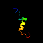
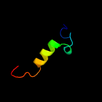
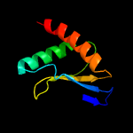
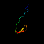
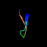
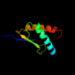
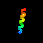
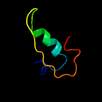
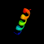
1 c3j61G_
78.3
33
PDB header: ribosomeChain: G: PDB Molecule: 60s ribosomal protein l8e;PDBTitle: localization of the large subunit ribosomal proteins into a 5.5 a2 cryo-em map of triticum aestivum translating 80s ribosome
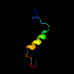
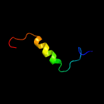
2 c3j39G_
72.3
40
PDB header: ribosomeChain: G: PDB Molecule: 60s ribosomal protein l7a;PDBTitle: structure of the d. melanogaster 60s ribosomal proteins
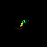
3 c3u5iG_
68.1
33
PDB header: ribosomeChain: G: PDB Molecule: 60s ribosomal protein l8-a;PDBTitle: the structure of the eukaryotic ribosome at 3.0 a resolution. this2 entry contains proteins of the 60s subunit, ribosome b
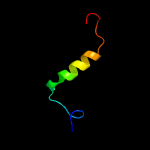
4 c3zf7x_
62.1
33
PDB header: ribosomeChain: X: PDB Molecule: 60s ribosomal protein l23a;PDBTitle: high-resolution cryo-electron microscopy structure of the trypanosoma2 brucei ribosome
5 c4a1eF_
57.2
33
PDB header: ribosomeChain: F: PDB Molecule: rpl7a;PDBTitle: t.thermophila 60s ribosomal subunit in complex with2 initiation factor 6. this file contains 5s rrna, 5.8s rrna3 and proteins of molecule 1
6 c3j3bG_
50.7
33
PDB header: ribosomeChain: G: PDB Molecule: 60s ribosomal protein l7a;PDBTitle: structure of the human 60s ribosomal proteins
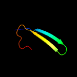
7 d2je8a2
37.2
13
Fold: Immunoglobulin-like beta-sandwichSuperfamily: beta-Galactosidase/glucuronidase domainFamily: beta-Galactosidase/glucuronidase domain
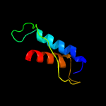
8 d2dy1a3
26.0
13
Fold: Ribosomal protein S5 domain 2-likeSuperfamily: Ribosomal protein S5 domain 2-likeFamily: Translational machinery components
9 d1j98a_
24.9
17
Fold: LuxS/MPP-like metallohydrolaseSuperfamily: LuxS/MPP-like metallohydrolaseFamily: Autoinducer-2 production protein LuxS
10 c2kglA_
20.3
31
PDB header: chaperoneChain: A: PDB Molecule: mesoderm development candidate 2;PDBTitle: nmr solution structure of mesd
11 d1sknp_
16.5
30
Fold: A DNA-binding domain in eukaryotic transcription factorsSuperfamily: A DNA-binding domain in eukaryotic transcription factorsFamily: A DNA-binding domain in eukaryotic transcription factors
12 d1jw2a_
15.2
38
Fold: Open three-helical up-and-down bundleSuperfamily: Hemolysin expression modulating protein HHAFamily: Hemolysin expression modulating protein HHA
13 d1vcpa_
15.1
27
Fold: Trypsin-like serine proteasesSuperfamily: Trypsin-like serine proteasesFamily: Viral proteases
14 d2gy9s1
13.7
46
Fold: Ribosomal protein S19Superfamily: Ribosomal protein S19Family: Ribosomal protein S19
15 d1vjea_
12.3
21
Fold: LuxS/MPP-like metallohydrolaseSuperfamily: LuxS/MPP-like metallohydrolaseFamily: Autoinducer-2 production protein LuxS
16 c1bb1A_
11.4
42
PDB header: de novo protein designChain: A: PDB Molecule: designed, thermostable heterotrimeric coiledPDBTitle: crystal structure of a designed, thermostable2 heterotrimeric coiled coil
17 c2lxrA_
11.3
33
PDB header: oxidoreductaseChain: A: PDB Molecule: nadh dehydrogenase i subunit e;PDBTitle: solution structure of hp1264 from helicobacter pylori
18 c4u5tB_
10.8
47
PDB header: transcription/transcription inhibitorChain: B: PDB Molecule: vbp leucine zipper;PDBTitle: crystal structure of vbp leucine zipper with bound arylstibonic acid
19 d2gnxa2
10.7
67
Fold: Gelsolin-likeSuperfamily: FLJ32549 C-terminal domain-likeFamily: FLJ32549 C-terminal domain-like
20 c4a1bB_
10.7
67
PDB header: ribosomeChain: B: PDB Molecule: rpl39;PDBTitle: t.thermophila 60s ribosomal subunit in complex with2 initiation factor 6. this file contains 26s rrna and3 proteins of molecule 3.
21 c3okqA_
not modelled
10.1
44
PDB header: protein bindingChain: A: PDB Molecule: bud site selection protein 6;PDBTitle: crystal structure of a core domain of yeast actin nucleation cofactor2 bud6
22 c2l42A_
not modelled
10.0
18
PDB header: protein bindingChain: A: PDB Molecule: dna-binding protein rap1;PDBTitle: the solution structure of rap1 brct domain from saccharomyces2 cerevisiae
23 d1oeda_
not modelled
9.8
15
Fold: Neurotransmitter-gated ion-channel transmembrane poreSuperfamily: Neurotransmitter-gated ion-channel transmembrane poreFamily: Neurotransmitter-gated ion-channel transmembrane pore
24 c3zf7q_
not modelled
9.8
83
PDB header: ribosomeChain: Q: PDB Molecule: ribosomal protein l15;PDBTitle: high-resolution cryo-electron microscopy structure of the trypanosoma2 brucei ribosome
25 c5xxuP_
not modelled
9.7
62
PDB header: ribosomeChain: P: PDB Molecule: ribosomal protein us19;PDBTitle: small subunit of toxoplasma gondii ribosome
26 c2mrlA_
not modelled
9.5
53
PDB header: unknown functionChain: A: PDB Molecule: uncharacterized protein bth i2711;PDBTitle: backbone 1h, 13c, and 15n chemical shift assignments and nmr structure2 for potential drug target from burkholderia thailandensis e264
27 c5e68A_
not modelled
9.4
22
PDB header: lyaseChain: A: PDB Molecule: s-ribosylhomocysteine lyase;PDBTitle: high resolution crystal structure of luxs - quorum sensor molecular2 complex from salmonella typhi at 1.58 angstroms
28 d1jeqa1
not modelled
9.4
67
Fold: LEM/SAP HeH motifSuperfamily: SAP domainFamily: SAP domain
29 d2uubs1
not modelled
9.2
62
Fold: Ribosomal protein S19Superfamily: Ribosomal protein S19Family: Ribosomal protein S19
30 d3eipa_
not modelled
9.1
31
Fold: FKBP-likeSuperfamily: Colicin E3 immunity proteinFamily: Colicin E3 immunity protein
31 c5xyiP_
not modelled
9.1
38
PDB header: ribosomeChain: P: PDB Molecule: ribosomal protein s19, putative;PDBTitle: small subunit of trichomonas vaginalis ribosome
32 c2xzmS_
not modelled
9.1
46
PDB header: ribosomeChain: S: PDB Molecule: rps15e;PDBTitle: crystal structure of the eukaryotic 40s ribosomal2 subunit in complex with initiation factor 1. this file3 contains the 40s subunit and initiation factor for4 molecule 1
33 c2zkr3_
not modelled
9.1
44
PDB header: ribosomal protein/rnaChain: 3: PDB Molecule: 60s ribosomal protein l39e;PDBTitle: structure of a mammalian ribosomal 60s subunit within an 80s complex2 obtained by docking homology models of the rna and proteins into an3 8.7 a cryo-em map
34 c3izso_
not modelled
8.8
67
PDB header: ribosomeChain: O: PDB Molecule: 60s ribosomal protein rpl28 (l15p);PDBTitle: localization of the large subunit ribosomal proteins into a 6.1 a2 cryo-em map of saccharomyces cerevisiae translating 80s ribosome
35 c2vxaL_
not modelled
8.6
33
PDB header: flavoproteinChain: L: PDB Molecule: dodecin;PDBTitle: h.halophila dodecin in complex with riboflavin
36 c3bbnS_
not modelled
8.5
31
PDB header: ribosomeChain: S: PDB Molecule: ribosomal protein s19;PDBTitle: homology model for the spinach chloroplast 30s subunit fitted to 9.4a2 cryo-em map of the 70s chlororibosome.
37 c5o5jS_
not modelled
8.3
23
PDB header: ribosomeChain: S: PDB Molecule: 30s ribosomal protein s19;PDBTitle: structure of the 30s small ribosomal subunit from mycobacterium2 smegmatis
38 c4xchB_
not modelled
8.3
19
PDB header: lyaseChain: B: PDB Molecule: s-ribosylhomocysteine lyase;PDBTitle: s-ribosylhomocysteinase from streptococcus suis
39 c3j20T_
not modelled
8.2
38
PDB header: ribosomeChain: T: PDB Molecule: 30s ribosomal protein s19p;PDBTitle: promiscuous behavior of proteins in archaeal ribosomes revealed by2 cryo-em: implications for evolution of eukaryotic ribosomes (30s3 ribosomal subunit)
40 d2i0ka1
not modelled
8.2
19
Fold: Ferredoxin-likeSuperfamily: FAD-linked oxidases, C-terminal domainFamily: Cholesterol oxidase
41 d2ux9a1
not modelled
8.1
22
Fold: Dodecin subunit-likeSuperfamily: Dodecin-likeFamily: Dodecin-like
42 c6r1eC_
not modelled
8.0
39
PDB header: flavoproteinChain: C: PDB Molecule: dodecin;PDBTitle: structure of dodecin from streptomyces coelicolor
43 c3c9dB_
not modelled
8.0
32
PDB header: chaperoneChain: B: PDB Molecule: vacuolar protein sorting-associated protein 75;PDBTitle: crystal structure of vps75
44 c6ehtE_
not modelled
7.9
56
PDB header: dna binding proteinChain: E: PDB Molecule: pcna-associated factor;PDBTitle: modulation of pcna sliding surface by p15paf suggests a suppressive2 mechanism for cisplatin-induced dna lesion bypass by pol eta3 holoenzyme
45 c2zkqs_
not modelled
7.7
46
PDB header: ribosomal protein/rnaChain: S: PDB Molecule: PDBTitle: structure of a mammalian ribosomal 40s subunit within an 80s complex2 obtained by docking homology models of the rna and proteins into an3 8.7 a cryo-em map
46 c3zeyI_
not modelled
7.6
46
PDB header: ribosomeChain: I: PDB Molecule: 40s ribosomal protein s15, putative;PDBTitle: high-resolution cryo-electron microscopy structure of the trypanosoma2 brucei ribosome
47 c2rqmA_
not modelled
7.5
54
PDB header: chaperoneChain: A: PDB Molecule: mesoderm development candidate 2;PDBTitle: nmr solution structure of mesoderm development (mesd) - open2 conformation
48 c1s1hS_
not modelled
7.5
38
PDB header: ribosomeChain: S: PDB Molecule: 40s ribosomal protein s15;PDBTitle: structure of the ribosomal 80s-eef2-sordarin complex from yeast2 obtained by docking atomic models for rna and protein components into3 a 11.7 a cryo-em map. this file, 1s1h, contains 40s subunit. the 60s4 ribosomal subunit is in file 1s1i.
49 c5lutK_
not modelled
7.5
67
PDB header: transferaseChain: K: PDB Molecule: blm helicase;PDBTitle: structures of dhbn domain of gallus gallus blm helicase
50 c3bxwB_
not modelled
7.3
28
PDB header: hydrolaseChain: B: PDB Molecule: chitinase domain-containing protein 1;PDBTitle: crystal structure of stabilin-1 interacting chitinase-like protein,2 si-clp
51 d1wi1a_
not modelled
7.3
32
Fold: PH domain-like barrelSuperfamily: PH domain-likeFamily: Pleckstrin-homology domain (PH domain)
52 c6gwsE_
not modelled
7.3
56
PDB header: replicationChain: E: PDB Molecule: pcna-associated factor;PDBTitle: crystal structure of human pcna in complex with three p15 peptides
53 c6ehtD_
not modelled
7.3
56
PDB header: dna binding proteinChain: D: PDB Molecule: pcna-associated factor;PDBTitle: modulation of pcna sliding surface by p15paf suggests a suppressive2 mechanism for cisplatin-induced dna lesion bypass by pol eta3 holoenzyme
54 c6gwsD_
not modelled
7.2
56
PDB header: replicationChain: D: PDB Molecule: pcna-associated factor;PDBTitle: crystal structure of human pcna in complex with three p15 peptides
55 c1sb3D_
not modelled
7.2
17
PDB header: oxidoreductaseChain: D: PDB Molecule: 4-hydroxybenzoyl-coa reductase alpha subunit;PDBTitle: structure of 4-hydroxybenzoyl-coa reductase from thauera2 aromatica
56 c4d2gE_
not modelled
7.2
56
PDB header: transcriptionChain: E: PDB Molecule: p15;PDBTitle: crystal structure of human pcna in complex with p15 peptide
57 c4d2gD_
not modelled
7.2
56
PDB header: transcriptionChain: D: PDB Molecule: p15;PDBTitle: crystal structure of human pcna in complex with p15 peptide
58 c2auhB_
not modelled
7.2
33
PDB header: transferase/signaling proteinChain: B: PDB Molecule: growth factor receptor-bound protein 14;PDBTitle: crystal structure of the grb14 bps region in complex with2 the insulin receptor tyrosine kinase
59 c2wj8N_
not modelled
7.2
41
PDB header: rna binding protein/rnaChain: N: PDB Molecule: nucleoprotein;PDBTitle: respiratory syncitial virus ribonucleoprotein
60 c3izbR_
not modelled
7.0
38
PDB header: ribosomeChain: R: PDB Molecule: 40s ribosomal protein rps15 (s19p);PDBTitle: localization of the small subunit ribosomal proteins into a 6.1 a2 cryo-em map of saccharomyces cerevisiae translating 80s ribosome
61 c3oqtP_
not modelled
6.7
17
PDB header: flavoproteinChain: P: PDB Molecule: rv1498a protein;PDBTitle: crystal structure of rv1498a protein from mycobacterium tuberculosis
62 c3onrI_
not modelled
6.7
21
PDB header: metal binding proteinChain: I: PDB Molecule: protein transport protein sece2;PDBTitle: crystal structure of the calcium chelating immunodominant antigen,2 calcium dodecin (rv0379),from mycobacterium tuberculosis with a novel3 calcium-binding site
63 d1t3qb2
not modelled
6.6
22
Fold: Molybdenum cofactor-binding domainSuperfamily: Molybdenum cofactor-binding domainFamily: Molybdenum cofactor-binding domain
64 c3j38P_
not modelled
6.6
54
PDB header: ribosomeChain: P: PDB Molecule: 40s ribosomal protein s15, isoform a;PDBTitle: structure of the d. melanogaster 40s ribosomal proteins
65 c4gh9A_
not modelled
6.5
50
PDB header: viral protein,rna binding proteinChain: A: PDB Molecule: polymerase cofactor vp35;PDBTitle: crystal structure of marburg virus vp35 rna binding domain
66 c4uebF_
not modelled
6.4
60
PDB header: translationChain: F: PDB Molecule: designed 4e-bp;PDBTitle: complex of d. melanogaster eif4e with a designed 4e-binding protein2 (form ii)
67 c1ueoA_
not modelled
6.1
86
PDB header: antibioticChain: A: PDB Molecule: penaeidin-3a;PDBTitle: solution structure of the [t8a]-penaeidin-3
68 c3gyvA_
not modelled
6.1
31
PDB header: chaperoneChain: A: PDB Molecule: nucleosome assembly protein 1, putative;PDBTitle: crystal structure of nucleosome assembly protein from plasmodium2 falciparum
69 d1g2qa_
not modelled
5.9
26
Fold: PRTase-likeSuperfamily: PRTase-likeFamily: Phosphoribosyltransferases (PRTases)
70 c1jjrA_
not modelled
5.8
67
PDB header: dna binding proteinChain: A: PDB Molecule: thyroid autoantigen;PDBTitle: the three-dimensional structure of the c-terminal dna2 binding domain of human ku70
71 c2ww9O_
not modelled
5.8
44
PDB header: ribosomeChain: O: PDB Molecule: 60s ribosomal protein l39;PDBTitle: cryo-em structure of the active yeast ssh1 complex bound to the yeast2 80s ribosome
72 d1euvb_
not modelled
5.8
14
Fold: beta-Grasp (ubiquitin-like)Superfamily: Ubiquitin-likeFamily: Ubiquitin-related
73 c3j21f_
not modelled
5.7
67
PDB header: ribosomeChain: F: PDB Molecule: 50s ribosomal protein l6p;PDBTitle: promiscuous behavior of proteins in archaeal ribosomes revealed by2 cryo-em: implications for evolution of eukaryotic ribosomes (50s3 ribosomal proteins)
74 d2gr8a1
not modelled
5.7
17
Fold: Pili subunitsSuperfamily: Pili subunitsFamily: YadA C-terminal domain-like
75 d2d8xa1
not modelled
5.6
54
Fold: Glucocorticoid receptor-like (DNA-binding domain)Superfamily: Glucocorticoid receptor-like (DNA-binding domain)Family: LIM domain
76 c3ks8D_
not modelled
5.3
70
PDB header: viral protein/rnaChain: D: PDB Molecule: polymerase cofactor vp35;PDBTitle: crystal structure of reston ebolavirus vp35 rna binding domain in2 complex with 18bp dsrna
77 c4axgC_
not modelled
5.3
60
PDB header: translationChain: C: PDB Molecule: protein cup;PDBTitle: structure of eif4e-cup complex
78 c2yewG_
not modelled
5.2
19
PDB header: virusChain: G: PDB Molecule: capsid protein;PDBTitle: modeling barmah forest virus structural proteins


