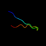
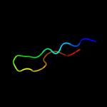
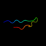
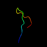
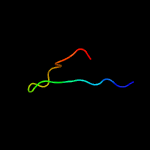
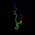
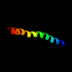
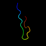
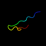
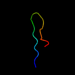
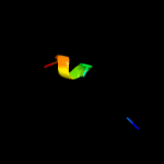
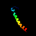
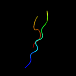
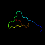
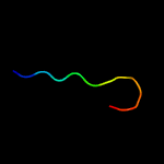
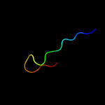
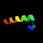
1 c1hymB_
61.6
44
PDB header: hydrolase (serine proteinase)Chain: B: PDB Molecule: hydrolyzed cucurbita maxima trypsin inhibitor v;PDBTitle: hydrolyzed trypsin inhibitor (cmti-v, minimized average nmr structure)
2 d1dwma_
47.6
33
Fold: CI-2 family of serine protease inhibitorsSuperfamily: CI-2 family of serine protease inhibitorsFamily: CI-2 family of serine protease inhibitors
3 c3rdyA_
45.5
50
PDB header: hydrolase inhibitorChain: A: PDB Molecule: bwi-1=protease inhibitor/trypsin inhibitor;PDBTitle: crystal structure of buckwheat trypsin inhibitor rbti at 1.84 angstrom2 resolution
4 c1vbwA_
45.0
41
PDB header: protein bindingChain: A: PDB Molecule: trypsin inhibitor bgit;PDBTitle: crystal structure of bitter gourd trypsin inhibitor
5 d2snii_
43.8
35
Fold: CI-2 family of serine protease inhibitorsSuperfamily: CI-2 family of serine protease inhibitorsFamily: CI-2 family of serine protease inhibitors
6 c2ci2I_
43.0
35
PDB header: proteinase inhibitor (chymotrypsin)Chain: I: PDB Molecule: chymotrypsin inhibitor 2;PDBTitle: crystal and molecular structure of the serine proteinase inhibitor ci-2 2 from barley seeds
7 c2kncB_
41.3
22
PDB header: cell adhesionChain: B: PDB Molecule: integrin beta-3;PDBTitle: platelet integrin alfaiib-beta3 transmembrane-cytoplasmic2 heterocomplex
8 d1to2i_
41.3
35
Fold: CI-2 family of serine protease inhibitorsSuperfamily: CI-2 family of serine protease inhibitorsFamily: CI-2 family of serine protease inhibitors
9 d1ypci_
40.7
35
Fold: CI-2 family of serine protease inhibitorsSuperfamily: CI-2 family of serine protease inhibitorsFamily: CI-2 family of serine protease inhibitors
10 c1tinA_
38.8
44
PDB header: serine protease inhibitorChain: A: PDB Molecule: trypsin inhibitor v;PDBTitle: three-dimensional structure in solution of cucurbita maxima trypsin2 inhibitor-v determined by nmr spectroscopy
11 c1hjiB_
24.9
44
PDB header: bacteriophage hk022Chain: B: PDB Molecule: nun-protein;PDBTitle: bacteriophage hk022 nun-protein-nutboxb-rna complex
12 c4tqvJ_
22.9
20
PDB header: transport proteinChain: J: PDB Molecule: algm2;PDBTitle: crystal structure of a bacterial abc transporter involved in the2 import of the acidic polysaccharide alginate
13 d1csei_
20.9
18
Fold: CI-2 family of serine protease inhibitorsSuperfamily: CI-2 family of serine protease inhibitorsFamily: CI-2 family of serine protease inhibitors
14 c3jt0B_
19.8
26
PDB header: structural proteinChain: B: PDB Molecule: lamin-b1;PDBTitle: crystal structure of the c-terminal fragment (426-558) lamin-b1 from2 homo sapiens, northeast structural genomics consortium target hr5546a
15 d2byoa1
17.0
17
Fold: LolA-like prokaryotic lipoproteins and lipoprotein localization factorsSuperfamily: Prokaryotic lipoproteins and lipoprotein localization factorsFamily: LppX-like
16 c4qa8A_
16.8
17
PDB header: lipid transportChain: A: PDB Molecule: putative lipoprotein lprf;PDBTitle: crystal structure of lprf from mycobacterium bovis
17 d1em9a_
14.5
28
Fold: Retrovirus capsid protein, N-terminal core domainSuperfamily: Retrovirus capsid protein, N-terminal core domainFamily: Retrovirus capsid protein, N-terminal core domain
18 d1egla_
13.9
18
Fold: CI-2 family of serine protease inhibitorsSuperfamily: CI-2 family of serine protease inhibitorsFamily: CI-2 family of serine protease inhibitors
19 c1uv7A_
13.4
17
PDB header: transportChain: A: PDB Molecule: general secretion pathway protein m;PDBTitle: periplasmic domain of epsm from vibrio cholerae
20 d1uv7a_
13.4
17
Fold: RRF/tRNA synthetase additional domain-likeSuperfamily: General secretion pathway protein M, EpsMFamily: General secretion pathway protein M, EpsM
21 d1p7na_
not modelled
13.1
28
Fold: Retrovirus capsid protein, N-terminal core domainSuperfamily: Retrovirus capsid protein, N-terminal core domainFamily: Retrovirus capsid protein, N-terminal core domain
22 c2lllA_
not modelled
12.9
26
PDB header: structural proteinChain: A: PDB Molecule: lamin-b2;PDBTitle: solution nmr structure of c-terminal globular domain of human lamin-2 b2, northeast structural genomics consortium target hr8546a
23 d1oe1a2
not modelled
12.7
21
Fold: Cupredoxin-likeSuperfamily: CupredoxinsFamily: Multidomain cupredoxins
24 c2e63A_
not modelled
12.0
50
PDB header: structural genomics, unknown functionChain: A: PDB Molecule: kiaa1787 protein;PDBTitle: solution structure of the neuz domain in kiaa1787 protein
25 c2hbpA_
not modelled
11.9
33
PDB header: endocytosis, protein bindingChain: A: PDB Molecule: cytoskeleton assembly control protein sla1;PDBTitle: solution structure of sla1 homology domain 1
26 c3mhaB_
not modelled
11.2
21
PDB header: lipid binding proteinChain: B: PDB Molecule: lipoprotein lprg;PDBTitle: crystal structure of lprg from mycobacterium tuberculosis bound to pim
27 c2bu8A_
not modelled
10.7
15
PDB header: transferaseChain: A: PDB Molecule: pyruvate dehydrogenase kinase isoenzyme 2;PDBTitle: crystal structures of human pyruvate dehydrogenase kinase 2 containing2 physiological and synthetic ligands
28 c2yueA_
not modelled
10.4
19
PDB header: rna binding proteinChain: A: PDB Molecule: protein neuralized;PDBTitle: solution structure of the neuz (nhr) domain in neuralized2 from drosophila melanogaster
29 d1qvpa_
not modelled
10.1
30
Fold: SH3-like barrelSuperfamily: C-terminal domain of transcriptional repressorsFamily: FeoA-like
30 c6d0gA_
not modelled
8.4
11
PDB header: oxidoreductaseChain: A: PDB Molecule: pirin family protein;PDBTitle: 1.78 angstrom resolution crystal structure of quercetin 2,3-2 dioxygenase from acinetobacter baumannii
31 c5lrvA_
not modelled
8.2
27
PDB header: hydrolaseChain: A: PDB Molecule: otu domain-containing protein 7b;PDBTitle: structure of cezanne/otud7b otu domain bound to lys11-linked2 diubiquitin
32 d1ivta_
not modelled
8.0
26
Fold: Immunoglobulin-like beta-sandwichSuperfamily: Lamin A/C globular tail domainFamily: Lamin A/C globular tail domain
33 c4kf9A_
not modelled
7.9
21
PDB header: transferaseChain: A: PDB Molecule: glutathione s-transferase protein;PDBTitle: crystal structure of a glutathione transferase family member from2 ralstonia solanacearum, target efi-501780, with bound gsh coordinated3 to a zinc ion, ordered active site
34 c1wq6A_
not modelled
7.8
20
PDB header: oncoproteinChain: A: PDB Molecule: aml1-eto;PDBTitle: the tetramer structure of the nervy homolgy two (nhr2) domain of aml1-2 eto is critical for aml1-eto's activity
35 c2jmbA_
not modelled
7.4
55
PDB header: structural genomics, unknown functionChain: A: PDB Molecule: hypothetical protein atu4866;PDBTitle: solution structure of the protein atu4866 from agrobacterium2 tumefaciens
36 c3ic8D_
not modelled
7.0
38
PDB header: structural genomics, unknown functionChain: D: PDB Molecule: uncharacterized gst-like proteinprotein;PDBTitle: the crystal structure of a gst-like protein from pseudomonas syringae2 to 2.4a
37 c4ardA_
not modelled
7.0
20
PDB header: viral proteinChain: A: PDB Molecule: capsid protein p27;PDBTitle: structure of the immature retroviral capsid at 8a resolution by cryo-2 electron microscopy
38 c5nv8A_
not modelled
7.0
50
PDB header: transferaseChain: A: PDB Molecule: ef-p arginine 32 rhamnosyl-transferase;PDBTitle: structural basis for earp-mediated arginine glycosylation of2 translation elongation factor ef-p
39 d1gvia2
not modelled
6.9
21
Fold: Glycosyl hydrolase domainSuperfamily: Glycosyl hydrolase domainFamily: alpha-Amylases, C-terminal beta-sheet domain
40 c3vgxD_
not modelled
6.9
25
PDB header: membrane proteinChain: D: PDB Molecule: envelope glycoprotein gp160;PDBTitle: structure of gp41 t21/cp621-652
41 c2vfjA_
not modelled
6.9
30
PDB header: hydrolaseChain: A: PDB Molecule: tumor necrosis factor;PDBTitle: structure of the a20 ovarian tumour (otu) domain
42 c6cp8B_
not modelled
6.8
25
PDB header: toxin/antitoxinChain: B: PDB Molecule: cdia;PDBTitle: contact-dependent growth inhibition toxin-immunity protein complex2 from from e. coli 3006
43 c2kgfA_
not modelled
6.7
20
PDB header: viral proteinChain: A: PDB Molecule: capsid protein p27;PDBTitle: n-terminal domain of capsid protein from the mason-pfizer2 monkey virus
44 c2v4xA_
not modelled
6.5
16
PDB header: viral proteinChain: A: PDB Molecule: capsid protein p27;PDBTitle: crystal structure of jaagsiekte sheep retrovirus capsid n-2 terminal domain
45 d1ufga_
not modelled
6.1
26
Fold: Immunoglobulin-like beta-sandwichSuperfamily: Lamin A/C globular tail domainFamily: Lamin A/C globular tail domain
46 c3dkbA_
not modelled
6.0
30
PDB header: hydrolaseChain: A: PDB Molecule: tumor necrosis factor, alpha-induced protein 3;PDBTitle: crystal structure of a20, 2.5 angstrom
47 c5xywD_
not modelled
5.5
13
PDB header: protein bindingChain: D: PDB Molecule: gd21652;PDBTitle: crystal structure of drosophila simulans rhino chromoshadow domain in2 complex with n-terminal domain
48 c1ep3B_
not modelled
5.3
15
PDB header: oxidoreductaseChain: B: PDB Molecule: dihydroorotate dehydrogenase b (pyrk subunit);PDBTitle: crystal structure of lactococcus lactis dihydroorotate dehydrogenase2 b. data collected under cryogenic conditions.
49 c3zv0D_
not modelled
5.3
15
PDB header: cell cycleChain: D: PDB Molecule: h/aca ribonucleoprotein complex subunit 4;PDBTitle: structure of the shq1p-cbf5p complex
50 d1ifra_
not modelled
5.3
26
Fold: Immunoglobulin-like beta-sandwichSuperfamily: Lamin A/C globular tail domainFamily: Lamin A/C globular tail domain


