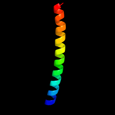
1 c3ogiD_
99.9
97
PDB header: structural genomics, unknown functionChain: D: PDB Molecule: putative esat-6-like protein 7;PDBTitle: crystal structure of the mycobacterium tuberculosis h37rv esxop2 complex (rv2346c-rv2347c)
2 d1wa8a1
76.2
21
Fold: Ferritin-likeSuperfamily: EsxAB dimer-likeFamily: ESAT-6 like
3 c4lwsA_
73.8
32
PDB header: unknown functionChain: A: PDB Molecule: uncharacterized protein;PDBTitle: esxa : esxb (semet) hetero-dimer from thermomonospora curvata
4 c3zbhC_
58.6
23
PDB header: unknown functionChain: C: PDB Molecule: esxa;PDBTitle: geobacillus thermodenitrificans esxa crystal form i
5 c3m0dC_
53.7
20
PDB header: signaling proteinChain: C: PDB Molecule: tnf receptor-associated factor 1;PDBTitle: crystal structure of the traf1:traf2:ciap2 complex
6 c3m06F_
42.0
23
PDB header: protein bindingChain: F: PDB Molecule: tnf receptor-associated factor 2;PDBTitle: crystal structure of traf2
7 c3gvmA_
38.2
17
PDB header: viral proteinChain: A: PDB Molecule: putative uncharacterized protein sag1039;PDBTitle: structure of the homodimeric wxg-100 family protein from streptococcus2 agalactiae
8 c2la2A_
34.7
27
PDB header: antimicrobial proteinChain: A: PDB Molecule: cecropin;PDBTitle: solution structure of papiliocin isolated from the swallowtail2 butterfly, papilio xuthus
9 c2vs0B_
30.9
11
PDB header: cell invasionChain: B: PDB Molecule: virulence factor esxa;PDBTitle: structural analysis of homodimeric staphylococcal aureus2 virulence factor esxa
10 d2j01h1
15.3
45
Fold: Ribosomal protein L6Superfamily: Ribosomal protein L6Family: Ribosomal protein L6
11 d2zjre2
14.8
45
Fold: Ribosomal protein L6Superfamily: Ribosomal protein L6Family: Ribosomal protein L6
12 d2qamg1
13.6
36
Fold: Ribosomal protein L6Superfamily: Ribosomal protein L6Family: Ribosomal protein L6
13 d1vqoe1
13.2
64
Fold: Ribosomal protein L6Superfamily: Ribosomal protein L6Family: Ribosomal protein L6
14 d1rl6a1
11.4
45
Fold: Ribosomal protein L6Superfamily: Ribosomal protein L6Family: Ribosomal protein L6
15 c487dJ_
11.3
42
PDB header: ribosomeChain: J: PDB Molecule: protein (50s l6 ribosomal protein);PDBTitle: seven ribosomal proteins fitted to a cryo-electron2 microscopic map of the large 50s subunit at 7.5 angstroms3 resolution
16 d2cqla1
10.7
45
Fold: Ribosomal protein L6Superfamily: Ribosomal protein L6Family: Ribosomal protein L6
17 c5o60G_
10.4
42
PDB header: ribosomeChain: G: PDB Molecule: 50s ribosomal protein l6;PDBTitle: structure of the 50s large ribosomal subunit from mycobacterium2 smegmatis
18 d1wa8b1
9.7
10
Fold: Ferritin-likeSuperfamily: EsxAB dimer-likeFamily: ESAT-6 like
19 c2hguH_
9.0
42
PDB header: ribosomeChain: H: PDB Molecule: 50s ribosomal protein l6;PDBTitle: 70s t.th. ribosome functional complex with mrna and e- and p-site2 trnas at 4.5a. this entry 2hgu contains 50s ribosomal subunit. the3 30s ribosomal subunit can be found in pdb entry 2hgr.
20 c2i2vG_
8.9
33
PDB header: ribosomeChain: G: PDB Molecule: 50s ribosomal protein l6;PDBTitle: crystal structure of ribosome with messenger rna and the anticodon2 stem-loop of p-site trna. this file contains the 50s subunit of one3 70s ribosome. the entire crystal structure contains two 70s ribosomes4 and is described in remark 400.
21 c1sm1E_
not modelled
8.4
42
PDB header: ribosome/antibioticChain: E: PDB Molecule: 50s ribosomal protein l6;PDBTitle: complex of the large ribosomal subunit from deinococcus radiodurans2 with quinupristin and dalfopristin
22 c1pnuE_
not modelled
8.4
42
PDB header: ribosomeChain: E: PDB Molecule: 50s ribosomal protein l6;PDBTitle: crystal structure of a streptomycin dependent ribosome from2 escherichia coli, 50s subunit of 70s ribosome. this file, 1pnu,3 contains only molecules of the 50s ribosomal subunit. the 30s4 subunit, mrna, p-site trna, and a-site trna are in the pdb file 1pns.
23 c1vw4F_
not modelled
8.4
42
PDB header: ribosomeChain: F: PDB Molecule: 54s ribosomal protein l6, mitochondrial;PDBTitle: structure of the yeast mitochondrial large ribosomal subunit
24 c2bbrA_
not modelled
8.0
44
PDB header: viral proteinChain: A: PDB Molecule: viral casp8 and fadd-like apoptosis regulator;PDBTitle: crystal structure of mc159 reveals molecular mechanism of2 disc assembly and vflip inhibition
25 c2zkre_
not modelled
8.0
42
PDB header: ribosomal protein/rnaChain: E: PDB Molecule: rna expansion segment es7 part ii;PDBTitle: structure of a mammalian ribosomal 60s subunit within an 80s complex2 obtained by docking homology models of the rna and proteins into an3 8.7 a cryo-em map
26 c3ccmE_
not modelled
7.9
64
PDB header: ribosomeChain: E: PDB Molecule: 50s ribosomal protein l6p;PDBTitle: structure of anisomycin resistant 50s ribosomal subunit: 23s rrna2 mutation g2611u
27 c5an9B_
not modelled
7.6
50
PDB header: translationChain: B: PDB Molecule: 60s ribosomal protein l9;PDBTitle: mechanism of eif6 release from the nascent 60s ribosomal subunit
28 c4iogD_
not modelled
7.4
26
PDB header: unknown functionChain: D: PDB Molecule: secreted protein esxb;PDBTitle: the crystal structure of a secreted protein esxb (wild-type, in p212 space group) from bacillus anthracis str. sterne
29 c4a1eE_
not modelled
7.1
55
PDB header: ribosomeChain: E: PDB Molecule: 60s ribosomal protein l9;PDBTitle: t.thermophila 60s ribosomal subunit in complex with2 initiation factor 6. this file contains 5s rrna, 5.8s rrna3 and proteins of molecule 1
30 c4wf9E_
not modelled
6.9
45
PDB header: ribosomeChain: E: PDB Molecule: 50s ribosomal protein l6;PDBTitle: the crystal structure of the large ribosomal subunit of staphylococcus2 aureus in complex with telithromycin
31 c3j39H_
not modelled
6.5
55
PDB header: ribosomeChain: H: PDB Molecule: 60s ribosomal protein l9;PDBTitle: structure of the d. melanogaster 60s ribosomal proteins
32 d1xbla_
not modelled
6.4
14
Fold: Long alpha-hairpinSuperfamily: Chaperone J-domainFamily: Chaperone J-domain
33 c3j21F_
not modelled
6.1
55
PDB header: ribosomeChain: F: PDB Molecule: 50s ribosomal protein l6p;PDBTitle: promiscuous behavior of proteins in archaeal ribosomes revealed by2 cryo-em: implications for evolution of eukaryotic ribosomes (50s3 ribosomal proteins)
34 c2bbzC_
not modelled
6.0
53
PDB header: viral proteinChain: C: PDB Molecule: viral casp8 and fadd-like apoptosis regulator;PDBTitle: crystal structure of mc159 reveals molecular mechanism of2 disc assembly and vflip inhibition
35 c3zf7y_
not modelled
5.7
45
PDB header: ribosomeChain: Y: PDB Molecule: 60s ribosomal protein l24, putative;PDBTitle: high-resolution cryo-electron microscopy structure of the trypanosoma2 brucei ribosome
36 c6fe8D_
not modelled
5.5
42
PDB header: dna binding proteinChain: D: PDB Molecule: centromere dna-binding protein complex cbf3 subunit c;PDBTitle: cryo-em structure of the core centromere binding factor 3 complex
37 c3bboI_
not modelled
5.5
45
PDB header: ribosomeChain: I: PDB Molecule: ribosomal protein l6;PDBTitle: homology model for the spinach chloroplast 50s subunit fitted to 9.4a2 cryo-em map of the 70s chlororibosome