Phyre Home Retrieve Phyre Job Id
Emailmdejesus@rockefeller.edu
DescriptionRVBD1804c_(-)_2047030_2047356
DateFri Aug 2 13:30:41 BST 2019
Unique Job IDd38c30233cab12da
SequenceMRVVSTLLSI ... Download FASTA
Job Typenormal
Job Expiry

Summary 

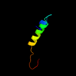
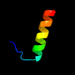
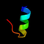
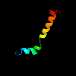
Top model
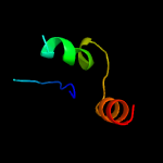
Image coloured by rainbow N → C terminus
Model dimensions (Å):X:33.307 Y:27.515 Z:37.406
Model (left) based on template c6c3rB_
Top template information
PDB header:viral protein
Chain: B: PDB Molecule:cricket paralysis virus 1a protein;
PDBTitle: cricket paralysis virus rnai suppressor protein crpv-1a
Confidence and coverage
Confidence: 61.7% Coverage: 34%
37 residues ( 34% of your sequence) have been modelled with 61.7% confidence by the single highest scoring template.
You may wish to submit your sequence to Phyrealarm. This will automatically scan your sequence every week for new potential templates as they appear in the Phyre2 library.
Please note: You must be registered and logged in to use Phyrealarm.
3D viewing
Interactive 3D view in JSmol
For other options to view your downloaded structure offline see the FAQ

Sequence analysis 

Download FASTA version

Secondary structure and disorder prediction [Show] 

Domain analysis [Show] 

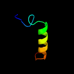
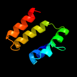
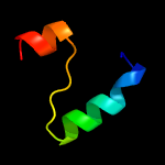
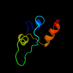
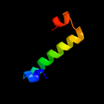
Detailed template information [Hide] 


#
Template Alignment Coverage3D Model Confidence
% i.d. Template Information
1c6c3rB_


61.7 14
PDB header:viral protein
Chain: B: PDB Molecule:cricket paralysis virus 1a protein;
PDBTitle: cricket paralysis virus rnai suppressor protein crpv-1a
2c4rl6A_


22.3 21
PDB header:oxidoreductase
Chain: A: PDB Molecule:saccharopine dehydrogenase;
PDBTitle: crystal structure of the q04l03_strp2 protein from streptococcus2 pneumoniae. northeast structural genomics consortium target spr105
3c3pvsA_


21.3 16
PDB header:recombination
Chain: A: PDB Molecule:replication-associated recombination protein a;
PDBTitle: structure and biochemical activities of escherichia coli mgsa
4d2csua3


20.4 25
Fold:Flavodoxin-like
Superfamily:Succinyl-CoA synthetase domains
Family:Succinyl-CoA synthetase domains
5d1mkea1


15.0 22
Fold:PH domain-like barrel
Superfamily:PH domain-like
Family:Enabled/VASP homology 1 domain (EVH1 domain)
6c4inaA_


14.5 21
PDB header:oxidoreductase
Chain: A: PDB Molecule:saccharopine dehydrogenase;
PDBTitle: crystal structure of the q7mss8_wolsu protein from wolinella2 succinogenes. northeast structural genomics consortium target wsr35
7c5k9xA_


14.2 21
PDB header:lyase
Chain: A: PDB Molecule:tryptophan synthase alpha chain;
PDBTitle: crystal structure of tryptophan synthase alpha chain from legionella2 pneumophila subsp. pneumophila
8c2ekcA_


13.3 21
PDB header:lyase
Chain: A: PDB Molecule:tryptophan synthase alpha chain;
PDBTitle: structural study of project id aq_1548 from aquifex aeolicus vf5
9c5uqdA_


11.8 38
PDB header:oxidoreductase
Chain: A: PDB Molecule:dumpy: shorter than wild-type;
PDBTitle: dpy-21 in complex with fe(ii) and alpha-ketoglutarate
10c5kinC_


10.5 20
PDB header:lyase
Chain: C: PDB Molecule:tryptophan synthase alpha chain;
PDBTitle: crystal structure of tryptophan synthase alpha beta complex from2 streptococcus pneumoniae
11c2lweA_


9.5 20
PDB header:signaling protein
Chain: A: PDB Molecule:probable atp-dependent rna helicase ddx58;
PDBTitle: solution structure of mutant (t170e) second card of human rig-i
12c3j9xJ_


9.0 28
PDB header:virus
Chain: J: PDB Molecule:coat protein;
PDBTitle: a virus that infects a hyperthermophile encapsidates a-form dna
13c5tchG_


9.0 24
PDB header:lyase
Chain: G: PDB Molecule:tryptophan synthase alpha chain;
PDBTitle: crystal structure of tryptophan synthase from m. tuberculosis -2 ligand-free form, trpa-g66v mutant
14c5l78A_


8.9 26
PDB header:oxidoreductase
Chain: A: PDB Molecule:alpha-aminoadipic semialdehyde synthase, mitochondrial;
PDBTitle: crystal structure of human aminoadipate semialdehyde synthase,2 saccharopine dehydrogenase domain (in nad+ bound form)
15c3ss4C_


8.5 31
PDB header:hydrolase
Chain: C: PDB Molecule:glutaminase c;
PDBTitle: crystal structure of mouse glutaminase c, phosphate-bound form
16d2j9ga2


8.3 22
Fold:PreATP-grasp domain
Superfamily:PreATP-grasp domain
Family:BC N-terminal domain-like
17d1zsqa2


7.8 25
Fold:(Phosphotyrosine protein) phosphatases II
Superfamily:(Phosphotyrosine protein) phosphatases II
Family:Myotubularin-like phosphatases
18d1ulza2


7.7 33
Fold:PreATP-grasp domain
Superfamily:PreATP-grasp domain
Family:BC N-terminal domain-like
19c2mulA_


7.1 58
PDB header:protein binding
Chain: A: PDB Molecule:e3 ubiquitin-protein ligase huwe1;
PDBTitle: solution structure of the ubm1 domain of human huwe1/arf-bp1
20c3f2eA_


6.6 23
PDB header:viral protein
Chain: A: PDB Molecule:sirv coat protein;
PDBTitle: crystal structure of yellowstone sirv coat protein c-terminus
21d1axib1


not modelled 6.4 27
Fold:Immunoglobulin-like beta-sandwich
Superfamily:Fibronectin type III
Family:Fibronectin type III
22c1vjqB_


not modelled 6.4 37
PDB header:structural genomics, de novo protein
Chain: B: PDB Molecule:designed protein;
PDBTitle: designed protein based on backbone conformation of2 procarboxypeptidase-a (1aye) with sidechains chosen for maximal3 predicted stability.
23d1xoda1


not modelled 6.1 27
Fold:PH domain-like barrel
Superfamily:PH domain-like
Family:Enabled/VASP homology 1 domain (EVH1 domain)
24c2gacD_


not modelled 6.0 24
PDB header:hydrolase
Chain: D: PDB Molecule:glycosylasparaginase;
PDBTitle: t152c mutant glycosylasparaginase from flavobacterium2 meningosepticum
25c5us3A_


not modelled 6.0 75
PDB header:de novo protein
Chain: A: PDB Molecule:heterogeneous-backbone variant of the sp1-3 zinc finger: n-
PDBTitle: heterogeneous-backbone foldamer mimic of the sp1-3 zinc finger
26c4y7iB_


not modelled 5.9 22
PDB header:hydrolase
Chain: B: PDB Molecule:myotubularin-related protein 8;
PDBTitle: crystal structure of mtmr8
27c2ifsA_


not modelled 5.6 19
PDB header:signaling protein
Chain: A: PDB Molecule:wiskott-aldrich syndrome protien interacting protein and
PDBTitle: structure of the n-wasp evh1 domain in complex with an extended wip2 peptide
28c1apzB_


not modelled 5.5 12
PDB header:complex (hydrolase/peptide)
Chain: B: PDB Molecule:aspartylglucosaminidase;
PDBTitle: human aspartylglucosaminidase complex with reaction product
29c3uo9B_


not modelled 5.4 31
PDB header:hydrolase/hydrolase inhibitor
Chain: B: PDB Molecule:glutaminase kidney isoform, mitochondrial;
PDBTitle: crystal structure of human gac in complex with glutamate and bptes
30c2l5yA_


not modelled 5.3 5
PDB header:signaling protein
Chain: A: PDB Molecule:stromal interaction molecule 2;
PDBTitle: nmr structure of calcium-loaded stim2 ef-sam.
31c2q5tA_


not modelled 5.3 26
PDB header:toxin
Chain: A: PDB Molecule:cholix toxin;
PDBTitle: full-length cholix toxin from vibrio cholerae
32c5uqeB_


not modelled 5.2 31
PDB header:hydrolase
Chain: B: PDB Molecule:glutaminase kidney isoform, mitochondrial;
PDBTitle: multidomain structure of human kidney-type glutaminase(kga/gls)
33c2li5B_


not modelled 5.2 56
PDB header:protein transport
Chain: B: PDB Molecule:ubiquitin-like modifier-activating enzyme atg7;
PDBTitle: nmr structure of atg8-atg7c30 complex
34c4naeA_


not modelled 5.1 28
PDB header:transferase
Chain: A: PDB Molecule:heptaprenylglyceryl phosphate synthase;
PDBTitle: pcrb from geobacillus kaustophilus, with bound g1p

Binding site prediction 

Due to computational demand, binding site predictions are not run for batch jobs

If you want to predict binding sites, please manually submit your model of choice to 3DLigandSite