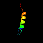
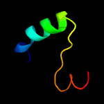
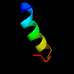
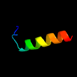
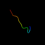
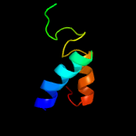
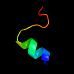
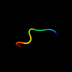
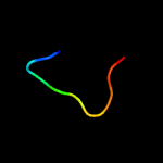
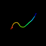
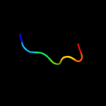
1 c6c3rB_
63.7
24
PDB header: viral proteinChain: B: PDB Molecule: cricket paralysis virus 1a protein;PDBTitle: cricket paralysis virus rnai suppressor protein crpv-1a
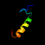
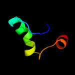
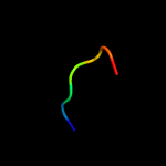
2 d2csua3
22.4
25
Fold: Flavodoxin-likeSuperfamily: Succinyl-CoA synthetase domainsFamily: Succinyl-CoA synthetase domains
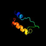
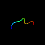
3 c1vjqB_
16.9
30
PDB header: structural genomics, de novo proteinChain: B: PDB Molecule: designed protein;PDBTitle: designed protein based on backbone conformation of2 procarboxypeptidase-a (1aye) with sidechains chosen for maximal3 predicted stability.
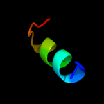
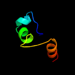
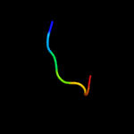
4 d1mkea1
16.6
22
Fold: PH domain-like barrelSuperfamily: PH domain-likeFamily: Enabled/VASP homology 1 domain (EVH1 domain)
5 c2mp6A_
13.4
67
PDB header: signaling proteinChain: A: PDB Molecule: suppressor of cytokine signaling 5;PDBTitle: structure and function of the jak interaction region in the2 intrinsically disordered n-terminus of socs5
6 c4inaA_
12.6
33
PDB header: oxidoreductaseChain: A: PDB Molecule: saccharopine dehydrogenase;PDBTitle: crystal structure of the q7mss8_wolsu protein from wolinella2 succinogenes. northeast structural genomics consortium target wsr35
7 c4rl6A_
12.5
24
PDB header: oxidoreductaseChain: A: PDB Molecule: saccharopine dehydrogenase;PDBTitle: crystal structure of the q04l03_strp2 protein from streptococcus2 pneumoniae. northeast structural genomics consortium target spr105
8 c5uqdA_
11.9
44
PDB header: oxidoreductaseChain: A: PDB Molecule: dumpy: shorter than wild-type;PDBTitle: dpy-21 in complex with fe(ii) and alpha-ketoglutarate
9 c5l78A_
11.7
32
PDB header: oxidoreductaseChain: A: PDB Molecule: alpha-aminoadipic semialdehyde synthase, mitochondrial;PDBTitle: crystal structure of human aminoadipate semialdehyde synthase,2 saccharopine dehydrogenase domain (in nad+ bound form)
10 c5k9xA_
11.2
24
PDB header: lyaseChain: A: PDB Molecule: tryptophan synthase alpha chain;PDBTitle: crystal structure of tryptophan synthase alpha chain from legionella2 pneumophila subsp. pneumophila
11 c2lweA_
10.3
13
PDB header: signaling proteinChain: A: PDB Molecule: probable atp-dependent rna helicase ddx58;PDBTitle: solution structure of mutant (t170e) second card of human rig-i
12 c2ekcA_
9.4
21
PDB header: lyaseChain: A: PDB Molecule: tryptophan synthase alpha chain;PDBTitle: structural study of project id aq_1548 from aquifex aeolicus vf5
13 c3ss4C_
9.4
38
PDB header: hydrolaseChain: C: PDB Molecule: glutaminase c;PDBTitle: crystal structure of mouse glutaminase c, phosphate-bound form
14 d1p9sa_
9.2
57
Fold: Trypsin-like serine proteasesSuperfamily: Trypsin-like serine proteasesFamily: Viral cysteine protease of trypsin fold
15 d2duca1
9.1
57
Fold: Trypsin-like serine proteasesSuperfamily: Trypsin-like serine proteasesFamily: Viral cysteine protease of trypsin fold
16 c3d23A_
8.7
57
PDB header: hydrolase/hydrolase inhibitorChain: A: PDB Molecule: 3c-like proteinase;PDBTitle: main protease of hcov-hku1
17 d1lvoa_
8.1
43
Fold: Trypsin-like serine proteasesSuperfamily: Trypsin-like serine proteasesFamily: Viral cysteine protease of trypsin fold
18 c4gicB_
8.1
50
PDB header: oxidoreductaseChain: B: PDB Molecule: histidinol dehydrogenase;PDBTitle: crystal structure of a putative histidinol dehydrogenase (target psi-2 014034) from methylococcus capsulatus
19 c2q6fB_
8.1
57
PDB header: hydrolaseChain: B: PDB Molecule: infectious bronchitis virus (ibv) main protease;PDBTitle: crystal structure of infectious bronchitis virus (ibv) main protease2 in complex with a michael acceptor inhibitor n3
20 c4xfqB_
7.8
43
PDB header: hydrolaseChain: B: PDB Molecule: pedv main protease;PDBTitle: crystal structure basis for pedv 3c like protease
21 c2ynbA_
not modelled
7.7
43
PDB header: hydrolaseChain: A: PDB Molecule: 3c-like proteinase;PDBTitle: crystal structure of the main protease of coronavirus hku4 in complex2 with a michael acceptor sg85
22 c2ifsA_
not modelled
7.7
22
PDB header: signaling proteinChain: A: PDB Molecule: wiskott-aldrich syndrome protien interacting protein andPDBTitle: structure of the n-wasp evh1 domain in complex with an extended wip2 peptide
23 c4v19S_
not modelled
7.6
18
PDB header: ribosomeChain: S: PDB Molecule: mitoribosomal protein ul18m, mrpl18;PDBTitle: structure of the large subunit of the mammalian mitoribosome, part 12 of 2
24 c3tloA_
not modelled
7.4
43
PDB header: hydrolaseChain: A: PDB Molecule: 3c-like proteinase;PDBTitle: crystal structure of hcov-nl63 3c-like protease
25 c2kktA_
not modelled
7.0
38
PDB header: transcriptionChain: A: PDB Molecule: ataxin-7-like protein 3;PDBTitle: solution structure of the sca7 domain of human ataxin-7-l3 protein
26 c5us3A_
not modelled
6.6
100
PDB header: de novo proteinChain: A: PDB Molecule: heterogeneous-backbone variant of the sp1-3 zinc finger: n-PDBTitle: heterogeneous-backbone foldamer mimic of the sp1-3 zinc finger
27 d1xoda1
not modelled
6.4
18
Fold: PH domain-like barrelSuperfamily: PH domain-likeFamily: Enabled/VASP homology 1 domain (EVH1 domain)
28 c1zi7C_
not modelled
6.4
40
PDB header: lipid binding proteinChain: C: PDB Molecule: kes1 protein;PDBTitle: structure of truncated yeast oxysterol binding protein osh4
29 c5vldC_
not modelled
6.2
60
PDB header: oxidoreductaseChain: C: PDB Molecule: histidinol dehydrogenase, chloroplastic;PDBTitle: crystal structure of medicago truncatula l-histidinol dehydrogenase in2 complex with l-histidine and nad+
30 c2v75A_
not modelled
6.2
25
PDB header: nuclear proteinChain: A: PDB Molecule: nuclear polyadenylated rna-binding protein nab2;PDBTitle: n-terminal domain of nab2
31 c3uo9B_
not modelled
6.2
38
PDB header: hydrolase/hydrolase inhibitorChain: B: PDB Molecule: glutaminase kidney isoform, mitochondrial;PDBTitle: crystal structure of human gac in complex with glutamate and bptes
32 d1k75a_
not modelled
6.1
60
Fold: ALDH-likeSuperfamily: ALDH-likeFamily: L-histidinol dehydrogenase HisD
33 c2djcA_
not modelled
6.1
60
PDB header: cytokineChain: A: PDB Molecule: growth-blocking peptide;PDBTitle: solution structure of growth-blocking peptide of the2 tobacco cutworm, spodoptera litura
34 c1b5nA_
not modelled
5.9
60
PDB header: signaling proteinChain: A: PDB Molecule: protein (plasmatocyte-spreading peptide);PDBTitle: nmr structure of psp1, plasmatocyte-spreading peptide from2 pseudoplusia includens
35 c1b1vA_
not modelled
5.9
60
PDB header: cytokineChain: A: PDB Molecule: protein (plasmatocyte-spreading peptide);PDBTitle: nmr structure of psp1, plasmatocyte-spreading peptide from2 pseudoplusia includens
36 c5uqeB_
not modelled
5.9
38
PDB header: hydrolaseChain: B: PDB Molecule: glutaminase kidney isoform, mitochondrial;PDBTitle: multidomain structure of human kidney-type glutaminase(kga/gls)
37 c1irrA_
not modelled
5.9
60
PDB header: cytokineChain: A: PDB Molecule: paralytic peptide;PDBTitle: solution structure of paralytic peptide of the silkworm,2 bombyx mori
38 c1bonB_
not modelled
5.8
50
PDB header: hormoneChain: B: PDB Molecule: bombyxin-ii,bombyxin a-6;PDBTitle: three-dimensional structure of bombyxin-ii, an insulin-2 related brain-secretory peptide of the silkmoth bombyx3 mori: comparison with insulin and relaxin
39 c2mulA_
not modelled
5.7
67
PDB header: protein bindingChain: A: PDB Molecule: e3 ubiquitin-protein ligase huwe1;PDBTitle: solution structure of the ubm1 domain of human huwe1/arf-bp1
40 d1zhxa1
not modelled
5.7
40
Fold: Oxysterol-binding protein-likeSuperfamily: Oxysterol-binding protein-likeFamily: Oxysterol-binding protein
41 c1bomB_
not modelled
5.6
50
PDB header: insulin-like brain-secretory peptideChain: B: PDB Molecule: bombyxin-ii,bombyxin a-6;PDBTitle: three-dimensional structure of bombyxin-ii, an insulin-2 related brain-secretory peptide of the silkmoth bombyx3 mori: comparison with insulin and relaxin
42 c6driA_
not modelled
5.6
67
PDB header: immune systemChain: A: PDB Molecule: acan1;PDBTitle: nmr solution structure of acan1 from the ancylostoma caninum hookworm
43 c4g07A_
not modelled
5.6
40
PDB header: oxidoreductaseChain: A: PDB Molecule: histidinol dehydrogenase;PDBTitle: the crystal structure of the c366s mutant of hdh from brucella suis
44 c3pvsA_
not modelled
5.6
17
PDB header: recombinationChain: A: PDB Molecule: replication-associated recombination protein a;PDBTitle: structure and biochemical activities of escherichia coli mgsa
45 c5tchG_
not modelled
5.4
26
PDB header: lyaseChain: G: PDB Molecule: tryptophan synthase alpha chain;PDBTitle: crystal structure of tryptophan synthase from m. tuberculosis -2 ligand-free form, trpa-g66v mutant
46 c2ffwA_
not modelled
5.3
50
PDB header: ligaseChain: A: PDB Molecule: midline-1;PDBTitle: solution structure of the rbcc/trim b-box1 domain of human2 mid1: b-box with a ring
47 c2dj9A_
not modelled
5.3
60
PDB header: cytokineChain: A: PDB Molecule: growth-blocking peptide;PDBTitle: solution structure of growth-blocking peptide of the2 cabbage armyworm, mamestra brassicae
48 c1hrlA_
not modelled
5.2
60
PDB header: toxinChain: A: PDB Molecule: paralytic peptide i;PDBTitle: structure of a paralytic peptide from an insect, manduca sexta
49 d2j9ga2
not modelled
5.2
11
Fold: PreATP-grasp domainSuperfamily: PreATP-grasp domainFamily: BC N-terminal domain-like
50 c6hv6A_
not modelled
5.2
17
PDB header: toxinChain: A: PDB Molecule: toxin pau_02230;PDBTitle: crystal structure of patoxp, a cysteine protease-like domain of2 photorhabdus asymbiotica toxin patox
51 d1ulza2
not modelled
5.2
22
Fold: PreATP-grasp domainSuperfamily: PreATP-grasp domainFamily: BC N-terminal domain-like
52 c6an0A_
not modelled
5.2
60
PDB header: oxidoreductaseChain: A: PDB Molecule: histidinol dehydrogenase;PDBTitle: crystal structure of histidinol dehydrogenase from elizabethkingia2 anophelis


