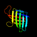
| 1 | c4xinB_
|
|
 |
100.0 |
28 |
PDB header:unknown function
Chain: B: PDB Molecule:lpqh orthologue;
PDBTitle: x-ray crystal structure of an lpqh orthologue from mycobacterium avium
|
|
|
|
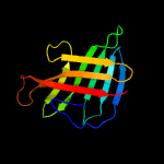
| 2 | c4zjmA_
|
|
 |
100.0 |
23 |
PDB header:unknown function
Chain: A: PDB Molecule:lipoprotein lpqh;
PDBTitle: crystal structure of mycobacterium tuberculosis lpqh (rv3763)
|
|
|
|
| 3 | d1h4ga_
|
|
 |
42.3 |
25 |
Fold:Concanavalin A-like lectins/glucanases
Superfamily:Concanavalin A-like lectins/glucanases
Family:Xylanase/endoglucanase 11/12 |
|
|
|
| 4 | d1hixa_
|
|
 |
39.7 |
22 |
Fold:Concanavalin A-like lectins/glucanases
Superfamily:Concanavalin A-like lectins/glucanases
Family:Xylanase/endoglucanase 11/12 |
|
|
|
| 5 | d1m4wa_
|
|
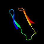 |
39.6 |
19 |
Fold:Concanavalin A-like lectins/glucanases
Superfamily:Concanavalin A-like lectins/glucanases
Family:Xylanase/endoglucanase 11/12 |
|
|
|
| 6 | c3uafA_
|
|
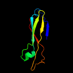 |
35.9 |
14 |
PDB header:protein binding
Chain: A: PDB Molecule:ttr-52;
PDBTitle: crystal structure of a ttr-52 mutant of c. elegans
|
|
|
|
| 7 | c3wp6A_
|
|
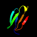 |
35.7 |
22 |
PDB header:hydrolase
Chain: A: PDB Molecule:cdbfv;
PDBTitle: the complex structure of cdbfv e109a with xylotriose
|
|
|
|
| 8 | d1xnka1
|
|
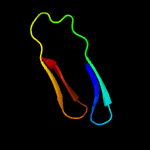 |
28.8 |
31 |
Fold:Concanavalin A-like lectins/glucanases
Superfamily:Concanavalin A-like lectins/glucanases
Family:Xylanase/endoglucanase 11/12 |
|
|
|
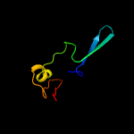
| 9 | c2l7yA_
|
|
 |
27.4 |
10 |
PDB header:structural protein
Chain: A: PDB Molecule:putative endo-beta-n-acetylglucosaminidase;
PDBTitle: solution structure of a putative surface protein
|
|
|
|
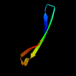
| 10 | c4uznA_
|
|
 |
25.7 |
31 |
PDB header:hydrolase
Chain: A: PDB Molecule:endo-beta-1,4-glucanase (celulase b);
PDBTitle: the native structure of the family 46 carbohydrate-binding2 module (cbm46) of endo-beta-1,4-glucanase b (cel5b) from3 bacillus halodurans
|
|
|
|
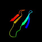
| 11 | c2vulA_
|
|
 |
22.5 |
25 |
PDB header:hydrolase
Chain: A: PDB Molecule:gh11 xylanase;
PDBTitle: thermostable mutant of environmentally isolated gh112 xylanase
|
|
|
|
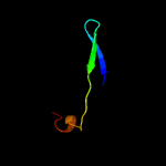
| 12 | c1vraA_
|
|
 |
20.3 |
31 |
PDB header:transferase
Chain: A: PDB Molecule:arginine biosynthesis bifunctional protein argj;
PDBTitle: crystal structure of arginine biosynthesis bifunctional protein argj2 (10175521) from bacillus halodurans at 2.00 a resolution
|
|
|
|
| 13 | d1ynaa_
|
|
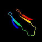 |
19.6 |
25 |
Fold:Concanavalin A-like lectins/glucanases
Superfamily:Concanavalin A-like lectins/glucanases
Family:Xylanase/endoglucanase 11/12 |
|
|
|
| 14 | c3mn8A_
|
|
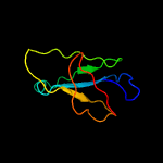 |
19.0 |
18 |
PDB header:hydrolase
Chain: A: PDB Molecule:lp15968p;
PDBTitle: structure of drosophila melanogaster carboxypeptidase d isoform 1b2 short
|
|
|
|
| 15 | c1uwyA_
|
|
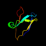 |
17.7 |
12 |
PDB header:hydrolase
Chain: A: PDB Molecule:carboxypeptidase m;
PDBTitle: crystal structure of human carboxypeptidase m
|
|
|
|
| 16 | c4f5cF_
|
|
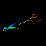 |
15.9 |
16 |
PDB header:hydrolase/viral protein
Chain: F: PDB Molecule:prcv spike protein;
PDBTitle: crystal structure of the spike receptor binding domain of a porcine2 respiratory coronavirus in complex with the pig aminopeptidase n3 ectodomain
|
|
|
|
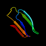
| 17 | c5vqjA_
|
|
 |
15.8 |
23 |
PDB header:hydrolase
Chain: A: PDB Molecule:exo-beta-1,4-xylanase;
PDBTitle: discovery of a first gh11 exo-1,4-beta-xylanase from a diverse2 microbial sugar cane bagasse composting community
|
|
|
|
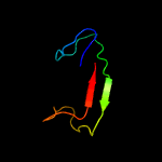
| 18 | c4q98A_
|
|
 |
14.8 |
13 |
PDB header:cell adhesion
Chain: A: PDB Molecule:major fimbrial subunit protein;
PDBTitle: crystal structure of a fimbrilin (fima) from porphyromonas gingivalis2 w83 at 1.30 a resolution (psi community target, nakayama)
|
|
|
|
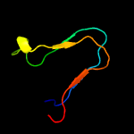
| 19 | c6qt9Y_
|
|
 |
14.2 |
24 |
PDB header:virus
Chain: Y: PDB Molecule:orf 31;
PDBTitle: cryo-em structure of sh1 full particle.
|
|
|
|
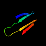
| 20 | c2dcjA_
|
|
 |
13.0 |
22 |
PDB header:hydrolase
Chain: A: PDB Molecule:xylanase j;
PDBTitle: a two-domain structure of alkaliphilic xynj from bacillus sp. 41m-1
|
|
|
|
| 21 | c5o6vC_ |
|
not modelled |
12.2 |
12 |
PDB header:virus
Chain: C: PDB Molecule:envelope protein;
PDBTitle: the cryo-em structure of tick-borne encephalitis virus complexed with2 fab fragment of neutralizing antibody 19/1786
|
|
|
| 22 | d1pvxa_ |
|
not modelled |
12.0 |
25 |
Fold:Concanavalin A-like lectins/glucanases
Superfamily:Concanavalin A-like lectins/glucanases
Family:Xylanase/endoglucanase 11/12 |
|
|
| 23 | d2dfba1 |
|
not modelled |
12.0 |
18 |
Fold:Concanavalin A-like lectins/glucanases
Superfamily:Concanavalin A-like lectins/glucanases
Family:Xylanase/endoglucanase 11/12 |
|
|
| 24 | d1te1b_ |
|
not modelled |
11.1 |
25 |
Fold:Concanavalin A-like lectins/glucanases
Superfamily:Concanavalin A-like lectins/glucanases
Family:Xylanase/endoglucanase 11/12 |
|
|
| 25 | d1uwya1 |
|
not modelled |
10.9 |
12 |
Fold:Prealbumin-like
Superfamily:Carboxypeptidase regulatory domain-like
Family:Carboxypeptidase regulatory domain |
|
|
| 26 | c4v2xA_ |
|
not modelled |
10.8 |
22 |
PDB header:hydrolase
Chain: A: PDB Molecule:endo-beta-1,4-glucanase (cellulase b);
PDBTitle: high resolution structure of the full length tri-modular2 endo-beta-1,4-glucanase b (cel5b) from bacillus halodurans
|
|
|
| 27 | d1svba2 |
|
not modelled |
10.7 |
14 |
Fold:Viral glycoprotein, central and dimerisation domains
Superfamily:Viral glycoprotein, central and dimerisation domains
Family:Viral glycoprotein, central and dimerisation domains |
|
|
| 28 | d1ljma_ |
|
not modelled |
10.6 |
28 |
Fold:Common fold of diphtheria toxin/transcription factors/cytochrome f
Superfamily:p53-like transcription factors
Family:RUNT domain |
|
|
| 29 | c3mxnB_ |
|
not modelled |
10.1 |
20 |
PDB header:replication
Chain: B: PDB Molecule:recq-mediated genome instability protein 2;
PDBTitle: crystal structure of the rmi core complex
|
|
|
| 30 | c5xxzB_ |
|
not modelled |
10.0 |
11 |
PDB header:lyase
Chain: B: PDB Molecule:chemokine protease c;
PDBTitle: crystal structure of a serine protease from streptococcus species
|
|
|
| 31 | c4m03A_ |
|
not modelled |
9.6 |
15 |
PDB header:calcium binding protein
Chain: A: PDB Molecule:serine-rich adhesin for platelets;
PDBTitle: c-terminal fragment(residues 576-751) of binding region of srap
|
|
|
| 32 | c2g16A_ |
|
not modelled |
9.0 |
20 |
PDB header:luminescent protein
Chain: A: PDB Molecule:green fluorescent protein;
PDBTitle: structure of s65a y66s gfp variant after backbone2 fragmentation
|
|
|
| 33 | c3ls1A_ |
|
not modelled |
8.5 |
21 |
PDB header:photosynthesis
Chain: A: PDB Molecule:sll1638 protein;
PDBTitle: crystal structure of cyanobacterial psbq from synechocystis2 sp. pcc 6803 complexed with zn2+
|
|
|
| 34 | d1ok8a2 |
|
not modelled |
8.3 |
6 |
Fold:Viral glycoprotein, central and dimerisation domains
Superfamily:Viral glycoprotein, central and dimerisation domains
Family:Viral glycoprotein, central and dimerisation domains |
|
|
| 35 | c1urzC_ |
|
not modelled |
8.1 |
13 |
PDB header:virus/viral protein
Chain: C: PDB Molecule:envelope protein;
PDBTitle: low ph induced, membrane fusion conformation of the2 envelope protein of tick-borne encephalitis virus
|
|
|
| 36 | c2ovsB_ |
|
not modelled |
8.1 |
10 |
PDB header:gene regulation, ligand binding protein
Chain: B: PDB Molecule:l0044;
PDBTitle: crystal strcuture of a type three secretion system protein
|
|
|
| 37 | d2go8a1 |
|
not modelled |
7.4 |
21 |
Fold:Ferredoxin-like
Superfamily:Dimeric alpha+beta barrel
Family:PG130-like |
|
|
| 38 | c1p58C_ |
|
not modelled |
7.2 |
7 |
PDB header:virus
Chain: C: PDB Molecule:major envelope protein e;
PDBTitle: complex organization of dengue virus membrane proteins as revealed by2 9.5 angstrom cryo-em reconstruction
|
|
|
| 39 | c4whiA_ |
|
not modelled |
7.1 |
11 |
PDB header:hydrolase
Chain: A: PDB Molecule:beta-lactamase;
PDBTitle: crystal structure of c-terminal domain of penicillin binding protein2 rv0907
|
|
|
| 40 | c4m02A_ |
|
not modelled |
6.7 |
13 |
PDB header:calcium binding protein
Chain: A: PDB Molecule:serine-rich adhesin for platelets;
PDBTitle: middle fragment(residues 494-663) of the binding region of srap
|
|
|
| 41 | d1ee6a_ |
|
not modelled |
6.6 |
17 |
Fold:Single-stranded right-handed beta-helix
Superfamily:Pectin lyase-like
Family:Pectate lyase-like |
|
|
| 42 | c6b6iD_ |
|
not modelled |
6.6 |
31 |
PDB header:viral protein,protease
Chain: D: PDB Molecule:3c-like protease;
PDBTitle: 2.4a resolution structure of human norovirus gii.4 protease
|
|
|
| 43 | c3b90A_ |
|
not modelled |
6.3 |
14 |
PDB header:lyase
Chain: A: PDB Molecule:endo-pectate lyase;
PDBTitle: crystal structure of the catalytic domain of pectate lyase peli from2 erwinia chrysanthemi
|
|
|
| 44 | d1cwva2 |
|
not modelled |
6.2 |
19 |
Fold:Immunoglobulin-like beta-sandwich
Superfamily:Invasin/intimin cell-adhesion fragments
Family:Invasin/intimin cell-adhesion fragments |
|
|
| 45 | c2of6C_ |
|
not modelled |
6.1 |
10 |
PDB header:virus
Chain: C: PDB Molecule:envelope glycoprotein e;
PDBTitle: structure of immature west nile virus
|
|
|
| 46 | d1xnda_ |
|
not modelled |
5.7 |
20 |
Fold:Concanavalin A-like lectins/glucanases
Superfamily:Concanavalin A-like lectins/glucanases
Family:Xylanase/endoglucanase 11/12 |
|
|
| 47 | d1wv3a1 |
|
not modelled |
5.5 |
44 |
Fold:SMAD/FHA domain
Superfamily:SMAD/FHA domain
Family:EssC N-terminal domain-like |
|
|
| 48 | d1igoa_ |
|
not modelled |
5.4 |
16 |
Fold:Concanavalin A-like lectins/glucanases
Superfamily:Concanavalin A-like lectins/glucanases
Family:Xylanase/endoglucanase 11/12 |
|
|
| 49 | c5u6fA_ |
|
not modelled |
5.3 |
6 |
PDB header:cell adhesion
Chain: A: PDB Molecule:lpxtg-motif cell wall anchor domain protein;
PDBTitle: bacterial adhesin from mobiluncus mulieris containing intramolecular2 disulfide, isopeptide, and ester bond cross-links (space group p21)
|
|
|
| 50 | c1ywkE_ |
|
not modelled |
5.3 |
14 |
PDB header:isomerase
Chain: E: PDB Molecule:4-deoxy-l-threo-5-hexosulose-uronate ketol-
PDBTitle: crystal structure of 4-deoxy-1-threo-5-hexosulose-uronate2 ketol-isomerase from enterococcus faecalis
|
|
|
| 51 | d1oa3a_ |
|
not modelled |
5.3 |
17 |
Fold:Concanavalin A-like lectins/glucanases
Superfamily:Concanavalin A-like lectins/glucanases
Family:Xylanase/endoglucanase 11/12 |
|
|
| 52 | d2jeka1 |
|
not modelled |
5.2 |
13 |
Fold:Rv1873-like
Superfamily:Rv1873-like
Family:Rv1873-like |
|
|
| 53 | c2c1fA_ |
|
not modelled |
5.1 |
22 |
PDB header:hydrolase
Chain: A: PDB Molecule:bifunctional endo-1,4-beta-xylanase a;
PDBTitle: the structure of the family 11 xylanase from neocallimastix2 patriciarum
|
|
|
| 54 | c1as5A_ |
|
not modelled |
5.1 |
33 |
PDB header:neurotoxin
Chain: A: PDB Molecule:conotoxin y-piiie;
PDBTitle: solution structure of conotoxin y-piiie from conus2 purpurascens, nmr, 14 structures
|
|
|


