 |
|
 |
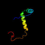
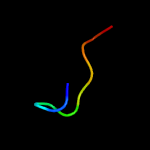
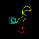
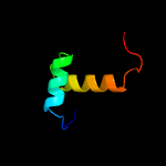
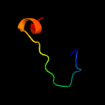
| Summary |
| Top model | |||||||||||||||||||||||
|
| ||||||||||||||||||||||

| Sequence analysis |
| Download FASTA version |
| Secondary structure and disorder prediction [Show] |


| Domain analysis [Show] |
|
|
|
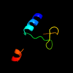
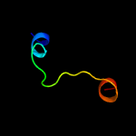
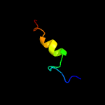
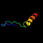
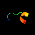
| Summary |
| Top model | |||||||||||||||||||||||
|
| ||||||||||||||||||||||
| Sequence analysis |
| Download FASTA version |
| Secondary structure and disorder prediction [Show] |
| Domain analysis [Show] |