| 1 |
|
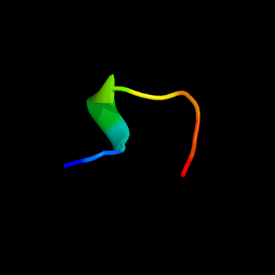
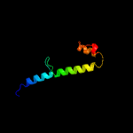
PDB 2cx1 chain A domain 2
Region: 165 - 173
Aligned: 9
Modelled: 9
Confidence: 15.7%
Identity: 89%
Fold: Cystatin-like
Superfamily: Pre-PUA domain
Family: Hypothetical protein APE0525, N-terminal domain
Phyre2
| 2 |
|
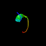
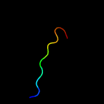
PDB 2j7q chain A domain 1
Region: 48 - 79
Aligned: 32
Modelled: 30
Confidence: 14.2%
Identity: 22%
Fold: Cysteine proteinases
Superfamily: Cysteine proteinases
Family: M48USP-like
Phyre2
| 3 |
|
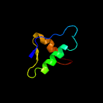
PDB 4b0f chain E
Region: 130 - 151
Aligned: 22
Modelled: 22
Confidence: 10.4%
Identity: 32%
PDB header:immune system
Chain: E: PDB Molecule:c4b-binding protein alpha chain;
PDBTitle: heptameric core complex structure of c4b-binding (c4bp) protein from2 human
Phyre2
| 4 |
|
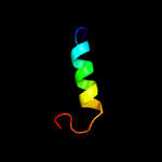
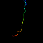
PDB 2p0t chain A
Region: 62 - 128
Aligned: 61
Modelled: 66
Confidence: 9.9%
Identity: 25%
PDB header:structural genomics, unknown function
Chain: A: PDB Molecule:upf0307 protein pspto_4464;
PDBTitle: structural genomics, the crystal structure of a conserved putative2 protein from pseudomonas syringae pv. tomato str. dc3000
Phyre2
| 5 |
|
PDB 2p0t chain A domain 1
Region: 62 - 128
Aligned: 61
Modelled: 66
Confidence: 9.9%
Identity: 25%
Fold: PSPTO4464-like
Superfamily: PSPTO4464-like
Family: PSPTO4464-like
Phyre2
| 6 |
|
PDB 4fay chain C
Region: 7 - 17
Aligned: 11
Modelled: 11
Confidence: 6.5%
Identity: 64%
PDB header:glycerol-binding protein
Chain: C: PDB Molecule:microcompartments protein;
PDBTitle: crystal structure of a trimeric bacterial microcompartment shell2 protein pdub with glycerol metabolites
Phyre2
| 7 |
|
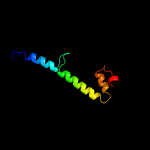
PDB 5xxs chain B
Region: 61 - 127
Aligned: 55
Modelled: 55
Confidence: 6.0%
Identity: 20%
PDB header:transferase
Chain: B: PDB Molecule:protein ribt;
PDBTitle: crystal structure of native ribt from bacillus subtilis
Phyre2
| 8 |
|
PDB 3io0 chain A
Region: 5 - 20
Aligned: 16
Modelled: 16
Confidence: 5.0%
Identity: 63%
PDB header:structural protein
Chain: A: PDB Molecule:etub protein;
PDBTitle: crystal structure of etub from clostridium kluyveri
Phyre2