1 c6ic4C_
99.9
25
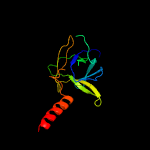
PDB header: protein transportChain: C: PDB Molecule: toluene tolerance efflux transporter (abc superfamily,PDBTitle: cryo-em structure of the a. baumannii mla complex at 8.7 a resolution
2 c5uw8C_
99.9
19
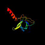
PDB header: transport proteinChain: C: PDB Molecule: probable phospholipid abc transporter-binding protein mlad;PDBTitle: structure of e. coli mce protein mlad, core mce domain
3 c5uvnE_
99.6
14
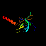
PDB header: transport proteinChain: E: PDB Molecule: paraquat-inducible protein b;PDBTitle: structure of e. coli mce protein pqib, periplasmic domain
4 c5uvnA_
99.6
14
PDB header: transport proteinChain: A: PDB Molecule: paraquat-inducible protein b;PDBTitle: structure of e. coli mce protein pqib, periplasmic domain
5 c5uvnD_
99.6
14
PDB header: transport proteinChain: D: PDB Molecule: paraquat-inducible protein b;PDBTitle: structure of e. coli mce protein pqib, periplasmic domain
6 c5uvnC_
99.6
14
PDB header: transport proteinChain: C: PDB Molecule: paraquat-inducible protein b;PDBTitle: structure of e. coli mce protein pqib, periplasmic domain
7 c5uvnB_
99.6
14
PDB header: transport proteinChain: B: PDB Molecule: paraquat-inducible protein b;PDBTitle: structure of e. coli mce protein pqib, periplasmic domain
8 c5uvnF_
99.6
14
PDB header: transport proteinChain: F: PDB Molecule: paraquat-inducible protein b;PDBTitle: structure of e. coli mce protein pqib, periplasmic domain
9 c1qu7A_
94.0
16
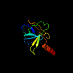
PDB header: signaling proteinChain: A: PDB Molecule: methyl-accepting chemotaxis protein i;PDBTitle: four helical-bundle structure of the cytoplasmic domain of a serine2 chemotaxis receptor
10 c2wpqA_
91.3
6
PDB header: membrane proteinChain: A: PDB Molecule: trimeric autotransporter adhesin fragment;PDBTitle: salmonella enterica sada 479-519 fused to gcn4 adaptors (sadak3, in-2 register fusion)
11 c2qf4A_
89.8
12
PDB header: structural proteinChain: A: PDB Molecule: cell shape determining protein mrec;PDBTitle: high resolution structure of the major periplasmic domain from the2 cell shape-determining filament mrec (orthorhombic form)
12 c3g67A_
87.3
11
PDB header: signaling proteinChain: A: PDB Molecule: methyl-accepting chemotaxis protein;PDBTitle: crystal structure of a soluble chemoreceptor from thermotoga2 maritima
13 d1eq1a_
85.7
15
Fold: Apolipophorin-IIISuperfamily: Apolipophorin-IIIFamily: Apolipophorin-III
14 c3zx6A_
85.4
16
PDB header: signalingChain: A: PDB Molecule: hamp, methyl-accepting chemotaxis protein i;PDBTitle: structure of hamp(af1503)-tsr fusion - hamp (a291v) mutant
15 c3cwgA_
83.5
12
PDB header: transcriptionChain: A: PDB Molecule: signal transducer and activator of transcriptionPDBTitle: unphosphorylated mouse stat3 core fragment
16 c3lnrA_
83.5
12
PDB header: signaling proteinChain: A: PDB Molecule: aerotaxis transducer aer2;PDBTitle: crystal structure of poly-hamp domains from the p. aeruginosa soluble2 receptor aer2
17 c2ch7A_
82.3
6
PDB header: chemotaxisChain: A: PDB Molecule: methyl-accepting chemotaxis protein;PDBTitle: crystal structure of the cytoplasmic domain of a bacterial2 chemoreceptor from thermotoga maritima
18 c2j5uB_
78.2
15
PDB header: cell shape regulationChain: B: PDB Molecule: mrec protein;PDBTitle: mrec lysteria monocytogenes
19 c1deqF_
78.0
8
PDB header: blood clottingChain: F: PDB Molecule: fibrinogen (gamma chain);PDBTitle: the crystal structure of modified bovine fibrinogen (at ~42 angstrom resolution)
20 c4rh7A_
74.9
12
PDB header: motor proteinChain: A: PDB Molecule: green fluorescent protein/cytoplasmic dynein 2 heavy chainPDBTitle: crystal structure of human cytoplasmic dynein 2 motor domain in2 complex with adp.vi
21 c4tkoB_
not modelled
73.7
18
PDB header: membrane proteinChain: B: PDB Molecule: emra;PDBTitle: structure of the periplasmic adaptor protein emra
22 c3vkhA_
not modelled
73.5
11
PDB header: motor proteinChain: A: PDB Molecule: dynein heavy chain, cytoplasmic;PDBTitle: x-ray structure of a functional full-length dynein motor domain
23 c1deqO_
not modelled
73.0
15
PDB header: blood clottingChain: O: PDB Molecule: fibrinogen (beta chain);PDBTitle: the crystal structure of modified bovine fibrinogen (at ~42 angstrom resolution)
24 c5xbjA_
not modelled
71.2
14
PDB header: biosynthetic proteinChain: A: PDB Molecule: flagellar hook-associated protein flgk;PDBTitle: the structure of the flagellar hook junction protein hap1 (flgk) from2 campylobacter jejuni
25 c2qihA_
not modelled
71.0
9
PDB header: cell adhesionChain: A: PDB Molecule: protein uspa1;PDBTitle: crystal structure of 527-665 fragment of uspa1 protein from moraxella2 catarrhalis
26 c1kmiZ_
not modelled
67.7
12
PDB header: signaling proteinChain: Z: PDB Molecule: chemotaxis protein chez;PDBTitle: crystal structure of an e.coli chemotaxis protein, chez
27 c5lp5F_
not modelled
64.6
22
PDB header: hydrolase/antibioticChain: F: PDB Molecule: rod shape-determining protein (mrec);PDBTitle: complex between penicillin-binding protein (pbp2) and mrec from2 helicobacter pylori
28 c3ojaB_
not modelled
61.7
8
PDB header: protein bindingChain: B: PDB Molecule: anopheles plasmodium-responsive leucine-rich repeat proteinPDBTitle: crystal structure of lrim1/apl1c complex
29 c6gajA_
not modelled
61.7
10
PDB header: viral proteinChain: A: PDB Molecule: outer capsid protein sigma-1;PDBTitle: crystal structure of the t1l reovirus sigma1 coiled coil tail (iodide)
30 c3ghgI_
not modelled
61.6
5
PDB header: blood clottingChain: I: PDB Molecule: fibrinogen gamma chain;PDBTitle: crystal structure of human fibrinogen
31 c5u0pU_
not modelled
61.4
16
PDB header: transcriptionChain: U: PDB Molecule: mediator complex subunit 21;PDBTitle: cryo-em structure of the transcriptional mediator
32 c2d4yA_
not modelled
60.3
14
PDB header: structural proteinChain: A: PDB Molecule: flagellar hook-associated protein 1;PDBTitle: crystal structure of a 49k fragment of hap1 (flgk)
33 d1st6a4
not modelled
59.3
14
Fold: Four-helical up-and-down bundleSuperfamily: alpha-catenin/vinculin-likeFamily: alpha-catenin/vinculin
34 c6b7nC_
not modelled
54.0
8
PDB header: viral proteinChain: C: PDB Molecule: spike protein;PDBTitle: cryo-electron microscopy structure of porcine delta coronavirus spike2 protein in the pre-fusion state
35 c1bf5A_
not modelled
53.7
9
PDB header: gene regulation/dnaChain: A: PDB Molecule: signal transducer and activator of transcription 1-PDBTitle: tyrosine phosphorylated stat-1/dna complex
36 c5cwsJ_
not modelled
53.2
10
PDB header: protein transportChain: J: PDB Molecule: nucleoporin nup49;PDBTitle: crystal structure of the intact chaetomium thermophilum nsp1-nup49-2 nup57 channel nucleoporin heterotrimer bound to its nic96 nuclear3 pore complex attachment site
37 c1ei3E_
not modelled
46.8
10
PDB header: blood clottingChain: E: PDB Molecule: fibrinogen;PDBTitle: crystal structure of native chicken fibrinogen
38 c1ei3C_
not modelled
46.5
9
PDB header: blood clottingChain: C: PDB Molecule: fibrinogen;PDBTitle: crystal structure of native chicken fibrinogen
39 c5lskD_
not modelled
44.2
16
PDB header: cell cycleChain: D: PDB Molecule: kinetochore-associated protein dsn1 homolog;PDBTitle: crystal structure of the human kinetochore mis12-cenp-c complex
40 c4ut1A_
not modelled
43.8
11
PDB header: motor proteinChain: A: PDB Molecule: flagellar hook-associated protein;PDBTitle: the structure of the flagellar hook junction protein flgk2 from burkholderia pseudomallei
41 c3zbhC_
not modelled
43.7
8
PDB header: unknown functionChain: C: PDB Molecule: esxa;PDBTitle: geobacillus thermodenitrificans esxa crystal form i
42 d1h9ra2
not modelled
42.3
32
Fold: OB-foldSuperfamily: MOP-likeFamily: BiMOP, duplicated molybdate-binding domain
43 c5n76C_
not modelled
41.0
25
PDB header: nickel-binding proteinChain: C: PDB Molecule: coot;PDBTitle: crystal structure of the apo-form of the co dehydrogenase accessory2 protein coot from rhodospirillum rubrum
44 c3gvmA_
not modelled
40.4
13
PDB header: viral proteinChain: A: PDB Molecule: putative uncharacterized protein sag1039;PDBTitle: structure of the homodimeric wxg-100 family protein from streptococcus2 agalactiae
45 d1st6a3
not modelled
39.8
14
Fold: Four-helical up-and-down bundleSuperfamily: alpha-catenin/vinculin-likeFamily: alpha-catenin/vinculin
46 c6gapB_
not modelled
39.5
10
PDB header: viral proteinChain: B: PDB Molecule: outer capsid protein sigma-1;PDBTitle: crystal structure of the t3d reovirus sigma1 coiled coil tail and body
47 c3ghgK_
not modelled
39.2
17
PDB header: blood clottingChain: K: PDB Molecule: fibrinogen beta chain;PDBTitle: crystal structure of human fibrinogen
48 c4zzkA_
not modelled
37.3
16
PDB header: motor proteinChain: A: PDB Molecule: basal-body rod modification protein flgd;PDBTitle: crystal structure of truncated flgd (monoclinic form) from the human2 pathogen helicobacter pylori
49 c4abxB_
not modelled
36.1
12
PDB header: dna binding proteinChain: B: PDB Molecule: dna repair protein recn;PDBTitle: crystal structure of deinococcus radiodurans recn coiled-2 coil domain
50 d1ykhb1
not modelled
35.7
11
Fold: Mediator hinge subcomplex-likeSuperfamily: Mediator hinge subcomplex-likeFamily: CSE2-like
51 d1h9ma2
not modelled
35.4
36
Fold: OB-foldSuperfamily: MOP-likeFamily: BiMOP, duplicated molybdate-binding domain
52 c5yfpG_
not modelled
34.7
13
PDB header: exocytosisChain: G: PDB Molecule: exocyst complex component exo70;PDBTitle: cryo-em structure of the exocyst complex
53 c6e6aB_
not modelled
34.2
12
PDB header: protein bindingChain: B: PDB Molecule: inclusion membrane protein a;PDBTitle: triclinic crystal form of inca g144a point mutant
54 c4lwsB_
not modelled
33.0
13
PDB header: unknown functionChain: B: PDB Molecule: uncharacterized protein;PDBTitle: esxa : esxb (semet) hetero-dimer from thermomonospora curvata
55 d1h9ma1
not modelled
31.0
28
Fold: OB-foldSuperfamily: MOP-likeFamily: BiMOP, duplicated molybdate-binding domain
56 c4wsrA_
not modelled
29.3
11
PDB header: viral proteinChain: A: PDB Molecule: hemagglutinin;PDBTitle: the crystal structure of hemagglutinin form a/chicken/new york/14677-2 13/1998
57 c4iogD_
not modelled
28.5
10
PDB header: unknown functionChain: D: PDB Molecule: secreted protein esxb;PDBTitle: the crystal structure of a secreted protein esxb (wild-type, in p212 space group) from bacillus anthracis str. sterne
58 c2vs0B_
not modelled
28.5
13
PDB header: cell invasionChain: B: PDB Molecule: virulence factor esxa;PDBTitle: structural analysis of homodimeric staphylococcal aureus2 virulence factor esxa
59 d1v5va1
not modelled
27.7
17
Fold: Elongation factor/aminomethyltransferase common domainSuperfamily: Aminomethyltransferase beta-barrel domainFamily: Aminomethyltransferase beta-barrel domain
60 c2nrjA_
not modelled
26.3
11
PDB header: toxinChain: A: PDB Molecule: hbl b protein;PDBTitle: crystal structure of hemolysin binding component from2 bacillus cereus
61 c6ewyA_
not modelled
26.2
9
PDB header: structural proteinChain: A: PDB Molecule: peptidoglycan endopeptidase ripa;PDBTitle: ripa peptidoglycan hydrolase (rv1477, mycobacterium tuberculosis) n-2 terminal domain
62 c4modB_
not modelled
25.4
17
PDB header: viral proteinChain: B: PDB Molecule: hr1 of s protein, linker, hr2 of s protein;PDBTitle: structure of the mers-cov fusion core
63 c5j65A_
not modelled
25.0
16
PDB header: toxinChain: A: PDB Molecule: pesticidal crystal protein cry6aa;PDBTitle: crystal structure of trypsin activated cry6aa
64 c3c12A_
not modelled
24.8
11
PDB header: biosynthetic proteinChain: A: PDB Molecule: flagellar protein;PDBTitle: crystal structure of flgd from xanthomonas campestris:2 insights into the hook capping essential for flagellar3 assembly
65 c5j2lB_
not modelled
24.8
17
PDB header: de novo proteinChain: B: PDB Molecule: protein design 2l4hc2_11;PDBTitle: de novo design of protein homo-oligomers with modular hydrogen bond2 network-mediated specificity
66 c2ieqC_
not modelled
22.7
10
PDB header: viral proteinChain: C: PDB Molecule: spike glycoprotein;PDBTitle: core structure of s2 from the human coronavirus nl63 spike2 glycoprotein
67 d2vzsa2
not modelled
22.5
18
Fold: Immunoglobulin-like beta-sandwichSuperfamily: beta-Galactosidase/glucuronidase domainFamily: beta-Galactosidase/glucuronidase domain
68 c4wa0A_
not modelled
21.3
19
PDB header: cell adhesionChain: A: PDB Molecule: possible adhesin;PDBTitle: the structure of a possible adhesin c-terminal domain from2 caldicellulosiruptor kronotskyensis
69 c6grjG_
not modelled
21.2
18
PDB header: toxinChain: G: PDB Molecule: ahlb;PDBTitle: structure of the ahlb pore of the tripartite alpha-pore forming toxin,2 ahl, from aeromonas hydrophila.
70 d1wosa1
not modelled
20.9
21
Fold: Elongation factor/aminomethyltransferase common domainSuperfamily: Aminomethyltransferase beta-barrel domainFamily: Aminomethyltransferase beta-barrel domain
71 c1qoyA_
not modelled
19.9
12
PDB header: toxinChain: A: PDB Molecule: hemolysin e;PDBTitle: e.coli hemolysin e (hlye, clya, shea)
72 c3pe0B_
not modelled
19.9
11
PDB header: structural proteinChain: B: PDB Molecule: plectin;PDBTitle: structure of the central region of the plakin domain of plectin
73 c1yvlB_
not modelled
19.8
9
PDB header: signaling proteinChain: B: PDB Molecule: signal transducer and activator of transcriptionPDBTitle: structure of unphosphorylated stat1
74 c5zuvB_
not modelled
19.6
19
PDB header: viral protein, inhibitorChain: B: PDB Molecule: spike glycoprotein,spike glycoprotein,inhibitor ek1;PDBTitle: crystal structure of the human coronavirus 229e hr1 motif in complex2 with pan-covs inhibitor ek1
75 d1hcia4
not modelled
19.6
11
Fold: Spectrin repeat-likeSuperfamily: Spectrin repeatFamily: Spectrin repeat
76 c5gasN_
not modelled
18.5
8
PDB header: hydrolaseChain: N: PDB Molecule: archaeal/vacuolar-type h+-atpase subunit i;PDBTitle: thermus thermophilus v/a-atpase, conformation 2
77 c3bt6B_
not modelled
18.5
11
PDB header: viral proteinChain: B: PDB Molecule: influenza b hemagglutinin (ha);PDBTitle: crystal structure of influenza b virus hemagglutinin
78 c5nmoA_
not modelled
18.3
12
PDB header: cell cycleChain: A: PDB Molecule: chromosome partition protein smc,chromosome partitionPDBTitle: structure of the bacillus subtilis smc joint domain
79 c5lskA_
not modelled
18.0
13
PDB header: cell cycleChain: A: PDB Molecule: protein mis12 homolog;PDBTitle: crystal structure of the human kinetochore mis12-cenp-c complex
80 c1cz5A_
not modelled
18.0
14
PDB header: hydrolaseChain: A: PDB Molecule: vcp-like atpase;PDBTitle: nmr structure of vat-n: the n-terminal domain of vat (vcp-2 like atpase of thermoplasma)
81 c4lwsA_
not modelled
17.7
9
PDB header: unknown functionChain: A: PDB Molecule: uncharacterized protein;PDBTitle: esxa : esxb (semet) hetero-dimer from thermomonospora curvata
82 c4xa3A_
not modelled
17.2
14
PDB header: motor proteinChain: A: PDB Molecule: gp7-myh7(1361-1425)-eb1 chimera protein;PDBTitle: crystal structure of the coiled-coil surrounding skip 2 of myh7
83 c2iakA_
not modelled
17.0
8
PDB header: cell adhesionChain: A: PDB Molecule: bullous pemphigoid antigen 1, isoform 5;PDBTitle: crystal structure of a protease resistant fragment of the plakin2 domain of bullous pemphigoid antigen1 (bpag1)
84 c3j6vL_
not modelled
17.0
19
PDB header: ribosomeChain: L: PDB Molecule: 28s ribosomal protein s12, mitochondrial;PDBTitle: cryo-em structure of the small subunit of the mammalian mitochondrial2 ribosome
85 c5szsC_
not modelled
16.7
15
PDB header: viral proteinChain: C: PDB Molecule: spike glycoprotein;PDBTitle: glycan shield and epitope masking of a coronavirus spike protein2 observed by cryo-electron microscopy
86 c1ha0A_
not modelled
16.7
11
PDB header: viral proteinChain: A: PDB Molecule: protein (hemagglutinin precursor);PDBTitle: hemagglutinin precursor ha0
87 c5wrgB_
not modelled
16.5
19
PDB header: virus like particleChain: B: PDB Molecule: spike glycoprotein;PDBTitle: sars-cov spike glycoprotein
88 c5zhyA_
not modelled
16.4
23
PDB header: viral proteinChain: A: PDB Molecule: spike glycoprotein, spike glycoprotein;PDBTitle: structural characterization of the hcov-229e fusion core
89 c6ezvX_
not modelled
16.4
9
PDB header: toxinChain: X: PDB Molecule: non-hemolytic enterotoxin lytic component l1;PDBTitle: the cytotoxin maka from vibrio cholerae
90 c2gl2B_
not modelled
16.3
11
PDB header: cell adhesionChain: B: PDB Molecule: adhesion a;PDBTitle: crystal structure of the tetra muntant (t66g,r67g,f68g,y69g) of2 bacterial adhesin fada
91 c4fiuC_
not modelled
16.0
10
PDB header: viral proteinChain: C: PDB Molecule: hemagglutinin;PDBTitle: the structure of hemagglutinin of h16 subtype influenza virus with2 v327g mutation
92 d1t01a1
not modelled
15.9
14
Fold: Four-helical up-and-down bundleSuperfamily: alpha-catenin/vinculin-likeFamily: alpha-catenin/vinculin
93 c2q13A_
not modelled
15.9
8
PDB header: protein transportChain: A: PDB Molecule: dcc-interacting protein 13 alpha;PDBTitle: crystal structure of bar-ph domain of appl1
94 d2uubl1
not modelled
15.7
29
Fold: OB-foldSuperfamily: Nucleic acid-binding proteinsFamily: Cold shock DNA-binding domain-like
95 c1v5vA_
not modelled
15.5
20
PDB header: transferaseChain: A: PDB Molecule: aminomethyltransferase;PDBTitle: crystal structure of a component of glycine cleavage system: t-protein2 from pyrococcus horikoshii ot3 at 1.5 a resolution
96 c3qr8A_
not modelled
15.2
10
PDB header: viral proteinChain: A: PDB Molecule: baseplate assembly protein v;PDBTitle: crystal structure of the bacteriophage p2 membrane-piercing protein2 gpv
97 c2wr2B_
not modelled
15.1
12
PDB header: viral proteinChain: B: PDB Molecule: hemagglutinin;PDBTitle: structure of influenza h2 avian hemagglutinin with avian2 receptor
98 c4e40A_
not modelled
15.1
11
PDB header: transport proteinChain: A: PDB Molecule: putative uncharacterized protein;PDBTitle: the haptoglobin-hemoglobin receptor of trypanosoma congolense
99 c6cfzD_
not modelled
14.9
10
PDB header: nuclear proteinChain: D: PDB Molecule: duo1;PDBTitle: structure of the dash/dam1 complex shows its role at the yeast2 kinetochore-microtubule interface