| 1 | c6a7vU_
|
|
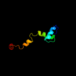 |
99.6 |
56 |
PDB header:toxin/antitoxin
Chain: U: PDB Molecule:antitoxin vapb11;
PDBTitle: crystal structure of mycobacterium tuberculosis vapbc11 toxin-2 antitoxin complex
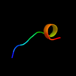
|
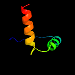
|
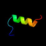
|
|
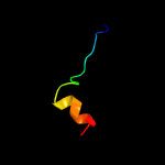
| 2 | c4chgJ_
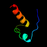
|
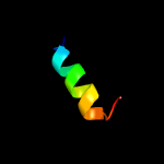
|
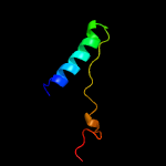
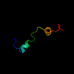 |
99.5 |
100 |
PDB header:toxin/antitoxin
Chain: J: PDB Molecule:antitoxin vapb15;
PDBTitle: crystal structure of vapbc15 complex from mycobacterium tuberculosis
|
|
|
|
| 3 | c4chgG_
|
|
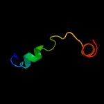 |
99.0 |
100 |
PDB header:toxin/antitoxin
Chain: G: PDB Molecule:antitoxin vapb15;
PDBTitle: crystal structure of vapbc15 complex from mycobacterium tuberculosis
|
|
|
|
| 4 | c5vgtA_
|
|
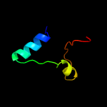 |
71.4 |
21 |
PDB header:viral protein
Chain: A: PDB Molecule:gene 7 protein;
PDBTitle: x-ray structure of bacteriophage sf6 tail adaptor protein gp7
|
|
|
|
| 5 | c2m4hA_
|
|
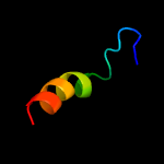 |
54.5 |
19 |
PDB header:viral protein
Chain: A: PDB Molecule:feline calicivirus vpg protein;
PDBTitle: solution structure of the core domain (10-76) of the feline2 calicivirus vpg protein
|
|
|
|
| 6 | c2mxdA_
|
|
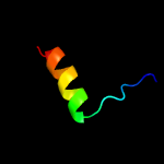 |
45.7 |
20 |
PDB header:viral protein
Chain: A: PDB Molecule:viral protein genome-linked;
PDBTitle: solution structure of vpg of porcine sapovirus
|
|
|
|
| 7 | c1xrxD_
|
|
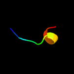 |
24.1 |
60 |
PDB header:replication inhibitor
Chain: D: PDB Molecule:seqa protein;
PDBTitle: crystal structure of a dna-binding protein
|
|
|
|
| 8 | d1xrxa1
|
|
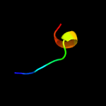 |
24.1 |
60 |
Fold:Ribbon-helix-helix
Superfamily:Ribbon-helix-helix
Family:SeqA N-terminal domain-like |
|
|
|
| 9 | c3fmtF_
|
|
 |
21.2 |
60 |
PDB header:replication inhibitor/dna
Chain: F: PDB Molecule:protein seqa;
PDBTitle: crystal structure of seqa bound to dna
|
|
|
|
| 10 | d2bj7a1
|
|
 |
18.3 |
15 |
Fold:Ribbon-helix-helix
Superfamily:Ribbon-helix-helix
Family:CopG-like |
|
|
|
| 11 | d1jjcb2
|
|
 |
15.6 |
18 |
Fold:Putative DNA-binding domain
Superfamily:Putative DNA-binding domain
Family:Domains B1 and B5 of PheRS-beta, PheT |
|
|
|
| 12 | c5odlA_
|
|
 |
11.3 |
23 |
PDB header:dna binding protein
Chain: A: PDB Molecule:single-stranded dna-binding protein;
PDBTitle: single-stranded dna-binding protein from bacteriophage enc34 in2 complex with ssdna
|
|
|
|
| 13 | c4e0fB_
|
|
 |
9.7 |
41 |
PDB header:transferase
Chain: B: PDB Molecule:riboflavin synthase subunit alpha;
PDBTitle: crystallographic structure of trimeric riboflavin synthase from2 brucella abortus in complex with riboflavin
|
|
|
|
| 14 | d2hzab1
|
|
 |
9.3 |
21 |
Fold:Ribbon-helix-helix
Superfamily:Ribbon-helix-helix
Family:CopG-like |
|
|
|
| 15 | d2ieaa3
|
|
 |
9.2 |
41 |
Fold:TK C-terminal domain-like
Superfamily:TK C-terminal domain-like
Family:Transketolase C-terminal domain-like |
|
|
|
| 16 | c5mu4A_
|
|
 |
8.6 |
32 |
PDB header:viral protein
Chain: A: PDB Molecule:tail tubular protein a;
PDBTitle: tail tubular protein a of klebsiella pneumoniae bacteriophage kp32
|
|
|
|
| 17 | c1i8dB_
|
|
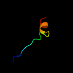 |
8.2 |
47 |
PDB header:transferase
Chain: B: PDB Molecule:riboflavin synthase;
PDBTitle: crystal structure of riboflavin synthase
|
|
|
|
| 18 | c1kzlA_
|
|
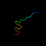 |
8.1 |
24 |
PDB header:transferase
Chain: A: PDB Molecule:riboflavin synthase;
PDBTitle: riboflavin synthase from s.pombe bound to2 carboxyethyllumazine
|
|
|
|
| 19 | c3i1lC_
|
|
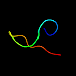 |
8.1 |
33 |
PDB header:hydrolase
Chain: C: PDB Molecule:hemagglutinin-esterase protein;
PDBTitle: structure of porcine torovirus hemagglutinin-esterase in complex with2 its receptor
|
|
|
|
| 20 | c1q5vB_
|
|
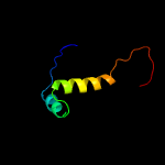 |
7.8 |
16 |
PDB header:transcription
Chain: B: PDB Molecule:nickel responsive regulator;
PDBTitle: apo-nikr
|
|
|
|
| 21 | d2hzaa1 |
|
not modelled |
7.7 |
21 |
Fold:Ribbon-helix-helix
Superfamily:Ribbon-helix-helix
Family:CopG-like |
|
|
| 22 | c6enoA_ |
|
not modelled |
7.6 |
14 |
PDB header:oxidoreductase
Chain: A: PDB Molecule:dehydratase family protein;
PDBTitle: double cubane cluster oxidoreductase
|
|
|
| 23 | c4klkA_ |
|
not modelled |
7.6 |
46 |
PDB header:unknown function
Chain: A: PDB Molecule:phage-related protein duf2815;
PDBTitle: phage-related protein duf2815 from enterococcus faecalis
|
|
|
| 24 | c3dpgA_ |
|
not modelled |
7.6 |
44 |
PDB header:hydrolase/dna
Chain: A: PDB Molecule:sgrair restriction enzyme;
PDBTitle: sgrai with noncognate dna bound
|
|
|
| 25 | d1i8da2 |
|
not modelled |
7.4 |
47 |
Fold:Reductase/isomerase/elongation factor common domain
Superfamily:Riboflavin synthase domain-like
Family:Riboflavin synthase |
|
|
| 26 | c3l4gL_ |
|
not modelled |
6.7 |
24 |
PDB header:ligase
Chain: L: PDB Molecule:phenylalanyl-trna synthetase beta chain;
PDBTitle: crystal structure of homo sapiens cytoplasmic phenylalanyl-trna2 synthetase
|
|
|
| 27 | c4jg2A_ |
|
not modelled |
6.4 |
38 |
PDB header:unknown function
Chain: A: PDB Molecule:phage-related protein;
PDBTitle: structure of phage-related protein from bacillus cereus atcc 10987
|
|
|
| 28 | c2vxzA_ |
|
not modelled |
6.2 |
23 |
PDB header:viral protein
Chain: A: PDB Molecule:pyrsv_gp04;
PDBTitle: crystal structure of hypothetical protein pyrsv_gp04 from pyrobaculum2 spherical virus
|
|
|
| 29 | c2bj3D_ |
|
not modelled |
5.8 |
15 |
PDB header:transcription
Chain: D: PDB Molecule:nickel responsive regulator;
PDBTitle: nikr-apo
|
|
|
| 30 | c5n6nC_ |
|
not modelled |
5.7 |
21 |
PDB header:signaling protein
Chain: C: PDB Molecule:neutral trehalase;
PDBTitle: crystal structure of the 14-3-3:neutral trehalase nth1 complex
|
|
|
| 31 | d1uerc2 |
|
not modelled |
5.6 |
36 |
Fold:Fe,Mn superoxide dismutase (SOD), C-terminal domain
Superfamily:Fe,Mn superoxide dismutase (SOD), C-terminal domain
Family:Fe,Mn superoxide dismutase (SOD), C-terminal domain |
|
|
| 32 | c5m4aA_ |
|
not modelled |
5.5 |
21 |
PDB header:hydrolase
Chain: A: PDB Molecule:neutral trehalase;
PDBTitle: neutral trehalase nth1 from saccharomyces cerevisiae in complex with2 trehalose
|
|
|
| 33 | c2m1bA_ |
|
not modelled |
5.4 |
40 |
PDB header:transcription regulator
Chain: A: PDB Molecule:transcriptional regulatory protein, c terminal family
PDBTitle: solution structure of the chxr dna-binding domain
|
|
|
| 34 | c2ns5A_ |
|
not modelled |
5.1 |
25 |
PDB header:signaling protein
Chain: A: PDB Molecule:partitioning-defective 3 homolog;
PDBTitle: the conserved n-terminal domain of par-3 adopts a novel pb1-2 like structure required for par-3 oligomerization and3 apical membrane localization
|
|
|
| 35 | d1kzla2 |
|
not modelled |
5.1 |
24 |
Fold:Reductase/isomerase/elongation factor common domain
Superfamily:Riboflavin synthase domain-like
Family:Riboflavin synthase |
|
|
| 36 | c1yx5A_ |
|
not modelled |
5.1 |
40 |
PDB header:hydrolase
Chain: A: PDB Molecule:26s proteasome non-atpase regulatory subunit 4;
PDBTitle: solution structure of s5a uim-1/ubiquitin complex
|
|
|
| 37 | d1ma1a2 |
|
not modelled |
5.1 |
44 |
Fold:Fe,Mn superoxide dismutase (SOD), C-terminal domain
Superfamily:Fe,Mn superoxide dismutase (SOD), C-terminal domain
Family:Fe,Mn superoxide dismutase (SOD), C-terminal domain |
|
|


