Phyre Home Retrieve Phyre Job Id
Emailmdejesus@rockefeller.edu
DescriptionRVBD2076c_(-)_2333045_2333296
DateMon Aug 5 13:25:19 BST 2019
Unique Job ID9abc223965213b38
SequenceVVVCLIGGVA ... Download FASTA
Job Typenormal
Job Expiry

Summary 

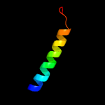
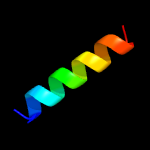
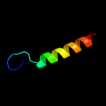
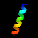
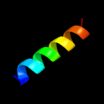
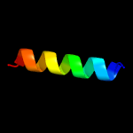
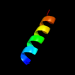
Top model
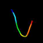
Image coloured by rainbow N → C terminus
Model dimensions (Å):X:35.964 Y:29.867 Z:28.373
Model (left) based on template c6n3qD_
Top template information
PDB header:transport protein
Chain: D: PDB Molecule:protein translocation protein sec63;
PDBTitle: cryo-em structure of the yeast sec complex
Confidence and coverage
Confidence: 47.3% Coverage: 34%
28 residues ( 34% of your sequence) have been modelled with 47.3% confidence by the single highest scoring template.
You may wish to submit your sequence to Phyrealarm. This will automatically scan your sequence every week for new potential templates as they appear in the Phyre2 library.
Please note: You must be registered and logged in to use Phyrealarm.
3D viewing
Interactive 3D view in JSmol
For other options to view your downloaded structure offline see the FAQ

Sequence analysis 

Download FASTA version

Secondary structure and disorder prediction [Show] 

Domain analysis [Show] 

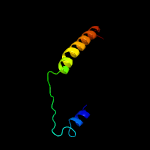
Detailed template information [Hide] 


#
Template Alignment Coverage3D Model Confidence
% i.d. Template Information
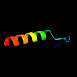
1c6n3qD_


47.3 14
PDB header:transport protein
Chain: D: PDB Molecule:protein translocation protein sec63;
PDBTitle: cryo-em structure of the yeast sec complex
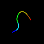
2c6nd1A_


42.1 17
PDB header:protein transport
Chain: A: PDB Molecule:protein translocation protein sec63;
PDBTitle: cryoem structure of the sec complex from yeast
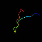
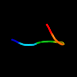
3c2dfxI_


33.1 30
PDB header:hydrolase
Chain: I: PDB Molecule:colicin-e5 immunity protein;
PDBTitle: crystal structure of the carboxy terminal domain of colicin2 e5 complexed with its inhibitor
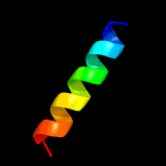
4c6g67A_


17.2 56
PDB header:de novo protein
Chain: A: PDB Molecule:cc-type2-ii;
PDBTitle: crystal structure of a parallel eight-helix coiled coil cc-type2-ii
5c6g67B_


17.2 56
PDB header:de novo protein
Chain: B: PDB Molecule:cc-type2-ii;
PDBTitle: crystal structure of a parallel eight-helix coiled coil cc-type2-ii
6c3gekA_


14.4 22
PDB header:structural genomics, unknown function
Chain: A: PDB Molecule:putative thioesterase yhda;
PDBTitle: crystal structure of putative thioesterase yhda from lactococcus2 lactis. northeast structural genomics consortium target kr113
7c6g66C_


13.4 56
PDB header:de novo protein
Chain: C: PDB Molecule:cc-type2-iv;
PDBTitle: crystal structure of a parallel seven-helix coiled coil cc-type2-iv
8c6g66D_


13.4 56
PDB header:de novo protein
Chain: D: PDB Molecule:cc-type2-iv;
PDBTitle: crystal structure of a parallel seven-helix coiled coil cc-type2-iv
9c6g66E_


13.4 56
PDB header:de novo protein
Chain: E: PDB Molecule:cc-type2-iv;
PDBTitle: crystal structure of a parallel seven-helix coiled coil cc-type2-iv
10c6g66B_


13.4 56
PDB header:de novo protein
Chain: B: PDB Molecule:cc-type2-iv;
PDBTitle: crystal structure of a parallel seven-helix coiled coil cc-type2-iv
11c6g66G_


13.4 56
PDB header:de novo protein
Chain: G: PDB Molecule:cc-type2-iv;
PDBTitle: crystal structure of a parallel seven-helix coiled coil cc-type2-iv
12c6g66A_


13.4 56
PDB header:de novo protein
Chain: A: PDB Molecule:cc-type2-iv;
PDBTitle: crystal structure of a parallel seven-helix coiled coil cc-type2-iv
13c6g66F_


13.4 56
PDB header:de novo protein
Chain: F: PDB Molecule:cc-type2-iv;
PDBTitle: crystal structure of a parallel seven-helix coiled coil cc-type2-iv
14d2f0xa1


12.8 15
Fold:Thioesterase/thiol ester dehydrase-isomerase
Superfamily:Thioesterase/thiol ester dehydrase-isomerase
Family:PaaI/YdiI-like
15c1zmmA_


11.6 100
PDB header:antimicrobial protein
Chain: A: PDB Molecule:neutrophil defensin 4;
PDBTitle: crystal structure of human alpha-defensin-4
16c1zmmC_


11.6 100
PDB header:antimicrobial protein
Chain: C: PDB Molecule:neutrophil defensin 4;
PDBTitle: crystal structure of human alpha-defensin-4
17c6dmqE_


11.5 100
PDB header:antimicrobial protein
Chain: E: PDB Molecule:neutrophil defensin 4;
PDBTitle: crystal structure of the t27a mutant of human alpha defensin hnp4.
18c6dmqH_


11.5 100
PDB header:antimicrobial protein
Chain: H: PDB Molecule:neutrophil defensin 4;
PDBTitle: crystal structure of the t27a mutant of human alpha defensin hnp4.
19c6dmqC_


11.5 100
PDB header:antimicrobial protein
Chain: C: PDB Molecule:neutrophil defensin 4;
PDBTitle: crystal structure of the t27a mutant of human alpha defensin hnp4.
20c6dmqG_


11.5 100
PDB header:antimicrobial protein
Chain: G: PDB Molecule:neutrophil defensin 4;
PDBTitle: crystal structure of the t27a mutant of human alpha defensin hnp4.
21c6dmqA_


not modelled 11.4 100
PDB header:antimicrobial protein
Chain: A: PDB Molecule:neutrophil defensin 4;
PDBTitle: crystal structure of the t27a mutant of human alpha defensin hnp4.
22c1zmmB_


not modelled 11.1 100
PDB header:antimicrobial protein
Chain: B: PDB Molecule:neutrophil defensin 4;
PDBTitle: crystal structure of human alpha-defensin-4
23c1zmmD_


not modelled 11.1 100
PDB header:antimicrobial protein
Chain: D: PDB Molecule:neutrophil defensin 4;
PDBTitle: crystal structure of human alpha-defensin-4
24c6dmqB_


not modelled 11.1 100
PDB header:antimicrobial protein
Chain: B: PDB Molecule:neutrophil defensin 4;
PDBTitle: crystal structure of the t27a mutant of human alpha defensin hnp4.
25c6dmqD_


not modelled 11.1 100
PDB header:antimicrobial protein
Chain: D: PDB Molecule:neutrophil defensin 4;
PDBTitle: crystal structure of the t27a mutant of human alpha defensin hnp4.
26c6dmqF_


not modelled 11.1 100
PDB header:antimicrobial protein
Chain: F: PDB Molecule:neutrophil defensin 4;
PDBTitle: crystal structure of the t27a mutant of human alpha defensin hnp4.
27c2l35B_


not modelled 10.7 47
PDB header:protein binding
Chain: B: PDB Molecule:tyro protein tyrosine kinase-binding protein;
PDBTitle: structure of the dap12-nkg2c transmembrane heterotrimer
28c4wolA_


not modelled 10.1 47
PDB header:signaling protein
Chain: A: PDB Molecule:tyro protein tyrosine kinase-binding protein;
PDBTitle: crystal structure of the dap12 transmembrane domain in lipidic cubic2 phase
29c2l34A_


not modelled 10.1 47
PDB header:protein binding
Chain: A: PDB Molecule:tyro protein tyrosine kinase-binding protein;
PDBTitle: structure of the dap12 transmembrane homodimer
30c2l34B_


not modelled 10.1 47
PDB header:protein binding
Chain: B: PDB Molecule:tyro protein tyrosine kinase-binding protein;
PDBTitle: structure of the dap12 transmembrane homodimer
31c4wolC_


not modelled 9.9 47
PDB header:signaling protein
Chain: C: PDB Molecule:tyro protein tyrosine kinase-binding protein;
PDBTitle: crystal structure of the dap12 transmembrane domain in lipidic cubic2 phase
32c4wolB_


not modelled 9.9 47
PDB header:signaling protein
Chain: B: PDB Molecule:tyro protein tyrosine kinase-binding protein;
PDBTitle: crystal structure of the dap12 transmembrane domain in lipidic cubic2 phase
33c4wo1B_


not modelled 9.9 47
PDB header:signaling protein
Chain: B: PDB Molecule:tyro protein tyrosine kinase-binding protein;
PDBTitle: crystal structure of the dap12 transmembrane domain in lipid cubic2 phase
34c4wo1D_


not modelled 9.9 47
PDB header:signaling protein
Chain: D: PDB Molecule:tyro protein tyrosine kinase-binding protein;
PDBTitle: crystal structure of the dap12 transmembrane domain in lipid cubic2 phase
35c4wo1C_


not modelled 9.9 47
PDB header:signaling protein
Chain: C: PDB Molecule:tyro protein tyrosine kinase-binding protein;
PDBTitle: crystal structure of the dap12 transmembrane domain in lipid cubic2 phase
36c4wo1A_


not modelled 9.9 47
PDB header:signaling protein
Chain: A: PDB Molecule:tyro protein tyrosine kinase-binding protein;
PDBTitle: crystal structure of the dap12 transmembrane domain in lipid cubic2 phase
37d1vh5a_


not modelled 9.4 19
Fold:Thioesterase/thiol ester dehydrase-isomerase
Superfamily:Thioesterase/thiol ester dehydrase-isomerase
Family:PaaI/YdiI-like
38d1j1ua_


not modelled 7.9 20
Fold:Adenine nucleotide alpha hydrolase-like
Superfamily:Nucleotidylyl transferase
Family:Class I aminoacyl-tRNA synthetases (RS), catalytic domain
39c5nmiK_


not modelled 7.7 36
PDB header:membrane protein
Chain: K: PDB Molecule:arg-asn-trp-val-pro-thr-ala-gln-leu-trp-gly-ala-val-gly-
PDBTitle: cytochrome bc1 bound to the inhibitor mjm170
40c5nmiX_


not modelled 7.7 36
PDB header:membrane protein
Chain: X: PDB Molecule:arg-asn-trp-val-pro-thr-ala-gln-leu-trp-gly-ala-val-gly-
PDBTitle: cytochrome bc1 bound to the inhibitor mjm170
41d1be3k_


not modelled 7.7 36
Fold:Single transmembrane helix
Superfamily:Subunit XI (6.4 kDa protein) of cytochrome bc1 complex (Ubiquinol-cytochrome c reductase)
Family:Subunit XI (6.4 kDa protein) of cytochrome bc1 complex (Ubiquinol-cytochrome c reductase)
42c5dm5E_


not modelled 7.1 19
PDB header:hydrolase
Chain: E: PDB Molecule:putative acyl-coa thioester hydrolase;
PDBTitle: crystal structure of the hexameric thioesterase y2039 from yersinia2 pestis
43c2ynmD_


not modelled 6.6 22
PDB header:oxidoreductase
Chain: D: PDB Molecule:light-independent protochlorophyllide reductase subunit b;
PDBTitle: structure of the adpxalf3-stabilized transition state of the2 nitrogenase-like dark-operative protochlorophyllide oxidoreductase3 complex from prochlorococcus marinus with its substrate4 protochlorophyllide a
44c6humQ_


not modelled 6.5 47
PDB header:proton transport
Chain: Q: PDB Molecule:proton-translocating nadh-quinone dehydrogenase subunit q
PDBTitle: structure of the photosynthetic complex i from thermosynechococcus2 elongatus
45c2qwzB_


not modelled 6.2 30
PDB header:hydrolase
Chain: B: PDB Molecule:phenylacetic acid degradation-related protein;
PDBTitle: crystal structure of a putative thioesterase (tm1040_1390) from2 silicibacter sp. tm1040 at 2.15 a resolution
46c6bqzA_


not modelled 6.1 13
PDB header:ligase
Chain: A: PDB Molecule:tyrosine--trna ligase;
PDBTitle: crystal structure of tyrosine-trna synthetase from acinetobacter2 baumannii with bound l-tyrosine
47d2gvha2


not modelled 6.0 32
Fold:Thioesterase/thiol ester dehydrase-isomerase
Superfamily:Thioesterase/thiol ester dehydrase-isomerase
Family:4HBT-like
48c2qg5D_


not modelled 5.9 40
PDB header:transferase
Chain: D: PDB Molecule:calcium/calmodulin-dependent protein kinase;
PDBTitle: cryptosporidium parvum calcium dependent protein kinase cgd7_1840
49c2ts1A_


not modelled 5.6 17
PDB header:ligase (synthetase)
Chain: A: PDB Molecule:tyrosyl-trna synthetase;
PDBTitle: structure of tyrosyl-t/rna synthetase refined at 2.3 angstroms2 resolution. interaction of the enzyme with the tyrosyl adenylate3 intermediate
50d2ts1a_


not modelled 5.6 17
Fold:Adenine nucleotide alpha hydrolase-like
Superfamily:Nucleotidylyl transferase
Family:Class I aminoacyl-tRNA synthetases (RS), catalytic domain
51c5vdkA_


not modelled 5.5 50
PDB header:transferase/transferase inhibitor
Chain: A: PDB Molecule:wee1-like protein kinase 2;
PDBTitle: crystal structure of human wee2 kinase domain in complex with mk1775
52d1vh9a_


not modelled 5.5 21
Fold:Thioesterase/thiol ester dehydrase-isomerase
Superfamily:Thioesterase/thiol ester dehydrase-isomerase
Family:PaaI/YdiI-like
53c3p1aA_


not modelled 5.5 55
PDB header:transferase
Chain: A: PDB Molecule:membrane-associated tyrosine- and threonine-specific cdc2-
PDBTitle: structure of human membrane-associated tyrosine- and threonine-2 specific cdc2-inhibitory kinase myt1 (pkmyt1)
54c3c0hB_


not modelled 5.3 30
PDB header:transferase
Chain: B: PDB Molecule:peripheral plasma membrane protein cask;
PDBTitle: cask cam-kinase domain- amppnp complex, p1 form
55c2jq1A_


not modelled 5.2 71
PDB header:antimicrobial protein
Chain: A: PDB Molecule:phylloseptin-3;
PDBTitle: phylloseptin-3

Binding site prediction 

Due to computational demand, binding site predictions are not run for batch jobs

If you want to predict binding sites, please manually submit your model of choice to 3DLigandSite

Transmembrane helix prediction 

Transmembrane helices have been predicted in your sequence to adopt the topology shown below