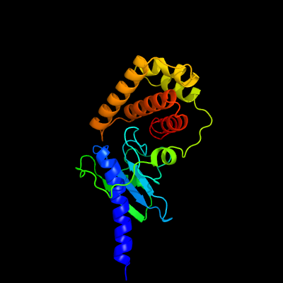
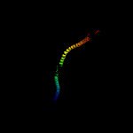
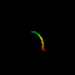
1 c5imuA_
100.0
63
PDB header: signaling proteinChain: A: PDB Molecule: tat (twin-arginine translocation) pathway signal sequencePDBTitle: a fragment of conserved hypothetical protein rv3899c (residues 184-2 410) from mycobacterium tuberculosis
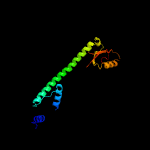
2 c3ojaB_
94.3
10
PDB header: protein bindingChain: B: PDB Molecule: anopheles plasmodium-responsive leucine-rich repeat proteinPDBTitle: crystal structure of lrim1/apl1c complex
3 c1ei3E_
92.9
4
PDB header: blood clottingChain: E: PDB Molecule: fibrinogen;PDBTitle: crystal structure of native chicken fibrinogen
4 c1ei3C_
91.9
8
PDB header: blood clottingChain: C: PDB Molecule: fibrinogen;PDBTitle: crystal structure of native chicken fibrinogen
5 c1deqO_
91.8
7
PDB header: blood clottingChain: O: PDB Molecule: fibrinogen (beta chain);PDBTitle: the crystal structure of modified bovine fibrinogen (at ~42 angstrom resolution)
6 c1deqF_
89.4
6
PDB header: blood clottingChain: F: PDB Molecule: fibrinogen (gamma chain);PDBTitle: the crystal structure of modified bovine fibrinogen (at ~42 angstrom resolution)
7 c3ghgI_
89.1
9
PDB header: blood clottingChain: I: PDB Molecule: fibrinogen gamma chain;PDBTitle: crystal structure of human fibrinogen
8 c6o7xa_
89.1
4
PDB header: membrane proteinChain: A: PDB Molecule: vacuolar atp synthase catalytic subunit a;PDBTitle: saccharomyces cerevisiae v-atpase stv1-v1vo state 3
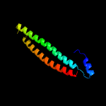
9 c5tbyA_
88.6
10
PDB header: contractile proteinChain: A: PDB Molecule: myosin-7;PDBTitle: human beta cardiac heavy meromyosin interacting-heads motif obtained2 by homology modeling (using swiss-model) of human sequence from3 aphonopelma homology model (pdb-3jbh), rigidly fitted to human beta-4 cardiac negatively stained thick filament 3d-reconstruction (emd-5 2240)
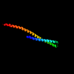
10 c3dtpA_
88.5
10
PDB header: contractile proteinChain: A: PDB Molecule: myosin 2 heavy chain chimera of smooth and cardiac muscle;PDBTitle: tarantula heavy meromyosin obtained by flexible docking to tarantula2 muscle thick filament cryo-em 3d-map
11 c3ghgK_
86.6
5
PDB header: blood clottingChain: K: PDB Molecule: fibrinogen beta chain;PDBTitle: crystal structure of human fibrinogen
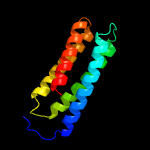
12 c6flnE_
85.9
8
PDB header: protein bindingChain: E: PDB Molecule: e3 ubiquitin/isg15 ligase trim25;PDBTitle: crystal structure of the human trim25 coiled-coil and pryspry domains
13 c5yz0C_
85.1
11
PDB header: cell cycleChain: C: PDB Molecule: atr-interacting protein;PDBTitle: cryo-em structure of human atr-atrip complex
14 c6o7ua_
82.8
5
PDB header: membrane proteinChain: A: PDB Molecule: PDBTitle: saccharomyces cerevisiae v-atpase stv1-vo
15 c2dfsA_
81.7
7
PDB header: contractile protein/transport proteinChain: A: PDB Molecule: myosin-5a;PDBTitle: 3-d structure of myosin-v inhibited state
16 c3lssA_
77.9
11
PDB header: ligaseChain: A: PDB Molecule: seryl-trna synthetase;PDBTitle: trypanosoma brucei seryl-trna synthetase in complex with atp
17 c1y4cA_
74.9
11
PDB header: de novo proteinChain: A: PDB Molecule: maltose binding protein fused with designedPDBTitle: designed helical protein fusion mbp
18 c5ew5C_
74.0
5
PDB header: hydrolaseChain: C: PDB Molecule: colicin-e9;PDBTitle: crystal structure of colicin e9 in complex with its immunity protein2 im9
19 c1bf5A_
71.2
7
PDB header: gene regulation/dnaChain: A: PDB Molecule: signal transducer and activator of transcription 1-PDBTitle: tyrosine phosphorylated stat-1/dna complex
20 c4cgkA_
70.4
8
PDB header: cell cycleChain: A: PDB Molecule: secreted 45 kda protein;PDBTitle: crystal structure of the essential protein pcsb from streptococcus2 pneumoniae
21 c6b3rE_
not modelled
68.1
9
PDB header: transport proteinChain: E: PDB Molecule: piezo-type mechanosensitive ion channel component 1;PDBTitle: structure of the mechanosensitive channel piezo1
22 c4cg4D_
not modelled
66.5
9
PDB header: actin-binding proteinChain: D: PDB Molecule: pyrin;PDBTitle: crystal structure of the chs-b30.2 domains of trim20
23 c5gasN_
not modelled
66.1
9
PDB header: hydrolaseChain: N: PDB Molecule: archaeal/vacuolar-type h+-atpase subunit i;PDBTitle: thermus thermophilus v/a-atpase, conformation 2
24 c6iu3A_
not modelled
65.1
9
PDB header: metal transportChain: A: PDB Molecule: vit1;PDBTitle: crystal structure of iron transporter vit1 with zinc ions
25 c4rh7A_
not modelled
63.9
9
PDB header: motor proteinChain: A: PDB Molecule: green fluorescent protein/cytoplasmic dynein 2 heavy chainPDBTitle: crystal structure of human cytoplasmic dynein 2 motor domain in2 complex with adp.vi
26 c3ojaA_
not modelled
63.0
6
PDB header: protein bindingChain: A: PDB Molecule: leucine-rich immune molecule 1;PDBTitle: crystal structure of lrim1/apl1c complex
27 c3cwgA_
not modelled
61.1
10
PDB header: transcriptionChain: A: PDB Molecule: signal transducer and activator of transcriptionPDBTitle: unphosphorylated mouse stat3 core fragment
28 c3jbhA_
not modelled
60.5
4
PDB header: contractile proteinChain: A: PDB Molecule: myosin 2 heavy chain striated muscle;PDBTitle: two heavy meromyosin interacting-heads motifs flexible docked into2 tarantula thick filament 3d-map allows in depth study of intra- and3 intermolecular interactions
29 c4ll8E_
not modelled
58.1
8
PDB header: motor protein/transport proteinChain: E: PDB Molecule: swi5-dependent ho expression protein 3;PDBTitle: complex of carboxy terminal domain of myo4p and she3p middle fragment
30 c3vkhA_
not modelled
57.1
9
PDB header: motor proteinChain: A: PDB Molecule: dynein heavy chain, cytoplasmic;PDBTitle: x-ray structure of a functional full-length dynein motor domain
31 c2zv4O_
not modelled
57.0
15
PDB header: structural proteinChain: O: PDB Molecule: major vault protein;PDBTitle: the structure of rat liver vault at 3.5 angstrom resolution
32 d2jf2a1
not modelled
52.9
20
Fold: Single-stranded left-handed beta-helixSuperfamily: Trimeric LpxA-like enzymesFamily: UDP N-acetylglucosamine acyltransferase
33 c6gapB_
not modelled
52.0
9
PDB header: viral proteinChain: B: PDB Molecule: outer capsid protein sigma-1;PDBTitle: crystal structure of the t3d reovirus sigma1 coiled coil tail and body
34 c3ipkA_
not modelled
50.3
4
PDB header: cell adhesionChain: A: PDB Molecule: agi/ii;PDBTitle: crystal structure of a3vp1 of agi/ii of streptococcus mutans
35 c1m1jA_
not modelled
50.3
7
PDB header: blood clottingChain: A: PDB Molecule: fibrinogen alpha subunit;PDBTitle: crystal structure of native chicken fibrinogen with two different2 bound ligands
36 c6gajA_
not modelled
49.5
10
PDB header: viral proteinChain: A: PDB Molecule: outer capsid protein sigma-1;PDBTitle: crystal structure of the t1l reovirus sigma1 coiled coil tail (iodide)
37 c6gaoC_
not modelled
45.9
9
PDB header: viral proteinChain: C: PDB Molecule: outer capsid protein sigma-1;PDBTitle: crystal structure of the t1l reovirus sigma1 coiled coil tail and body
38 c5dfzD_
not modelled
44.4
11
PDB header: transferaseChain: D: PDB Molecule: vacuolar protein sorting-associated protein 30;PDBTitle: structure of vps34 complex ii from s. cerevisiae.
39 c4pd3B_
not modelled
43.3
10
PDB header: contractile proteinChain: B: PDB Molecule: nonmuscle myosin heavy chain b, alpha-actinin a chimeraPDBTitle: crystal structure of rigor-like human nonmuscle myosin-2b
40 c1jchC_
not modelled
40.2
7
PDB header: ribosome inhibitor, hydrolaseChain: C: PDB Molecule: colicin e3;PDBTitle: crystal structure of colicin e3 in complex with its immunity protein
41 c5mg8B_
not modelled
39.3
8
PDB header: recombinationChain: B: PDB Molecule: structural maintenance of chromosomes protein 6;PDBTitle: crystal structure of the s.pombe smc5/6 hinge domain
42 c1d7mA_
not modelled
39.3
13
PDB header: contractile proteinChain: A: PDB Molecule: cortexillin i;PDBTitle: coiled-coil dimerization domain from cortexillin i
43 c3vkgB_
not modelled
39.1
11
PDB header: motor proteinChain: B: PDB Molecule: dynein heavy chain, cytoplasmic;PDBTitle: x-ray structure of an mtbd truncation mutant of dynein motor domain
44 c1i84V_
not modelled
38.5
6
PDB header: contractile proteinChain: V: PDB Molecule: smooth muscle myosin heavy chain;PDBTitle: cryo-em structure of the heavy meromyosin subfragment of2 chicken gizzard smooth muscle myosin with regulatory light3 chain in the dephosphorylated state. only c alphas4 provided for regulatory light chain. only backbone atoms5 provided for s2 fragment.
45 c1kmiZ_
not modelled
37.6
8
PDB header: signaling proteinChain: Z: PDB Molecule: chemotaxis protein chez;PDBTitle: crystal structure of an e.coli chemotaxis protein, chez
46 c5voxb_
not modelled
36.5
4
PDB header: hydrolaseChain: B: PDB Molecule: v-type proton atpase subunit b;PDBTitle: yeast v-atpase in complex with legionella pneumophila effector sidk2 (rotational state 1)
47 c2i1kA_
not modelled
36.4
9
PDB header: cell adhesion, membrane proteinChain: A: PDB Molecule: moesin;PDBTitle: moesin from spodoptera frugiperda reveals the coiled-coil domain at2 3.0 angstrom resolution
48 c5nf8A_
not modelled
36.2
15
PDB header: membrane proteinChain: A: PDB Molecule: respiratory supercomplex factor 1, mitochondrial;PDBTitle: solution structure of detergent-solubilized rcf1, a yeast2 mitochondrial inner membrane protein involved in respiratory complex3 iii/iv supercomplex formation
49 c6ogdB_
not modelled
34.2
8
PDB header: toxinChain: B: PDB Molecule: toxin subunit yena2;PDBTitle: cryo-em structure of yentca in its prepore state
50 c5jxfA_
not modelled
32.6
5
PDB header: hydrolaseChain: A: PDB Molecule: asp/glu-specific dipeptidyl-peptidase;PDBTitle: crystal structure of flavobacterium psychrophilum dpp11 in complex2 with dipeptide arg-asp
51 c2xzrA_
not modelled
32.6
9
PDB header: cell adhesionChain: A: PDB Molecule: immunoglobulin-binding protein eibd;PDBTitle: escherichia coli immunoglobulin-binding protein eibd 391-438 fused2 to gcn4 adaptors
52 c3qo8A_
not modelled
32.5
7
PDB header: ligaseChain: A: PDB Molecule: seryl-trna synthetase, cytoplasmic;PDBTitle: crystal structure of seryl-trna synthetase from candida albicans
53 c2ncaA_
not modelled
29.8
10
PDB header: chaperoneChain: A: PDB Molecule: hsp90 co-chaperone cdc37;PDBTitle: structural model for the n-terminal domain of human cdc37
54 c2gl2B_
not modelled
29.3
13
PDB header: cell adhesionChain: B: PDB Molecule: adhesion a;PDBTitle: crystal structure of the tetra muntant (t66g,r67g,f68g,y69g) of2 bacterial adhesin fada
55 c6cfzC_
not modelled
27.3
21
PDB header: nuclear proteinChain: C: PDB Molecule: dad2;PDBTitle: structure of the dash/dam1 complex shows its role at the yeast2 kinetochore-microtubule interface
56 c3vkgA_
not modelled
26.3
12
PDB header: motor proteinChain: A: PDB Molecule: dynein heavy chain, cytoplasmic;PDBTitle: x-ray structure of an mtbd truncation mutant of dynein motor domain
57 c6ezvX_
not modelled
26.1
11
PDB header: toxinChain: X: PDB Molecule: non-hemolytic enterotoxin lytic component l1;PDBTitle: the cytotoxin maka from vibrio cholerae
58 c2qzvB_
not modelled
25.4
13
PDB header: structural proteinChain: B: PDB Molecule: major vault protein;PDBTitle: draft crystal structure of the vault shell at 9 angstroms2 resolution
59 c3lvgD_
not modelled
25.4
7
PDB header: structural proteinChain: D: PDB Molecule: clathrin light chain b;PDBTitle: crystal structure of a clathrin heavy chain and clathrin light chain2 complex
60 c5tvbB_
not modelled
24.6
13
PDB header: transferaseChain: B: PDB Molecule: nucleoprotein tpr;PDBTitle: structure of the tpr oligomerization domain
61 c1wleB_
not modelled
24.4
16
PDB header: ligaseChain: B: PDB Molecule: seryl-trna synthetase;PDBTitle: crystal structure of mammalian mitochondrial seryl-trna2 synthetase complexed with seryl-adenylate
62 c3u59C_
not modelled
24.0
11
PDB header: contractile proteinChain: C: PDB Molecule: tropomyosin beta chain;PDBTitle: n-terminal 98-aa fragment of smooth muscle tropomyosin beta
63 c3vkhB_
not modelled
23.6
11
PDB header: motor proteinChain: B: PDB Molecule: dynein heavy chain, cytoplasmic;PDBTitle: x-ray structure of a functional full-length dynein motor domain
64 c5jxpA_
not modelled
23.0
10
PDB header: hydrolaseChain: A: PDB Molecule: asp/glu-specific dipeptidyl-peptidase;PDBTitle: crystal structure of porphyromonas endodontalis dpp11 in alternate2 conformation
65 c6nr84_
not modelled
23.0
21
PDB header: chaperoneChain: 4: PDB Molecule: prefoldin subunit 4;PDBTitle: htric-hpfd class6
66 c5bu8A_
not modelled
22.2
13
PDB header: viral proteinChain: A: PDB Molecule: dna stabilization protein;PDBTitle: hk620 tail needle crystallized at ph 7.5 and derivatized with xenon
67 c6bwfB_
not modelled
22.1
10
PDB header: membrane proteinChain: B: PDB Molecule: trpm7;PDBTitle: 4.1 angstrom mg2+-unbound structure of mouse trpm7
68 c6bwfA_
not modelled
22.1
10
PDB header: membrane proteinChain: A: PDB Molecule: trpm7;PDBTitle: 4.1 angstrom mg2+-unbound structure of mouse trpm7
69 c6bwfD_
not modelled
22.1
10
PDB header: membrane proteinChain: D: PDB Molecule: trpm7;PDBTitle: 4.1 angstrom mg2+-unbound structure of mouse trpm7
70 c6bwfC_
not modelled
22.1
10
PDB header: membrane proteinChain: C: PDB Molecule: trpm7;PDBTitle: 4.1 angstrom mg2+-unbound structure of mouse trpm7
71 c5mqfK_
not modelled
21.1
11
PDB header: splicingChain: K: PDB Molecule: pre-mrna-splicing factor spf27;PDBTitle: cryo-em structure of a human spliceosome activated for step 2 of2 splicing (c* complex)
72 c3iv1F_
not modelled
20.9
8
PDB header: hydrolaseChain: F: PDB Molecule: tumor susceptibility gene 101 protein;PDBTitle: coiled-coil domain of tumor susceptibility gene 101
73 c3rx6A_
not modelled
20.3
17
PDB header: transcription regulatorChain: A: PDB Molecule: polarity suppression protein;PDBTitle: crystal structure of polarity suppression protein from enterobacteria2 phage p4
74 c2ycuA_
not modelled
20.2
13
PDB header: motor proteinChain: A: PDB Molecule: non muscle myosin 2c, alpha-actinin;PDBTitle: crystal structure of human non muscle myosin 2c in pre-power stroke2 state
75 c2y3aB_
not modelled
20.2
9
PDB header: transferaseChain: B: PDB Molecule: phosphatidylinositol 3-kinase regulatory subunit beta;PDBTitle: crystal structure of p110beta in complex with icsh2 of p85beta and the2 drug gdc-0941
76 c4xa1D_
not modelled
20.0
9
PDB header: motor proteinChain: D: PDB Molecule: gp7-myh7(1173-1238)-eb1 chimera protein;PDBTitle: crystal structure of the coiled-coil surrounding skip 1 of myh7
77 c4v1av_
not modelled
19.1
20
PDB header: ribosomeChain: V: PDB Molecule: PDBTitle: structure of the large subunit of the mammalian mitoribosome, part 22 of 2
78 c5vgzC_
not modelled
18.5
16
PDB header: hydrolaseChain: C: PDB Molecule: 26s proteasome regulatory subunit 8;PDBTitle: conformational landscape of the p28-bound human proteasome regulatory2 particle
79 c3j99M_
not modelled
18.5
14
PDB header: hydrolaseChain: M: PDB Molecule: synaptosomal-associated protein 25;PDBTitle: structure of 20s supercomplex determined by single particle2 cryoelectron microscopy (state iiib)
80 c3ghgD_
not modelled
18.5
8
PDB header: blood clottingChain: D: PDB Molecule: fibrinogen alpha chain;PDBTitle: crystal structure of human fibrinogen
81 c1a92B_
not modelled
18.1
14
PDB header: leucine zipperChain: B: PDB Molecule: delta antigen;PDBTitle: oligomerization domain of hepatitis delta antigen
82 c1vw2H_
not modelled
18.1
11
PDB header: toxinChain: H: PDB Molecule: tcda1;PDBTitle: crystal structure of tcda1
83 c5jxxC_
not modelled
18.1
10
PDB header: transferaseChain: C: PDB Molecule: acyl-[acyl-carrier-protein]--udp-n-acetylglucosamine o-PDBTitle: crystal structure of udp-n-acetylglucosamine o-acyltransferase (lpxa)2 from moraxella catarrhalis rh4.
84 c6bwdA_
not modelled
17.3
18
PDB header: membrane proteinChain: A: PDB Molecule: transient receptor potential cation channel subfamily mPDBTitle: 3.7 angstrom cryoem structure of truncated mouse trpm7
85 c6bwdD_
not modelled
17.3
18
PDB header: membrane proteinChain: D: PDB Molecule: transient receptor potential cation channel subfamily mPDBTitle: 3.7 angstrom cryoem structure of truncated mouse trpm7
86 c6bwdC_
not modelled
17.3
18
PDB header: membrane proteinChain: C: PDB Molecule: transient receptor potential cation channel subfamily mPDBTitle: 3.7 angstrom cryoem structure of truncated mouse trpm7
87 c6bwdB_
not modelled
17.3
18
PDB header: membrane proteinChain: B: PDB Molecule: transient receptor potential cation channel subfamily mPDBTitle: 3.7 angstrom cryoem structure of truncated mouse trpm7
88 c2v71A_
not modelled
17.1
11
PDB header: nuclear proteinChain: A: PDB Molecule: nuclear distribution protein nude-like 1;PDBTitle: coiled-coil region of nudel
89 c2pohA_
not modelled
16.7
14
PDB header: viral proteinChain: A: PDB Molecule: head completion protein;PDBTitle: structure of phage p22 tail needle gp26
90 c3ni0A_
not modelled
16.6
13
PDB header: immune systemChain: A: PDB Molecule: bone marrow stromal antigen 2;PDBTitle: crystal structure of mouse bst-2/tetherin ectodomain
91 c5cwsE_
not modelled
16.6
10
PDB header: protein transportChain: E: PDB Molecule: nucleoporin nup57;PDBTitle: crystal structure of the intact chaetomium thermophilum nsp1-nup49-2 nup57 channel nucleoporin heterotrimer bound to its nic96 nuclear3 pore complex attachment site
92 c6mi3A_
not modelled
16.5
6
PDB header: transcriptionChain: A: PDB Molecule: nf-kb essential modulator,nf-kappa-b essential modulator,PDBTitle: structure of nemo(51-112) with n- and c-terminal coiled-coil adaptors.
93 d1pd3a_
not modelled
16.5
14
Fold: ROP-likeSuperfamily: Nonstructural protein ns2, Nep, M1-binding domainFamily: Nonstructural protein ns2, Nep, M1-binding domain
94 c5xyiG_
not modelled
15.0
20
PDB header: ribosomeChain: G: PDB Molecule: 40s ribosomal protein s6;PDBTitle: small subunit of trichomonas vaginalis ribosome
95 c4nqjB_
not modelled
14.8
8
PDB header: ligaseChain: B: PDB Molecule: e3 ubiquitin-protein ligase trim69;PDBTitle: structure of coiled-coil domain
96 c5td8B_
not modelled
14.7
15
PDB header: replicationChain: B: PDB Molecule: kinetochore protein nuf2;PDBTitle: crystal structure of an extended dwarf ndc80 complex
97 c3tweA_
not modelled
14.5
28
PDB header: unknown functionChain: A: PDB Molecule: alpha4h;PDBTitle: crystal structure of the de novo designed peptide alpha4h
98 c5y06A_
not modelled
14.5
15
PDB header: unknown functionChain: A: PDB Molecule: msmeg_4306;PDBTitle: structural characterization of msmeg_4306 from mycobacterium smegmatis
99 c1ytzI_
not modelled
14.4
17
PDB header: contractile proteinChain: I: PDB Molecule: troponin i;PDBTitle: crystal structure of skeletal muscle troponin in the ca2+-2 activated state