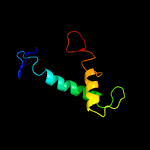
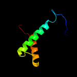
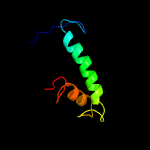
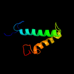
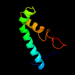
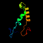
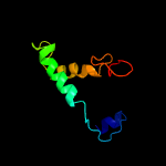
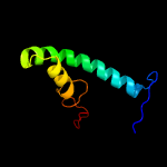
1 c3f9kV_
98.0
11
PDB header: viral protein, recombinationChain: V: PDB Molecule: integrase;PDBTitle: two domain fragment of hiv-2 integrase in complex with ledgf ibd
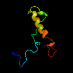
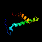
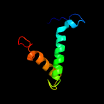
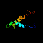
2 d1bcoa2
98.0
17
Fold: Ribonuclease H-like motifSuperfamily: Ribonuclease H-likeFamily: mu transposase, core domain
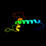
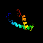
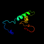
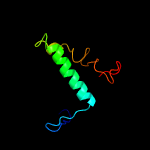
3 c5cz1B_
97.7
11
PDB header: hydrolaseChain: B: PDB Molecule: integrase;PDBTitle: crystal structure of the catalytic core domain of mmtv integrase
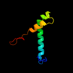
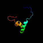
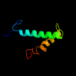
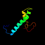
4 c3nf9A_
96.9
8
PDB header: hydrolase/hydrolase inhibitorChain: A: PDB Molecule: integrase;PDBTitle: structural basis for a new mechanism of inhibition of hiv integrase2 identified by fragment screening and structure based design
5 d1asua_
96.4
12
Fold: Ribonuclease H-like motifSuperfamily: Ribonuclease H-likeFamily: Retroviral integrase, catalytic domain
6 c4mq3A_
96.2
19
PDB header: viral proteinChain: A: PDB Molecule: integrase;PDBTitle: the 1.1 angstrom structure of catalytic core domain of fiv integrase
7 d1c0ma2
96.1
14
Fold: Ribonuclease H-like motifSuperfamily: Ribonuclease H-likeFamily: Retroviral integrase, catalytic domain
8 c3kksB_
95.9
14
PDB header: dna binding proteinChain: B: PDB Molecule: integrase;PDBTitle: crystal structure of catalytic core domain of biv integrase in crystal2 form ii
9 c1bcoA_
95.6
14
PDB header: transposaseChain: A: PDB Molecule: bacteriophage mu transposase;PDBTitle: bacteriophage mu transposase core domain
10 c5u1cA_
95.4
8
PDB header: viral proteinChain: A: PDB Molecule: hiv-1 integrase, sso7d chimera;PDBTitle: structure of tetrameric hiv-1 strand transfer complex intasome
11 d1hyva_
95.4
9
Fold: Ribonuclease H-like motifSuperfamily: Ribonuclease H-likeFamily: Retroviral integrase, catalytic domain
12 d1exqa_
95.0
18
Fold: Ribonuclease H-like motifSuperfamily: Ribonuclease H-likeFamily: Retroviral integrase, catalytic domain
13 c1k6yB_
94.5
10
PDB header: transferaseChain: B: PDB Molecule: integrase;PDBTitle: crystal structure of a two-domain fragment of hiv-1 integrase
14 c3jcaE_
94.4
19
PDB header: viral proteinChain: E: PDB Molecule: integrase;PDBTitle: core model of the mouse mammary tumor virus intasome
15 c1ex4A_
94.2
8
PDB header: viral proteinChain: A: PDB Molecule: integrase;PDBTitle: hiv-1 integrase catalytic core and c-terminal domain
16 c1c0mA_
94.1
14
PDB header: transferaseChain: A: PDB Molecule: protein (integrase);PDBTitle: crystal structure of rsv two-domain integrase
17 c5m0rF_
94.0
16
PDB header: hydrolaseChain: F: PDB Molecule: integrase;PDBTitle: cryo-em reconstruction of the maedi-visna virus (mvv) strand transfer2 complex
18 d1c6va_
93.3
17
Fold: Ribonuclease H-like motifSuperfamily: Ribonuclease H-likeFamily: Retroviral integrase, catalytic domain
19 c3l2tB_
92.5
9
PDB header: recombination/dnaChain: B: PDB Molecule: integrase;PDBTitle: crystal structure of the prototype foamy virus (pfv) intasome in2 complex with magnesium and mk0518 (raltegravir)
20 c3hpgC_
90.1
18
PDB header: transferaseChain: C: PDB Molecule: integrase;PDBTitle: visna virus integrase (residues 1-219) in complex with ledgf2 ibd: examples of open integrase dimer-dimer interfaces
21 d1cxqa_
not modelled
83.8
13
Fold: Ribonuclease H-like motifSuperfamily: Ribonuclease H-likeFamily: Retroviral integrase, catalytic domain
22 c2f7tA_
not modelled
79.9
10
PDB header: dna binding proteinChain: A: PDB Molecule: mos1 transposase;PDBTitle: crystal structure of the catalytic domain of mos1 mariner2 transposase
23 c3f2kB_
not modelled
76.1
17
PDB header: transferaseChain: B: PDB Molecule: histone-lysine n-methyltransferase setmar;PDBTitle: structure of the transposase domain of human histone-lysine2 n-methyltransferase setmar
24 c5cr4B_
not modelled
71.7
12
PDB header: hydrolaseChain: B: PDB Molecule: sleeping beauty transposase, sb100x;PDBTitle: crystal structure of the sleeping beauty transposase catalytic domain
25 c3hosA_
not modelled
70.4
10
PDB header: transferase, dna binding protein/dnaChain: A: PDB Molecule: transposable element mariner, complete cds;PDBTitle: crystal structure of the mariner mos1 paired end complex with mg
26 c4fcyA_
not modelled
53.4
10
PDB header: dna binding protein/dnaChain: A: PDB Molecule: transposase;PDBTitle: crystal structure of the bacteriophage mu transpososome
27 c3dlrA_
not modelled
52.0
8
PDB header: transferaseChain: A: PDB Molecule: integrase;PDBTitle: crystal structure of the catalytic core domain from pfv integrase
28 c6n1cB_
not modelled
50.8
5
PDB header: hydrolaseChain: B: PDB Molecule: inorganic pyrophosphatase;PDBTitle: crystal structure of inorganic pyrophosphatase from legionella2 pneumophila philadelphia 1
29 c4lugA_
not modelled
44.9
0
PDB header: hydrolaseChain: A: PDB Molecule: inorganic pyrophosphatase;PDBTitle: crystal structure of inorganic pyrophosphatase ppa1 from arabidopsis2 thaliana
30 c3fq3H_
not modelled
44.0
8
PDB header: hydrolaseChain: H: PDB Molecule: inorganic pyrophosphatase:bacterial/archaeal inorganicPDBTitle: crystal structure of inorganic phosphatase from brucella melitensis
31 c3g0tA_
not modelled
37.7
20
PDB header: transferaseChain: A: PDB Molecule: putative aminotransferase;PDBTitle: crystal structure of putative aspartate aminotransferase (np_905498.1)2 from porphyromonas gingivalis w83 at 1.75 a resolution
32 d2prda_
not modelled
37.1
5
Fold: OB-foldSuperfamily: Inorganic pyrophosphataseFamily: Inorganic pyrophosphatase
33 d1i40a_
not modelled
36.3
8
Fold: OB-foldSuperfamily: Inorganic pyrophosphataseFamily: Inorganic pyrophosphatase
34 d1twla_
not modelled
30.5
8
Fold: OB-foldSuperfamily: Inorganic pyrophosphataseFamily: Inorganic pyrophosphatase
35 d1udea_
not modelled
29.9
8
Fold: OB-foldSuperfamily: Inorganic pyrophosphataseFamily: Inorganic pyrophosphatase
36 c2mzyA_
not modelled
29.5
14
PDB header: iron binding proteinChain: A: PDB Molecule: probable fe(2+)-trafficking protein;PDBTitle: 1h, 13c, and 15n chemical shift assignments and structure of probable2 fe(2+)-trafficking protein from burkholderia pseudomallei 1710b.
37 c6cfzD_
not modelled
27.4
17
PDB header: nuclear proteinChain: D: PDB Molecule: duo1;PDBTitle: structure of the dash/dam1 complex shows its role at the yeast2 kinetochore-microtubule interface
38 d1w0ba_
not modelled
27.3
8
Fold: Spectrin repeat-likeSuperfamily: Alpha-hemoglobin stabilizing protein AHSPFamily: Alpha-hemoglobin stabilizing protein AHSP
39 c3ld3A_
not modelled
27.3
8
PDB header: hydrolaseChain: A: PDB Molecule: inorganic pyrophosphatase;PDBTitle: crystal structure of inorganic phosphatase from anaplasma2 phagocytophilum at 1.75a resolution
40 c3d63B_
not modelled
26.4
5
PDB header: hydrolaseChain: B: PDB Molecule: inorganic pyrophosphatase;PDBTitle: crystal structure of inorganic pyrophosphatase from burkholderia2 pseudomallei
41 d1z8ua1
not modelled
24.7
8
Fold: Spectrin repeat-likeSuperfamily: Alpha-hemoglobin stabilizing protein AHSPFamily: Alpha-hemoglobin stabilizing protein AHSP
42 c2uxsA_
not modelled
24.1
13
PDB header: hydrolaseChain: A: PDB Molecule: inorganic pyrophosphatase;PDBTitle: 2.7a crystal structure of inorganic pyrophosphatase (rv3628)2 from mycobacterium tuberculosis at ph 7.5
43 c1ygzC_
not modelled
18.0
0
PDB header: hydrolaseChain: C: PDB Molecule: inorganic pyrophosphatase;PDBTitle: crystal structure of inorganic pyrophosphatase from helicobacter2 pylori
44 c3tr4C_
not modelled
17.4
3
PDB header: hydrolaseChain: C: PDB Molecule: inorganic pyrophosphatase;PDBTitle: structure of an inorganic pyrophosphatase (ppa) from coxiella burnetii
45 d1b7ea_
not modelled
16.0
17
Fold: Ribonuclease H-like motifSuperfamily: Ribonuclease H-likeFamily: Transposase inhibitor (Tn5 transposase)
46 c5tj5P_
not modelled
15.6
23
PDB header: motor proteinChain: P: PDB Molecule: v-type proton atpase subunit d;PDBTitle: atomic model for the membrane-embedded motor of a eukaryotic v-atpase
47 d2gtaa1
not modelled
14.6
25
Fold: all-alpha NTP pyrophosphatasesSuperfamily: all-alpha NTP pyrophosphatasesFamily: MazG-like
48 c5teaF_
not modelled
13.1
5
PDB header: hydrolaseChain: F: PDB Molecule: inorganic pyrophosphatase;PDBTitle: crystal structure of an inorganic pyrophosphatase from neisseria2 gonorrhoeae
49 c1xb4C_
not modelled
11.9
19
PDB header: unknown functionChain: C: PDB Molecule: hypothetical 23.6 kda protein in yuh1-ura8 intergenicPDBTitle: crystal structure of subunit vps25 of the endosomal trafficking2 complex escrt-ii
50 d1xs8a_
not modelled
11.5
13
Fold: YggX-likeSuperfamily: YggX-likeFamily: YggX-like
51 c3u21B_
not modelled
11.5
17
PDB header: transcription regulation, dna bindingChain: B: PDB Molecule: nuclear factor related to kappa-b-binding protein;PDBTitle: crystal structure of a fragment of nuclear factor related to kappa-b-2 binding protein (residues 370-495) (nfrkb) from homo sapiens at 2.183 a resolution
52 c2kdtA_
not modelled
11.3
27
PDB header: protein transportChain: A: PDB Molecule: neuroendocrine convertase 1;PDBTitle: pc1/3 dcsg sorting domain structure in dpc
53 c2ke3A_
not modelled
11.2
27
PDB header: hydrolaseChain: A: PDB Molecule: neuroendocrine convertase 1;PDBTitle: pc1/3 dcsg sorting domain in chaps
54 c5wrtB_
not modelled
10.8
13
PDB header: hydrolaseChain: B: PDB Molecule: soluble inorganic pyrophosphatase;PDBTitle: crystal structure of type i inorganic pyrophosphatase from toxoplasma2 gondii.
55 c2f42A_
not modelled
10.2
15
PDB header: chaperoneChain: A: PDB Molecule: stip1 homology and u-box containing protein 1;PDBTitle: dimerization and u-box domains of zebrafish c-terminal of hsp702 interacting protein
56 d2bcqa2
not modelled
9.8
25
Fold: SAM domain-likeSuperfamily: PsbU/PolX domain-likeFamily: DNA polymerase beta-like, second domain
57 c3h3hA_
not modelled
9.7
13
PDB header: unknown functionChain: A: PDB Molecule: uncharacterized snoal-like protein;PDBTitle: crystal structure of a snoal-like protein of unknown function2 (bth_ii0226) from burkholderia thailandensis e264 at 1.60 a3 resolution
58 c2cj0A_
not modelled
9.4
13
PDB header: oxidoreductaseChain: A: PDB Molecule: chloroperoxidase;PDBTitle: chloroperoxidase complexed with nitrate
59 d1qeza_
not modelled
9.3
5
Fold: OB-foldSuperfamily: Inorganic pyrophosphataseFamily: Inorganic pyrophosphatase
60 c3emjL_
not modelled
9.0
5
PDB header: hydrolaseChain: L: PDB Molecule: inorganic pyrophosphatase;PDBTitle: 2.2 a crystal structure of inorganic pyrophosphatase from2 rickettsia prowazekii (p21 form)
61 c6nmcC_
not modelled
8.1
23
PDB header: unknown function/rnaChain: C: PDB Molecule: acrva1;PDBTitle: cryoem structure of the lbcas12a-crrna-2xacrva1 complex
62 d1jmsa3
not modelled
7.7
25
Fold: SAM domain-likeSuperfamily: PsbU/PolX domain-likeFamily: DNA polymerase beta-like, second domain
63 c3lo0A_
not modelled
7.5
4
PDB header: hydrolaseChain: A: PDB Molecule: inorganic pyrophosphatase;PDBTitle: crystal structure of inorganic pyrophosphatase from2 ehrlichia chaffeensis
64 d2gtad1
not modelled
7.3
12
Fold: all-alpha NTP pyrophosphatasesSuperfamily: all-alpha NTP pyrophosphatasesFamily: MazG-like
65 c3k6qB_
not modelled
7.3
5
PDB header: ligand binding proteinChain: B: PDB Molecule: putative ligand binding protein;PDBTitle: crystal structure of an antitoxin part of a putative toxin/antitoxin2 system (swol_0700) from syntrophomonas wolfei subsp. wolfei at 1.80 a3 resolution
66 c5ejkG_
not modelled
7.2
7
PDB header: transferase/dnaChain: G: PDB Molecule: gag-pro-pol polyprotein;PDBTitle: crystal structure of the rous sarcoma virus intasome
67 d2vana1
not modelled
7.0
33
Fold: SAM domain-likeSuperfamily: PsbU/PolX domain-likeFamily: DNA polymerase beta-like, second domain
68 d3b77a1
not modelled
6.9
17
Fold: PH domain-like barrelSuperfamily: PH domain-likeFamily: BPHL domain
69 c4wz1B_
not modelled
6.5
11
PDB header: ligaseChain: B: PDB Molecule: e3 ubiquitin-protein ligase lubx;PDBTitle: crystal structure of u-box 2 of lubx / legu2 / lpp2887 from legionella2 pneumophila str. paris, wild-type
70 c5yrqE_
not modelled
6.4
13
PDB header: dna binding proteinChain: E: PDB Molecule: dna repair protein rad5,dna repair protein rev1;PDBTitle: crystal structure of rad5 and rev1
71 c1bmxA_
not modelled
6.2
13
PDB header: viral proteinChain: A: PDB Molecule: human immunodeficiency virus type 1 capsid;PDBTitle: hiv-1 capsid protein major homology region peptide analog,2 nmr, 8 structures
72 d2fmpa2
not modelled
5.9
45
Fold: SAM domain-likeSuperfamily: PsbU/PolX domain-likeFamily: DNA polymerase beta-like, second domain
73 c3cuqC_
not modelled
5.9
19
PDB header: protein transportChain: C: PDB Molecule: vacuolar protein-sorting-associated protein 25;PDBTitle: integrated structural and functional model of the human escrt-ii2 complex
74 c2kkmA_
not modelled
5.8
13
PDB header: translationChain: A: PDB Molecule: translation machinery-associated protein 16;PDBTitle: solution nmr structure of yeast protein yor252w [residues2 38-178]: northeast structural genomics consortium target3 yt654
75 c4rnxA_
not modelled
5.7
14
PDB header: oxidoreductaseChain: A: PDB Molecule: nadph dehydrogenase 1;PDBTitle: k154 circular permutation of old yellow enzyme


