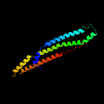
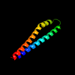
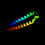
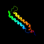
1 c5xfsB_
100.0
43
PDB header: protein transportChain: B: PDB Molecule: ppe family protein ppe15;PDBTitle: crystal structure of pe8-ppe15 in complex with espg5 from m.2 tuberculosis
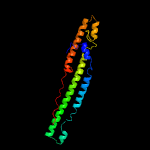
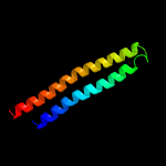
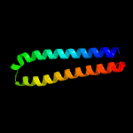
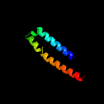
2 c2g38B_
100.0
34
PDB header: structural genomics, unknown functionChain: B: PDB Molecule: ppe family protein;PDBTitle: a pe/ppe protein complex from mycobacterium tuberculosis
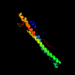
3 d2g38b1
100.0
34
Fold: Ferritin-likeSuperfamily: PE/PPE dimer-likeFamily: PPE
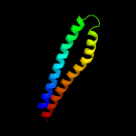
4 c4xy3A_
100.0
16
PDB header: protein transportChain: A: PDB Molecule: esx-1 secretion-associated protein espb;PDBTitle: structure of esx-1 secreted protein espb
5 c4wj2A_
98.5
15
PDB header: unknown functionChain: A: PDB Molecule: antigen mtb48;PDBTitle: mycobacterial protein
6 c2vs0B_
97.9
16
PDB header: cell invasionChain: B: PDB Molecule: virulence factor esxa;PDBTitle: structural analysis of homodimeric staphylococcal aureus2 virulence factor esxa
7 c3gvmA_
97.8
18
PDB header: viral proteinChain: A: PDB Molecule: putative uncharacterized protein sag1039;PDBTitle: structure of the homodimeric wxg-100 family protein from streptococcus2 agalactiae
8 c4iogD_
97.7
12
PDB header: unknown functionChain: D: PDB Molecule: secreted protein esxb;PDBTitle: the crystal structure of a secreted protein esxb (wild-type, in p212 space group) from bacillus anthracis str. sterne
9 c3zbhC_
97.6
23
PDB header: unknown functionChain: C: PDB Molecule: esxa;PDBTitle: geobacillus thermodenitrificans esxa crystal form i
10 d1wa8a1
96.9
12
Fold: Ferritin-likeSuperfamily: EsxAB dimer-likeFamily: ESAT-6 like
11 c4lwsA_
96.5
19
PDB header: unknown functionChain: A: PDB Molecule: uncharacterized protein;PDBTitle: esxa : esxb (semet) hetero-dimer from thermomonospora curvata
12 c4lwsB_
96.2
13
PDB header: unknown functionChain: B: PDB Molecule: uncharacterized protein;PDBTitle: esxa : esxb (semet) hetero-dimer from thermomonospora curvata
13 d1wa8b1
96.0
17
Fold: Ferritin-likeSuperfamily: EsxAB dimer-likeFamily: ESAT-6 like
14 c4i0xA_
95.4
16
PDB header: structural genomics, unknown functionChain: A: PDB Molecule: esat-6-like protein mab_3112;PDBTitle: crystal structure of the mycobacterum abscessus esxef (mab_3112-2 mab_3113) complex
15 c2kg7B_
91.4
13
PDB header: unknown functionChain: B: PDB Molecule: esat-6-like protein esxh;PDBTitle: structure and features of the complex formed by the tuberculosis2 virulence factors rv0287 and rv0288
16 c4i0xJ_
75.3
19
PDB header: structural genomics, unknown functionChain: J: PDB Molecule: esat-6-like protein mab_3113;PDBTitle: crystal structure of the mycobacterum abscessus esxef (mab_3112-2 mab_3113) complex
17 c3txaA_
44.7
57
PDB header: cell adhesionChain: A: PDB Molecule: cell wall surface anchor family protein;PDBTitle: structural analysis of adhesive tip pilin, gbs104 from group b2 streptococcus agalactiae
18 d1zyea1
20.5
18
Fold: Thioredoxin foldSuperfamily: Thioredoxin-likeFamily: Glutathione peroxidase-like
19 d2f8aa1
19.8
18
Fold: Thioredoxin foldSuperfamily: Thioredoxin-likeFamily: Glutathione peroxidase-like
20 d1gp1a_
17.1
17
Fold: Thioredoxin foldSuperfamily: Thioredoxin-likeFamily: Glutathione peroxidase-like
21 c3drnB_
not modelled
16.9
38
PDB header: oxidoreductaseChain: B: PDB Molecule: peroxiredoxin, bacterioferritin comigratory proteinPDBTitle: the crystal structure of bcp1 from sulfolobus sulfataricus
22 d1st9a_
not modelled
16.8
31
Fold: Thioredoxin foldSuperfamily: Thioredoxin-likeFamily: Glutathione peroxidase-like
23 d1jfma_
not modelled
16.6
14
Fold: MHC antigen-recognition domainSuperfamily: MHC antigen-recognition domainFamily: MHC antigen-recognition domain
24 c1bkvA_
not modelled
16.4
50
PDB header: structural proteinChain: A: PDB Molecule: t3-785;PDBTitle: collagen
25 c1bkvC_
not modelled
15.4
50
PDB header: structural proteinChain: C: PDB Molecule: t3-785;PDBTitle: collagen
26 c1bkvB_
not modelled
15.4
50
PDB header: structural proteinChain: B: PDB Molecule: t3-785;PDBTitle: collagen
27 c2p5qA_
not modelled
15.2
22
PDB header: oxidoreductaseChain: A: PDB Molecule: glutathione peroxidase 5;PDBTitle: crystal structure of the poplar glutathione peroxidase 5 in the2 reduced form
28 c3tw0D_
not modelled
14.8
33
PDB header: cell adhesionChain: D: PDB Molecule: cell wall surface anchor family protein;PDBTitle: structural analysis of adhesive tip pilin, gbs104 from group b2 streptococcus agalactiae
29 c5enuB_
not modelled
13.8
25
PDB header: oxidoreductaseChain: B: PDB Molecule: alkyl hydroperoxide reductase/ thiol specific antioxidant/PDBTitle: crystal structure of an alkyl hyroperoxide reductase from burkholderia2 ambifaria
30 c2n5uA_
not modelled
13.4
21
PDB header: photosynthesisChain: A: PDB Molecule: tsr0524 protein;PDBTitle: solution structure of the cyanobacterial cytochrome b6f complex2 subunit petp
31 c3lorB_
not modelled
13.3
38
PDB header: isomeraseChain: B: PDB Molecule: thiol-disulfide isomerase and thioredoxins;PDBTitle: the crystal structure of a thiol-disulfide isomerase from2 corynebacterium glutamicum to 2.2a
32 c2hyxA_
not modelled
12.1
33
PDB header: unknown functionChain: A: PDB Molecule: protein dipz;PDBTitle: structure of the c-terminal domain of dipz from mycobacterium2 tuberculosis
33 c2v1mA_
not modelled
11.9
25
PDB header: oxidoreductaseChain: A: PDB Molecule: glutathione peroxidase;PDBTitle: crystal structure of schistosoma mansoni glutathione peroxidase
34 c4ka0C_
not modelled
11.5
29
PDB header: oxidoreductaseChain: C: PDB Molecule: putative thiol-disulfide oxidoreductase;PDBTitle: crystal structure of a putative thiol-disulfide oxidoreductase from2 bacteroides vulgatus (target nysgrc-011676), space group p21221
35 c2wpyA_
not modelled
11.4
50
PDB header: transcriptionChain: A: PDB Molecule: general control protein gcn4;PDBTitle: gcn4 leucine zipper mutant with one vxxnxxx motif2 coordinating chloride
36 d1qxha_
not modelled
10.9
9
Fold: Thioredoxin foldSuperfamily: Thioredoxin-likeFamily: Glutathione peroxidase-like
37 c3pmdA_
not modelled
10.8
29
PDB header: lipid binding proteinChain: A: PDB Molecule: conserved domain protein;PDBTitle: crystal structure of the sporulation inhibitor pxo1-118 from bacillus2 anthracis
38 c2yp6A_
not modelled
10.7
24
PDB header: oxidoreductaseChain: A: PDB Molecule: thioredoxin family protein;PDBTitle: crystal structure of the pneumoccocal exposed lipoprotein2 thioredoxin sp_1000 (etrx2) from streptococcus pneumoniae3 strain tigr4 in complex with cyclofos 3 tm
39 c3eytA_
not modelled
10.4
31
PDB header: structural genomics, unknown functionChain: A: PDB Molecule: uncharacterized protein spoa0173;PDBTitle: crystal structure of thioredoxin-like superfamily protein spoa0173
40 c2h66G_
not modelled
10.4
12
PDB header: structural genomics/oxidoreductaseChain: G: PDB Molecule: pv-pf14_0368;PDBTitle: the crystal structure of plasmodium vivax 2-cys2 peroxiredoxin
41 c3mp6A_
not modelled
10.3
53
PDB header: histone binding proteinChain: A: PDB Molecule: maltose-binding periplasmic protein,linker,saga-associatedPDBTitle: complex structure of sgf29 and dimethylated h3k4
42 c4g7xB_
not modelled
10.2
17
PDB header: protein binding/protein bindingChain: B: PDB Molecule: tola protein;PDBTitle: crystal structure of a complex between the ctxphi piii n-terminal2 domain and the vibrio cholerae tola c-terminal domain
43 c2iu1A_
not modelled
10.2
14
PDB header: transcriptionChain: A: PDB Molecule: eukaryotic translation initiation factor 5;PDBTitle: crystal structure of eif5 c-terminal domain
44 c3ixrA_
not modelled
10.1
8
PDB header: oxidoreductaseChain: A: PDB Molecule: bacterioferritin comigratory protein;PDBTitle: crystal structure of xylella fastidiosa prxq c47s mutant
45 c4je1A_
not modelled
9.6
14
PDB header: oxidoreductaseChain: A: PDB Molecule: probable thiol peroxidase;PDBTitle: crystal structure of thiol peroxidase from burkholderia cenocepacia2 j2315
46 d3e9va1
not modelled
9.6
27
Fold: BTG domain-likeSuperfamily: BTG domain-likeFamily: BTG domain-like
47 c4yk3B_
not modelled
9.4
20
PDB header: protein bindingChain: B: PDB Molecule: bepe protein;PDBTitle: crystal structure of the bid domain of bepe from bartonella henselae
48 c4yk2B_
not modelled
9.1
14
PDB header: protein bindingChain: B: PDB Molecule: bartonella effector protein (bep) substrate of virb t4ss;PDBTitle: crystal structure of the bid domain of bep9 from bartonella2 clarridgeiae
49 c2eqmA_
not modelled
9.1
25
PDB header: transcriptionChain: A: PDB Molecule: phd finger protein 20-like 1;PDBTitle: solution structure of the tudor domain of phd finger2 protein 20-like 1 [homo sapiens]
50 c2l5oA_
not modelled
8.9
47
PDB header: oxidoreductaseChain: A: PDB Molecule: putative thioredoxin;PDBTitle: solution structure of a putative thioredoxin from neisseria2 meningitidis
51 d1knga_
not modelled
8.7
44
Fold: Thioredoxin foldSuperfamily: Thioredoxin-likeFamily: Glutathione peroxidase-like
52 d1e6yb1
not modelled
8.6
26
Fold: Methyl-coenzyme M reductase alpha and beta chain C-terminal domainSuperfamily: Methyl-coenzyme M reductase alpha and beta chain C-terminal domainFamily: Methyl-coenzyme M reductase alpha and beta chain C-terminal domain
53 d1rqba1
not modelled
8.6
48
Fold: RuvA C-terminal domain-likeSuperfamily: post-HMGL domain-likeFamily: Conserved carboxylase domain
54 d2bzba1
not modelled
8.6
35
Fold: ROP-likeSuperfamily: BAS1536-likeFamily: BAS1536-like
55 c3rmrA_
not modelled
8.4
32
PDB header: protein bindingChain: A: PDB Molecule: avirulence protein;PDBTitle: crystal structure of hyaloperonospora arabidopsidis atr1 effector2 domain
56 c2r37A_
not modelled
8.4
19
PDB header: oxidoreductaseChain: A: PDB Molecule: glutathione peroxidase 3;PDBTitle: crystal structure of human glutathione peroxidase 3 (selenocysteine to2 glycine mutant)
57 c2x5rA_
not modelled
8.3
37
PDB header: viral proteinChain: A: PDB Molecule: hypothetical protein orf126;PDBTitle: crystal structure of the hypothetical protein orf126 from pyrobaculum2 spherical virus
58 c3gknA_
not modelled
8.3
15
PDB header: oxidoreductaseChain: A: PDB Molecule: bacterioferritin comigratory protein;PDBTitle: insights into the alkyl peroxide reduction activity of xanthomonas2 campestris bacterioferritin comigratory protein from the trapped3 intermediate/ligand complex structures
59 d1hbnb1
not modelled
8.3
38
Fold: Methyl-coenzyme M reductase alpha and beta chain C-terminal domainSuperfamily: Methyl-coenzyme M reductase alpha and beta chain C-terminal domainFamily: Methyl-coenzyme M reductase alpha and beta chain C-terminal domain
60 c3cmiA_
not modelled
8.2
31
PDB header: oxidoreductaseChain: A: PDB Molecule: peroxiredoxin hyr1;PDBTitle: crystal structure of glutathione-dependent phospholipid peroxidase2 hyr1 from the yeast saccharomyces cerevisiae
61 c4luqD_
not modelled
8.1
100
PDB header: protein binding/toxin inhibitorChain: D: PDB Molecule: uncharacterized protein;PDBTitle: crystal structure of virulence effector tse3 in complex with2 neutralizer tsi3
62 c4mm1E_
not modelled
8.1
24
PDB header: transferaseChain: E: PDB Molecule: geranylgeranylglyceryl phosphate synthase;PDBTitle: gggps from methanothermobacter thermautotrophicus
63 c6mn5A_
not modelled
8.1
25
PDB header: transferase/antibioticChain: A: PDB Molecule: aminoglycoside n(3)-acetyltransferase, aac(3)-iva;PDBTitle: crystal structure of aminoglycoside acetyltransferase aac(3)-iva,2 h154a mutant, in complex with gentamicin c1a
64 c2fulE_
not modelled
8.0
28
PDB header: translationChain: E: PDB Molecule: eukaryotic translation initiation factor 5;PDBTitle: crystal structure of the c-terminal domain of s. cerevisiae eif5
65 d1zupa1
not modelled
7.8
33
Fold: Ribonuclease H-like motifSuperfamily: Ribonuclease H-likeFamily: TM1739-like
66 c4gyxC_
not modelled
7.8
45
PDB header: structural protein, blood clottingChain: C: PDB Molecule: type iii collagen fragment in a host peptide stabilized byPDBTitle: the von willebrand factor a3 domain binding region of type iii2 collagen stabilized by the cysteine knot
67 c6a97A_
not modelled
7.8
19
PDB header: immune systemChain: A: PDB Molecule: mhc i-like leukocyte 2 long form;PDBTitle: crystal structure of mhc-like mill2
68 c2lndA_
not modelled
7.8
44
PDB header: de novo proteinChain: A: PDB Molecule: de novo designed protein, pfk fold;PDBTitle: solution nmr structure of de novo designed protein, pfk fold,2 northeast structural genomics consortium target or134
69 c3hfxA_
not modelled
7.8
23
PDB header: transport proteinChain: A: PDB Molecule: l-carnitine/gamma-butyrobetaine antiporter;PDBTitle: crystal structure of carnitine transporter
70 c2b1kA_
not modelled
7.8
31
PDB header: oxidoreductaseChain: A: PDB Molecule: thiol:disulfide interchange protein dsbe;PDBTitle: crystal structure of e. coli ccmg protein
71 c5frgA_
not modelled
7.8
63
PDB header: protein bindingChain: A: PDB Molecule: formin-binding protein 1-like;PDBTitle: the nmr structure of the cdc42-interacting region of toca1
72 c3hm6X_
not modelled
7.7
17
PDB header: signaling proteinChain: X: PDB Molecule: plexin-b1;PDBTitle: crystal structure of the cytoplasmic domain of human plexin b1
73 d2b0ja2
not modelled
7.7
70
Fold: NAD(P)-binding Rossmann-fold domainsSuperfamily: NAD(P)-binding Rossmann-fold domainsFamily: 6-phosphogluconate dehydrogenase-like, N-terminal domain
74 c2wpzA_
not modelled
7.7
50
PDB header: transcriptionChain: A: PDB Molecule: general control protein gcn4;PDBTitle: gcn4 leucine zipper mutant with two vxxnxxx motifs2 coordinating chloride
75 c5j4fB_
not modelled
7.6
22
PDB header: unknown functionChain: B: PDB Molecule: uncharacterized protein;PDBTitle: crystal structure of the n-terminally his6-tagged hp0902, an2 uncharacterized protein from helicobacter pylori 26695
76 c2y5tG_
not modelled
7.5
83
PDB header: immune systemChain: G: PDB Molecule: c1;PDBTitle: crystal structure of the pathogenic autoantibody ciic1 in complex with2 the triple-helical c1 peptide
77 c6qm7V_
not modelled
7.5
14
PDB header: hydrolaseChain: V: PDB Molecule: proteasome beta1 chain;PDBTitle: leishmania tarentolae proteasome 20s subunit complexed with gsk3494245
78 c4gyxB_
not modelled
7.5
45
PDB header: structural protein, blood clottingChain: B: PDB Molecule: type iii collagen fragment in a host peptide stabilized byPDBTitle: the von willebrand factor a3 domain binding region of type iii2 collagen stabilized by the cysteine knot
79 c4gyxA_
not modelled
7.5
45
PDB header: structural protein, blood clottingChain: A: PDB Molecule: type iii collagen fragment in a host peptide stabilized byPDBTitle: the von willebrand factor a3 domain binding region of type iii2 collagen stabilized by the cysteine knot
80 c5e6pA_
not modelled
7.4
24
PDB header: signaling proteinChain: A: PDB Molecule: plexin-b2;PDBTitle: plexinb2 cytoplasmic region/pdz-rhogef pdz domain complex
81 c3dwvB_
not modelled
7.3
13
PDB header: oxidoreductaseChain: B: PDB Molecule: glutathione peroxidase-like protein;PDBTitle: glutathione peroxidase-type tryparedoxin peroxidase, oxidized form
82 d2b5xa1
not modelled
7.3
31
Fold: Thioredoxin foldSuperfamily: Thioredoxin-likeFamily: Glutathione peroxidase-like
83 d1ui5a2
not modelled
7.2
17
Fold: Tetracyclin repressor-like, C-terminal domainSuperfamily: Tetracyclin repressor-like, C-terminal domainFamily: Tetracyclin repressor-like, C-terminal domain
84 c5v6rB_
not modelled
7.2
17
PDB header: protein bindingChain: B: PDB Molecule: plexin-d1;PDBTitle: structure of plexin d1 intracellular domain
85 c2y5tE_
not modelled
7.2
83
PDB header: immune systemChain: E: PDB Molecule: c1;PDBTitle: crystal structure of the pathogenic autoantibody ciic1 in complex with2 the triple-helical c1 peptide
86 c3lwaA_
not modelled
7.2
31
PDB header: isomeraseChain: A: PDB Molecule: secreted thiol-disulfide isomerase;PDBTitle: the crystal structure of a secreted thiol-disulfide isomerase from2 corynebacterium glutamicum to 1.75a
87 c2wpzB_
not modelled
7.2
50
PDB header: transcriptionChain: B: PDB Molecule: general control protein gcn4;PDBTitle: gcn4 leucine zipper mutant with two vxxnxxx motifs2 coordinating chloride
88 c3ig3A_
not modelled
7.1
24
PDB header: signaling protein, membrane proteinChain: A: PDB Molecule: plxna3 protein;PDBTitle: crystal structure of mouse plexin a3 intracellular domain
89 c2c0dA_
not modelled
7.1
24
PDB header: oxidoreductaseChain: A: PDB Molecule: thioredoxin peroxidase 2;PDBTitle: structure of the mitochondrial 2-cys peroxiredoxin from2 plasmodium falciparum
90 c4h5bB_
not modelled
7.1
34
PDB header: unknown functionChain: B: PDB Molecule: dr_1245 protein;PDBTitle: crystal structure of dr_1245 from deinococcus radiodurans
91 c1de4A_
not modelled
7.1
16
PDB header: metal transport inhibitor/receptorChain: A: PDB Molecule: hemochromatosis protein;PDBTitle: hemochromatosis protein hfe complexed with transferrin receptor
92 c3ia1A_
not modelled
7.0
31
PDB header: oxidoreductaseChain: A: PDB Molecule: thio-disulfide isomerase/thioredoxin;PDBTitle: crystal structure of thio-disulfide isomerase from thermus2 thermophilus
93 c2wpzC_
not modelled
7.0
50
PDB header: transcriptionChain: C: PDB Molecule: general control protein gcn4;PDBTitle: gcn4 leucine zipper mutant with two vxxnxxx motifs2 coordinating chloride
94 d1jfua_
not modelled
7.0
22
Fold: Thioredoxin foldSuperfamily: Thioredoxin-likeFamily: Glutathione peroxidase-like
95 c3su8X_
not modelled
6.9
17
PDB header: apoptosis/signaling proteinChain: X: PDB Molecule: plexin-b1;PDBTitle: crystal structure of a truncated intracellular domain of plexin-b1 in2 complex with rac1
96 c4dmtC_
not modelled
6.9
38
PDB header: structural proteinChain: C: PDB Molecule: collagen iii derived peptide;PDBTitle: crystal structure of a vwf binding collagen iii derived triple helical2 peptide
97 c4dmtB_
not modelled
6.9
38
PDB header: structural proteinChain: B: PDB Molecule: collagen iii derived peptide;PDBTitle: crystal structure of a vwf binding collagen iii derived triple helical2 peptide
98 c4dmtA_
not modelled
6.9
38
PDB header: structural proteinChain: A: PDB Molecule: collagen iii derived peptide;PDBTitle: crystal structure of a vwf binding collagen iii derived triple helical2 peptide
99 d1we0a1
not modelled
6.7
17
Fold: Thioredoxin foldSuperfamily: Thioredoxin-likeFamily: Glutathione peroxidase-like