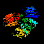
| 1 | c3uagA_
|
|
 |
100.0 |
32 |
PDB header:ligase
Chain: A: PDB Molecule:protein (udp-n-acetylmuramoyl-l-alanine:d-
PDBTitle: udp-n-acetylmuramoyl-l-alanine:d-glutamate ligase
|
|
|
|
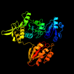
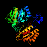
| 2 | c3lk7A_
|
|
 |
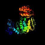
100.0 |
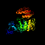
30 |
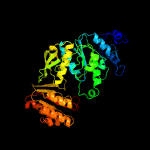
PDB header:ligase
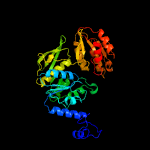
Chain: A: PDB Molecule:udp-n-acetylmuramoylalanine--d-glutamate ligase;
PDBTitle: the crystal structure of udp-n-acetylmuramoylalanine-d-glutamate2 (murd) ligase from streptococcus agalactiae to 1.5a
|
|
|
|
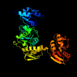
| 3 | c4bucA_
|
|
 |
100.0 |
27 |
PDB header:ligase
Chain: A: PDB Molecule:udp-n-acetylmuramoylalanine--d-glutamate ligase;
PDBTitle: crystal structure of murd ligase from thermotoga maritima in apo form
|
|
|
|
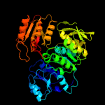
| 4 | c2f00A_
|
|
 |
100.0 |
20 |
PDB header:ligase
Chain: A: PDB Molecule:udp-n-acetylmuramate--l-alanine ligase;
PDBTitle: escherichia coli murc
|
|
|
|
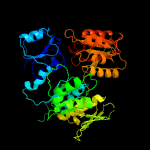
| 5 | c3hn7A_
|
|
 |
100.0 |
17 |
PDB header:ligase
Chain: A: PDB Molecule:udp-n-acetylmuramate-l-alanine ligase;
PDBTitle: crystal structure of a murein peptide ligase mpl (psyc_0032) from2 psychrobacter arcticus 273-4 at 1.65 a resolution
|
|
|
|
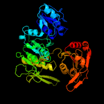
| 6 | c1j6uA_
|
|
 |
100.0 |
19 |
PDB header:ligase
Chain: A: PDB Molecule:udp-n-acetylmuramate-alanine ligase murc;
PDBTitle: crystal structure of udp-n-acetylmuramate-alanine ligase murc (tm0231)2 from thermotoga maritima at 2.3 a resolution
|
|
|
|
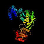
| 7 | c1gqqA_
|
|
 |
100.0 |
20 |
PDB header:cell wall biosynthesis
Chain: A: PDB Molecule:udp-n-acetylmuramate-l-alanine ligase;
PDBTitle: murc - crystal structure of the apo-enzyme from haemophilus influenzae
|
|
|
|
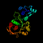
| 8 | c5vvwA_
|
|
 |
100.0 |
21 |
PDB header:ligase
Chain: A: PDB Molecule:udp-n-acetylmuramate--l-alanine ligase;
PDBTitle: structure of murc from pseudomonas aeruginosa
|
|
|
|
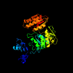
| 9 | c2xjaD_
|
|
 |
100.0 |
27 |
PDB header:ligase
Chain: D: PDB Molecule:udp-n-acetylmuramoyl-l-alanyl-d-glutamate--2,6-
PDBTitle: structure of mure from m.tuberculosis with dipeptide and adp
|
|
|
|
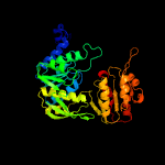
| 10 | c4qdiA_
|
|
 |
100.0 |
19 |
PDB header:ligase
Chain: A: PDB Molecule:udp-n-acetylmuramoyl-tripeptide--d-alanyl-d-alanine ligase;
PDBTitle: crystal structure ii of murf from acinetobacter baumannii
|
|
|
|
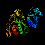
| 11 | c3eagA_
|
|
 |
100.0 |
19 |
PDB header:ligase
Chain: A: PDB Molecule:udp-n-acetylmuramate:l-alanyl-gamma-d-glutamyl-meso-
PDBTitle: the crystal structure of udp-n-acetylmuramate:l-alanyl-gamma-d-2 glutamyl-meso-diaminopimelate ligase (mpl) from neisseria3 meningitides
|
|
|
|
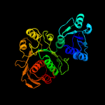
| 12 | c6cauA_
|
|
 |
100.0 |
24 |
PDB header:ligase
Chain: A: PDB Molecule:udp-n-acetylmuramate--l-alanine ligase;
PDBTitle: udp-n-acetylmuramate--alanine ligase from acinetobacter baumannii2 ab5075-uw with amppnp
|
|
|
|
| 13 | c2am1A_
|
|
 |
100.0 |
21 |
PDB header:ligase
Chain: A: PDB Molecule:udp-n-acetylmuramoylalanine-d-glutamyl-lysine-d-alanyl-d-
PDBTitle: sp protein ligand 1
|
|
|
|
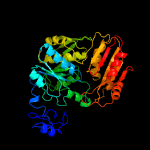
| 14 | c4c13A_
|
|
 |
100.0 |
21 |
PDB header:ligase
Chain: A: PDB Molecule:udp-n-acetylmuramoyl-l-alanyl-d-glutamate--l-lysine ligase;
PDBTitle: x-ray crystal structure of staphylococcus aureus mure with udp-murnac-2 ala-glu-lys
|
|
|
|
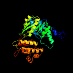
| 15 | c1e8cB_
|
|
 |
100.0 |
24 |
PDB header:ligase
Chain: B: PDB Molecule:udp-n-acetylmuramoylalanyl-d-glutamate--2,6-diaminopimelate
PDBTitle: structure of mure the udp-n-acetylmuramyl tripeptide synthetase from2 e. coli
|
|
|
|
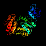
| 16 | c4bubA_
|
|
 |
100.0 |
21 |
PDB header:ligase
Chain: A: PDB Molecule:udp-n-acetylmuramoyl-l-alanyl-d-glutamate--ld-lysine
PDBTitle: crystal structure of mure ligase from thermotoga maritima2 in complex with adp
|
|
|
|
| 17 | c4cvkA_
|
|
 |
100.0 |
24 |
PDB header:ligase
Chain: A: PDB Molecule:udp-n-acetylmuramoyl-tripeptide--d-alanyl-d-alanine
PDBTitle: pamurf in complex with udp-murnac-tripeptide (mdap)
|
|
|
|
| 18 | c2wtzC_
|
|
 |
100.0 |
30 |
PDB header:ligase
Chain: C: PDB Molecule:udp-n-acetylmuramoyl-l-alanyl-d-glutamate-
PDBTitle: mure ligase of mycobacterium tuberculosis
|
|
|
|
| 19 | c1gg4A_
|
|
 |
100.0 |
24 |
PDB header:ligase
Chain: A: PDB Molecule:udp-n-acetylmuramoylalanyl-d-glutamyl-2,6-diaminopimelate-
PDBTitle: crystal structure of escherichia coli udpmurnac-tripeptide d-alanyl-d-2 alanine-adding enzyme (murf) at 2.3 angstrom resolution
|
|
|
|
| 20 | c3zl8A_
|
|
 |
100.0 |
18 |
PDB header:ligase
Chain: A: PDB Molecule:udp-n-acetylmuramoyl-tripeptide--d-alanyl-d-alanine
PDBTitle: crystal structure of murf ligase from thermotoga maritima2 in complex with adp
|
|
|
|
| 21 | c2vosA_ |
|
not modelled |
100.0 |
17 |
PDB header:ligase
Chain: A: PDB Molecule:folylpolyglutamate synthase protein folc;
PDBTitle: mycobacterium tuberculosis folylpolyglutamate synthase2 complexed with adp
|
|
|
| 22 | c2gc6A_ |
|
not modelled |
100.0 |
20 |
PDB header:ligase
Chain: A: PDB Molecule:folylpolyglutamate synthase;
PDBTitle: s73a mutant of l. casei fpgs
|
|
|
| 23 | c1w78A_ |
|
not modelled |
100.0 |
26 |
PDB header:synthase
Chain: A: PDB Molecule:folc bifunctional protein;
PDBTitle: e.coli folc in complex with dhpp and adp
|
|
|
| 24 | c1o5zA_ |
|
not modelled |
100.0 |
15 |
PDB header:ligase
Chain: A: PDB Molecule:folylpolyglutamate synthase/dihydrofolate synthase;
PDBTitle: crystal structure of folylpolyglutamate synthase (tm0166) from2 thermotoga maritima at 2.10 a resolution
|
|
|
| 25 | c6gs2B_ |
|
not modelled |
100.0 |
18 |
PDB header:biosynthetic protein
Chain: B: PDB Molecule:sa1708 protein;
PDBTitle: crystal structure of the gatd/murt enzyme complex from staphylococcus2 aureus
|
|
|
| 26 | c3n2aA_ |
|
not modelled |
100.0 |
20 |
PDB header:ligase
Chain: A: PDB Molecule:bifunctional folylpolyglutamate synthase/dihydrofolate
PDBTitle: crystal structure of bifunctional folylpolyglutamate2 synthase/dihydrofolate synthase from yersinia pestis co92
|
|
|
| 27 | d2jfga3 |
|
not modelled |
100.0 |
31 |
Fold:Ribokinase-like
Superfamily:MurD-like peptide ligases, catalytic domain
Family:MurCDEF |
|
|
| 28 | c6fqbD_ |
|
not modelled |
100.0 |
19 |
PDB header:ligase
Chain: D: PDB Molecule:mur ligase family protein;
PDBTitle: murt/gatd peptidoglycan amidotransferase complex from streptococcus2 pneumoniae r6
|
|
|
| 29 | d1p3da3 |
|
not modelled |
100.0 |
22 |
Fold:Ribokinase-like
Superfamily:MurD-like peptide ligases, catalytic domain
Family:MurCDEF |
|
|
| 30 | d1e8ca3 |
|
not modelled |
100.0 |
27 |
Fold:Ribokinase-like
Superfamily:MurD-like peptide ligases, catalytic domain
Family:MurCDEF |
|
|
| 31 | d2gc6a2 |
|
not modelled |
100.0 |
16 |
Fold:Ribokinase-like
Superfamily:MurD-like peptide ligases, catalytic domain
Family:Folylpolyglutamate synthetase |
|
|
| 32 | d1j6ua3 |
|
not modelled |
100.0 |
21 |
Fold:Ribokinase-like
Superfamily:MurD-like peptide ligases, catalytic domain
Family:MurCDEF |
|
|
| 33 | d1gg4a4 |
|
not modelled |
100.0 |
23 |
Fold:Ribokinase-like
Superfamily:MurD-like peptide ligases, catalytic domain
Family:MurCDEF |
|
|
| 34 | d2jfga2 |
|
not modelled |
100.0 |
33 |
Fold:MurD-like peptide ligases, peptide-binding domain
Superfamily:MurD-like peptide ligases, peptide-binding domain
Family:MurCDEF C-terminal domain |
|
|
| 35 | d1o5za2 |
|
not modelled |
100.0 |
14 |
Fold:Ribokinase-like
Superfamily:MurD-like peptide ligases, catalytic domain
Family:Folylpolyglutamate synthetase |
|
|
| 36 | d1p3da1 |
|
not modelled |
99.7 |
16 |
Fold:MurCD N-terminal domain
Superfamily:MurCD N-terminal domain
Family:MurCD N-terminal domain |
|
|
| 37 | d1j6ua1 |
|
not modelled |
99.7 |
15 |
Fold:MurCD N-terminal domain
Superfamily:MurCD N-terminal domain
Family:MurCD N-terminal domain |
|
|
| 38 | d2jfga1 |
|
not modelled |
99.7 |
30 |
Fold:MurCD N-terminal domain
Superfamily:MurCD N-terminal domain
Family:MurCD N-terminal domain |
|
|
| 39 | d1p3da2 |
|
not modelled |
99.6 |
13 |
Fold:MurD-like peptide ligases, peptide-binding domain
Superfamily:MurD-like peptide ligases, peptide-binding domain
Family:MurCDEF C-terminal domain |
|
|
| 40 | d1e8ca2 |
|
not modelled |
99.6 |
22 |
Fold:MurD-like peptide ligases, peptide-binding domain
Superfamily:MurD-like peptide ligases, peptide-binding domain
Family:MurCDEF C-terminal domain |
|
|
| 41 | d1gg4a1 |
|
not modelled |
99.6 |
22 |
Fold:MurD-like peptide ligases, peptide-binding domain
Superfamily:MurD-like peptide ligases, peptide-binding domain
Family:MurCDEF C-terminal domain |
|
|
| 42 | c3mvnA_ |
|
not modelled |
99.5 |
24 |
PDB header:ligase
Chain: A: PDB Molecule:udp-n-acetylmuramate:l-alanyl-gamma-d-glutamyl-medo-
PDBTitle: crystal structure of a domain from a putative udp-n-acetylmuramate:l-2 alanyl-gamma-d-glutamyl-medo-diaminopimelate ligase from haemophilus3 ducreyi 35000hp
|
|
|
| 43 | d1j6ua2 |
|
not modelled |
99.2 |
20 |
Fold:MurD-like peptide ligases, peptide-binding domain
Superfamily:MurD-like peptide ligases, peptide-binding domain
Family:MurCDEF C-terminal domain |
|
|
| 44 | d1o5za1 |
|
not modelled |
99.0 |
16 |
Fold:MurD-like peptide ligases, peptide-binding domain
Superfamily:MurD-like peptide ligases, peptide-binding domain
Family:Folylpolyglutamate synthetase, C-terminal domain |
|
|
| 45 | d2gc6a1 |
|
not modelled |
98.9 |
25 |
Fold:MurD-like peptide ligases, peptide-binding domain
Superfamily:MurD-like peptide ligases, peptide-binding domain
Family:Folylpolyglutamate synthetase, C-terminal domain |
|
|
| 46 | d1pjqa1 |
|
not modelled |
98.3 |
18 |
Fold:NAD(P)-binding Rossmann-fold domains
Superfamily:NAD(P)-binding Rossmann-fold domains
Family:Siroheme synthase N-terminal domain-like |
|
|
| 47 | c4wb1B_ |
|
not modelled |
98.2 |
22 |
PDB header:oxidoreductase
Chain: B: PDB Molecule:cals8;
PDBTitle: crystal structure of cals8 from micromonospora echinospora (p294s2 mutant)
|
|
|
| 48 | c3d4oA_ |
|
not modelled |
98.1 |
22 |
PDB header:oxidoreductase
Chain: A: PDB Molecule:dipicolinate synthase subunit a;
PDBTitle: crystal structure of dipicolinate synthase subunit a (np_243269.1)2 from bacillus halodurans at 2.10 a resolution
|
|
|
| 49 | c1mv8A_ |
|
not modelled |
98.1 |
15 |
PDB header:oxidoreductase
Chain: A: PDB Molecule:gdp-mannose 6-dehydrogenase;
PDBTitle: 1.55 a crystal structure of a ternary complex of gdp-mannose2 dehydrogenase from psuedomonas aeruginosa
|
|
|
| 50 | c3gg2B_ |
|
not modelled |
98.0 |
16 |
PDB header:oxidoreductase
Chain: B: PDB Molecule:sugar dehydrogenase, udp-glucose/gdp-mannose dehydrogenase
PDBTitle: crystal structure of udp-glucose 6-dehydrogenase from porphyromonas2 gingivalis bound to product udp-glucuronate
|
|
|
| 51 | c3cumA_ |
|
not modelled |
98.0 |
20 |
PDB header:oxidoreductase
Chain: A: PDB Molecule:probable 3-hydroxyisobutyrate dehydrogenase;
PDBTitle: crystal structure of a possible 3-hydroxyisobutyrate dehydrogenase2 from pseudomonas aeruginosa pao1
|
|
|
| 52 | c4a7pA_ |
|
not modelled |
98.0 |
17 |
PDB header:oxidoreductase
Chain: A: PDB Molecule:udp-glucose dehydrogenase;
PDBTitle: se-met derivatized ugdg, udp-glucose dehydrogenase from sphingomonas2 elodea
|
|
|
| 53 | c2rirA_ |
|
not modelled |
98.0 |
21 |
PDB header:oxidoreductase
Chain: A: PDB Molecule:dipicolinate synthase, a chain;
PDBTitle: crystal structure of dipicolinate synthase, a chain, from bacillus2 subtilis
|
|
|
| 54 | c4r16A_ |
|
not modelled |
98.0 |
17 |
PDB header:oxidoreductase
Chain: A: PDB Molecule:418aa long hypothetical udp-n-acetyl-d-mannosaminuronic
PDBTitle: structure of udp-d-mannac dehdrogeanse from pyrococcus horikoshii
|
|
|
| 55 | c3vtfA_ |
|
not modelled |
98.0 |
24 |
PDB header:oxidoreductase
Chain: A: PDB Molecule:udp-glucose 6-dehydrogenase;
PDBTitle: structure of a udp-glucose dehydrogenase from the hyperthermophilic2 archaeon pyrobaculum islandicum
|
|
|
| 56 | c6aqjB_ |
|
not modelled |
97.9 |
18 |
PDB header:isomerase
Chain: B: PDB Molecule:ketol-acid reductoisomerase (nadp(+));
PDBTitle: crystal structures of staphylococcus aureus ketol-acid2 reductoisomerase in complex with two transition state analogs that3 have biocidal activity.
|
|
|
| 57 | c4xdzB_ |
|
not modelled |
97.9 |
16 |
PDB header:oxidoreductase
Chain: B: PDB Molecule:ketol-acid reductoisomerase;
PDBTitle: holo structure of ketol-acid reductoisomerase from ignisphaera2 aggregans
|
|
|
| 58 | c4dioB_ |
|
not modelled |
97.9 |
21 |
PDB header:oxidoreductase
Chain: B: PDB Molecule:nad(p) transhydrogenase subunit alpha part 1;
PDBTitle: the crystal structure of transhydrogenase from sinorhizobium meliloti
|
|
|
| 59 | c2o3jC_ |
|
not modelled |
97.9 |
21 |
PDB header:oxidoreductase
Chain: C: PDB Molecule:udp-glucose 6-dehydrogenase;
PDBTitle: structure of caenorhabditis elegans udp-glucose dehydrogenase
|
|
|
| 60 | c5g6sD_ |
|
not modelled |
97.9 |
21 |
PDB header:oxidoreductase
Chain: D: PDB Molecule:imine reductase;
PDBTitle: imine reductase from aspergillus oryzae in complex with nadp(h) and2 (r)-rasagiline
|
|
|
| 61 | d3cuma2 |
|
not modelled |
97.9 |
20 |
Fold:NAD(P)-binding Rossmann-fold domains
Superfamily:NAD(P)-binding Rossmann-fold domains
Family:6-phosphogluconate dehydrogenase-like, N-terminal domain |
|
|
| 62 | c2y0dB_ |
|
not modelled |
97.9 |
20 |
PDB header:oxidoreductase
Chain: B: PDB Molecule:udp-glucose dehydrogenase;
PDBTitle: bcec mutation y10k
|
|
|
| 63 | c3ojlA_ |
|
not modelled |
97.9 |
12 |
PDB header:oxidoreductase
Chain: A: PDB Molecule:cap5o;
PDBTitle: native structure of the udp-n-acetyl-mannosamine dehydrogenase cap5o2 from staphylococcus aureus
|
|
|
| 64 | d1l7da1 |
|
not modelled |
97.9 |
18 |
Fold:NAD(P)-binding Rossmann-fold domains
Superfamily:NAD(P)-binding Rossmann-fold domains
Family:Formate/glycerate dehydrogenases, NAD-domain |
|
|
| 65 | c4oqzA_ |
|
not modelled |
97.9 |
21 |
PDB header:oxidoreductase
Chain: A: PDB Molecule:putative oxidoreductase yfjr;
PDBTitle: streptomyces aurantiacus imine reductase
|
|
|
| 66 | c6jczL_ |
|
not modelled |
97.9 |
17 |
PDB header:isomerase
Chain: L: PDB Molecule:putative ketol-acid reductoisomerase 2;
PDBTitle: cryo-em structure of sulfolobus solfataricus ketol-acid2 reductoisomerase (sso-kari) in complex with mg2+, nadph, and cpd at3 ph7.5
|
|
|
| 67 | c3g79A_ |
|
not modelled |
97.9 |
19 |
PDB header:oxidoreductase
Chain: A: PDB Molecule:ndp-n-acetyl-d-galactosaminuronic acid dehydrogenase;
PDBTitle: crystal structure of ndp-n-acetyl-d-galactosaminuronic acid2 dehydrogenase from methanosarcina mazei go1
|
|
|
| 68 | d1li4a1 |
|
not modelled |
97.8 |
24 |
Fold:NAD(P)-binding Rossmann-fold domains
Superfamily:NAD(P)-binding Rossmann-fold domains
Family:Formate/glycerate dehydrogenases, NAD-domain |
|
|
| 69 | c5ocmA_ |
|
not modelled |
97.8 |
19 |
PDB header:oxidoreductase
Chain: A: PDB Molecule:nad_gly3p_dh, nad-dependent glycerol-3-phosphate
PDBTitle: imine reductase from streptosporangium roseum in complex with nadp+2 and 2,2,2-trifluoroacetophenone hydrate
|
|
|
| 70 | c3prjB_ |
|
not modelled |
97.8 |
14 |
PDB header:oxidoreductase
Chain: B: PDB Molecule:udp-glucose 6-dehydrogenase;
PDBTitle: role of packing defects in the evolution of allostery and induced fit2 in human udp-glucose dehydrogenase.
|
|
|
| 71 | c5b37A_ |
|
not modelled |
97.8 |
14 |
PDB header:oxidoreductase
Chain: A: PDB Molecule:tryptophan dehydrogenase;
PDBTitle: crystal structure of l-tryptophan dehydrogenase from nostoc2 punctiforme
|
|
|
| 72 | d1bg6a2 |
|
not modelled |
97.8 |
14 |
Fold:NAD(P)-binding Rossmann-fold domains
Superfamily:NAD(P)-binding Rossmann-fold domains
Family:6-phosphogluconate dehydrogenase-like, N-terminal domain |
|
|
| 73 | c4kqxB_ |
|
not modelled |
97.8 |
19 |
PDB header:oxidoreductase/oxidoreductase inhibitor
Chain: B: PDB Molecule:ketol-acid reductoisomerase;
PDBTitle: mutant slackia exigua kari ddv in complex with nad and an inhibitor
|
|
|
| 74 | c2g5cD_ |
|
not modelled |
97.8 |
16 |
PDB header:oxidoreductase
Chain: D: PDB Molecule:prephenate dehydrogenase;
PDBTitle: crystal structure of prephenate dehydrogenase from aquifex aeolicus
|
|
|
| 75 | c3w6uA_ |
|
not modelled |
97.8 |
18 |
PDB header:oxidoreductase
Chain: A: PDB Molecule:6-phosphogluconate dehydrogenase, nad-binding protein;
PDBTitle: crystal structure of nadp bound l-serine 3-dehydrogenase from2 hyperthermophilic archaeon pyrobaculum calidifontis
|
|
|
| 76 | c4ypoB_ |
|
not modelled |
97.8 |
22 |
PDB header:oxidoreductase
Chain: B: PDB Molecule:ketol-acid reductoisomerase;
PDBTitle: crystal structure of mycobacterium tuberculosis ketol-acid2 reductoisomerase in complex with mg2+
|
|
|
| 77 | c3x2fA_ |
|
not modelled |
97.8 |
24 |
PDB header:hydrolase
Chain: A: PDB Molecule:adenosylhomocysteinase;
PDBTitle: a thermophilic s-adenosylhomocysteine hydrolase
|
|
|
| 78 | c3ktdC_ |
|
not modelled |
97.7 |
16 |
PDB header:oxidoreductase
Chain: C: PDB Molecule:prephenate dehydrogenase;
PDBTitle: crystal structure of a putative prephenate dehydrogenase (cgl0226)2 from corynebacterium glutamicum atcc 13032 at 2.60 a resolution
|
|
|
| 79 | c4d3fB_ |
|
not modelled |
97.7 |
21 |
PDB header:oxidoreductase
Chain: B: PDB Molecule:imine reductase;
PDBTitle: bcsired from bacillus cereus in complex with nadph
|
|
|
| 80 | c5a9tA_ |
|
not modelled |
97.7 |
24 |
PDB header:oxidoreductase
Chain: A: PDB Molecule:putative dehydrogenase;
PDBTitle: imine reductase from amycolatopsis orientalis in complex2 with (r)-methyltetrahydroisoquinoline
|
|
|
| 81 | c1np3B_ |
|
not modelled |
97.7 |
23 |
PDB header:oxidoreductase
Chain: B: PDB Molecule:ketol-acid reductoisomerase;
PDBTitle: crystal structure of class i acetohydroxy acid isomeroreductase from2 pseudomonas aeruginosa
|
|
|
| 82 | c1bg6A_ |
|
not modelled |
97.7 |
16 |
PDB header:oxidoreductase
Chain: A: PDB Molecule:n-(1-d-carboxylethyl)-l-norvaline dehydrogenase;
PDBTitle: crystal structure of the n-(1-d-carboxylethyl)-l-norvaline2 dehydrogenase from arthrobacter sp. strain 1c
|
|
|
| 83 | d1a4ia1 |
|
not modelled |
97.7 |
20 |
Fold:NAD(P)-binding Rossmann-fold domains
Superfamily:NAD(P)-binding Rossmann-fold domains
Family:Aminoacid dehydrogenase-like, C-terminal domain |
|
|
| 84 | c4oqyA_ |
|
not modelled |
97.7 |
17 |
PDB header:oxidoreductase
Chain: A: PDB Molecule:(s)-imine reductase;
PDBTitle: streptomyces sp. gf3546 imine reductase
|
|
|
| 85 | c5butG_ |
|
not modelled |
97.7 |
14 |
PDB header:membrane protein
Chain: G: PDB Molecule:ktr system potassium uptake protein a,ktr system potassium
PDBTitle: crystal structure of inactive conformation of ktrab k+ transporter
|
|
|
| 86 | c5u5gC_ |
|
not modelled |
97.7 |
18 |
PDB header:oxidoreductase
Chain: C: PDB Molecule:6-phosphogluconate dehydrogenase;
PDBTitle: psf3 in complex with nadp+ and 2-opp
|
|
|
| 87 | c4tskA_ |
|
not modelled |
97.7 |
12 |
PDB header:oxidoreductase,isomerase
Chain: A: PDB Molecule:ketol-acid reductoisomerase;
PDBTitle: ketol-acid reductoisomerase from alicyclobacillus acidocaldarius
|
|
|
| 88 | c5yeqB_ |
|
not modelled |
97.7 |
17 |
PDB header:isomerase
Chain: B: PDB Molecule:ketol-acid reductoisomerase (nadp(+));
PDBTitle: the structure of sac-kari protein
|
|
|
| 89 | d1uxja1 |
|
not modelled |
97.7 |
16 |
Fold:NAD(P)-binding Rossmann-fold domains
Superfamily:NAD(P)-binding Rossmann-fold domains
Family:LDH N-terminal domain-like |
|
|
| 90 | c3g0oA_ |
|
not modelled |
97.7 |
20 |
PDB header:oxidoreductase
Chain: A: PDB Molecule:3-hydroxyisobutyrate dehydrogenase;
PDBTitle: crystal structure of 3-hydroxyisobutyrate dehydrogenase2 (ygbj) from salmonella typhimurium
|
|
|
| 91 | c3gvpB_ |
|
not modelled |
97.7 |
19 |
PDB header:hydrolase
Chain: B: PDB Molecule:adenosylhomocysteinase 3;
PDBTitle: human sahh-like domain of human adenosylhomocysteinase 3
|
|
|
| 92 | d1v8ba1 |
|
not modelled |
97.7 |
20 |
Fold:NAD(P)-binding Rossmann-fold domains
Superfamily:NAD(P)-binding Rossmann-fold domains
Family:Formate/glycerate dehydrogenases, NAD-domain |
|
|
| 93 | c3pefA_ |
|
not modelled |
97.7 |
20 |
PDB header:oxidoreductase
Chain: A: PDB Molecule:6-phosphogluconate dehydrogenase, nad-binding;
PDBTitle: crystal structure of gamma-hydroxybutyrate dehydrogenase from2 geobacter metallireducens in complex with nadp+
|
|
|
| 94 | c4gbjB_ |
|
not modelled |
97.7 |
21 |
PDB header:oxidoreductase
Chain: B: PDB Molecule:6-phosphogluconate dehydrogenase nad-binding;
PDBTitle: crystal structure of nad-binding 6-phosphogluconate dehydrogenase from2 dyadobacter fermentans
|
|
|
| 95 | d1e5qa1 |
|
not modelled |
97.6 |
15 |
Fold:NAD(P)-binding Rossmann-fold domains
Superfamily:NAD(P)-binding Rossmann-fold domains
Family:Glyceraldehyde-3-phosphate dehydrogenase-like, N-terminal domain |
|
|
| 96 | c3l6dB_ |
|
not modelled |
97.6 |
17 |
PDB header:oxidoreductase
Chain: B: PDB Molecule:putative oxidoreductase;
PDBTitle: crystal structure of putative oxidoreductase from pseudomonas putida2 kt2440
|
|
|
| 97 | c3dhyC_ |
|
not modelled |
97.6 |
22 |
PDB header:hydrolase
Chain: C: PDB Molecule:adenosylhomocysteinase;
PDBTitle: crystal structures of mycobacterium tuberculosis s-adenosyl-l-2 homocysteine hydrolase in ternary complex with substrate and3 inhibitors
|
|
|
| 98 | d1mv8a2 |
|
not modelled |
97.6 |
13 |
Fold:NAD(P)-binding Rossmann-fold domains
Superfamily:NAD(P)-binding Rossmann-fold domains
Family:6-phosphogluconate dehydrogenase-like, N-terminal domain |
|
|
| 99 | c4e21B_ |
|
not modelled |
97.6 |
12 |
PDB header:oxidoreductase
Chain: B: PDB Molecule:6-phosphogluconate dehydrogenase (decarboxylating);
PDBTitle: the crystal structure of 6-phosphogluconate dehydrogenase from2 geobacter metallireducens
|
|
|
| 100 | c5v96A_ |
|
not modelled |
97.6 |
20 |
PDB header:hydrolase
Chain: A: PDB Molecule:s-adenosyl-l-homocysteine hydrolase;
PDBTitle: crystal structure of s-adenosyl-l-homocysteine hydrolase from2 naegleria fowleri with bound nad and adenosine
|
|
|
| 101 | c1e5lA_ |
|
not modelled |
97.6 |
17 |
PDB header:oxidoreductase
Chain: A: PDB Molecule:saccharopine reductase;
PDBTitle: apo saccharopine reductase from magnaporthe grisea
|
|
|
| 102 | c2hk8B_ |
|
not modelled |
97.6 |
18 |
PDB header:oxidoreductase
Chain: B: PDB Molecule:shikimate dehydrogenase;
PDBTitle: crystal structure of shikimate dehydrogenase from aquifex2 aeolicus at 2.35 angstrom resolution
|
|
|
| 103 | c1d4fD_ |
|
not modelled |
97.6 |
23 |
PDB header:hydrolase
Chain: D: PDB Molecule:s-adenosylhomocysteine hydrolase;
PDBTitle: crystal structure of recombinant rat-liver d244e mutant s-2 adenosylhomocysteine hydrolase
|
|
|
| 104 | c3d64A_ |
|
not modelled |
97.6 |
26 |
PDB header:hydrolase
Chain: A: PDB Molecule:adenosylhomocysteinase;
PDBTitle: crystal structure of s-adenosyl-l-homocysteine hydrolase from2 burkholderia pseudomallei
|
|
|
| 105 | d2cvza2 |
|
not modelled |
97.6 |
17 |
Fold:NAD(P)-binding Rossmann-fold domains
Superfamily:NAD(P)-binding Rossmann-fold domains
Family:6-phosphogluconate dehydrogenase-like, N-terminal domain |
|
|
| 106 | c4dllB_ |
|
not modelled |
97.6 |
17 |
PDB header:oxidoreductase
Chain: B: PDB Molecule:2-hydroxy-3-oxopropionate reductase;
PDBTitle: crystal structure of a 2-hydroxy-3-oxopropionate reductase from2 polaromonas sp. js666
|
|
|
| 107 | d1vpda2 |
|
not modelled |
97.6 |
16 |
Fold:NAD(P)-binding Rossmann-fold domains
Superfamily:NAD(P)-binding Rossmann-fold domains
Family:6-phosphogluconate dehydrogenase-like, N-terminal domain |
|
|
| 108 | c2f1kD_ |
|
not modelled |
97.6 |
13 |
PDB header:oxidoreductase
Chain: D: PDB Molecule:prephenate dehydrogenase;
PDBTitle: crystal structure of synechocystis arogenate dehydrogenase
|
|
|
| 109 | d1f0ya2 |
|
not modelled |
97.6 |
21 |
Fold:NAD(P)-binding Rossmann-fold domains
Superfamily:NAD(P)-binding Rossmann-fold domains
Family:6-phosphogluconate dehydrogenase-like, N-terminal domain |
|
|
| 110 | c2q3eH_ |
|
not modelled |
97.6 |
15 |
PDB header:oxidoreductase
Chain: H: PDB Molecule:udp-glucose 6-dehydrogenase;
PDBTitle: structure of human udp-glucose dehydrogenase complexed with nadh and2 udp-glucose
|
|
|
| 111 | c1vpdA_ |
|
not modelled |
97.6 |
17 |
PDB header:oxidoreductase
Chain: A: PDB Molecule:tartronate semialdehyde reductase;
PDBTitle: x-ray crystal structure of tartronate semialdehyde reductase2 [salmonella typhimurium lt2]
|
|
|
| 112 | c3b1fA_ |
|
not modelled |
97.6 |
11 |
PDB header:oxidoreductase
Chain: A: PDB Molecule:putative prephenate dehydrogenase;
PDBTitle: crystal structure of prephenate dehydrogenase from streptococcus2 mutans
|
|
|
| 113 | c6grlA_ |
|
not modelled |
97.6 |
20 |
PDB header:oxidoreductase
Chain: A: PDB Molecule:beta-hydroxyacid dehydrogenase, 3-hydroxyisobutyrate
PDBTitle: structure of imine reductase (apo form) at 1.6 a resolution from2 saccharomonospora xinjiangensis
|
|
|
| 114 | c6f3oC_ |
|
not modelled |
97.6 |
20 |
PDB header:hydrolase
Chain: C: PDB Molecule:adenosylhomocysteinase;
PDBTitle: crystal structure of s-adenosyl-l-homocysteine hydrolase from2 pseudomonas aeruginosa complexed with adenine, k+ and zn2+ cations
|
|
|
| 115 | d1np3a2 |
|
not modelled |
97.6 |
23 |
Fold:NAD(P)-binding Rossmann-fold domains
Superfamily:NAD(P)-binding Rossmann-fold domains
Family:6-phosphogluconate dehydrogenase-like, N-terminal domain |
|
|
| 116 | d2naca1 |
|
not modelled |
97.6 |
15 |
Fold:NAD(P)-binding Rossmann-fold domains
Superfamily:NAD(P)-binding Rossmann-fold domains
Family:Formate/glycerate dehydrogenases, NAD-domain |
|
|
| 117 | c3n58D_ |
|
not modelled |
97.6 |
26 |
PDB header:hydrolase
Chain: D: PDB Molecule:adenosylhomocysteinase;
PDBTitle: crystal structure of s-adenosyl-l-homocysteine hydrolase from brucella2 melitensis in ternary complex with nad and adenosine, orthorhombic3 form
|
|
|
| 118 | c6apeA_ |
|
not modelled |
97.6 |
23 |
PDB header:oxidoreductase, hydrolase
Chain: A: PDB Molecule:bifunctional protein fold;
PDBTitle: crystal structure of bifunctional protein fold from helicobacter2 pylori
|
|
|
| 119 | c6aphA_ |
|
not modelled |
97.6 |
23 |
PDB header:hydrolase
Chain: A: PDB Molecule:adenosylhomocysteinase;
PDBTitle: crystal structure of adenosylhomocysteinase from elizabethkingia2 anophelis nuhp1 in complex with nad and adenosine
|
|
|
| 120 | c3k96B_ |
|
not modelled |
97.6 |
20 |
PDB header:oxidoreductase
Chain: B: PDB Molecule:glycerol-3-phosphate dehydrogenase [nad(p)+];
PDBTitle: 2.1 angstrom resolution crystal structure of glycerol-3-phosphate2 dehydrogenase (gpsa) from coxiella burnetii
|
|
|


