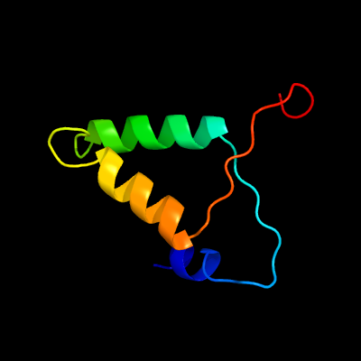
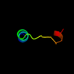
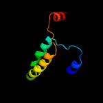
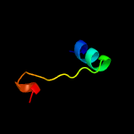
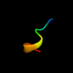
1 c4xgrG_
69.5
19
PDB header: toxin/antitoxinChain: G: PDB Molecule: ribonuclease vapc30;PDBTitle: crystal structure of addiction module from mycobacterial species
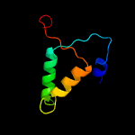
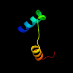
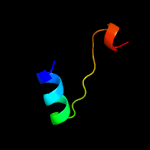
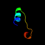
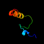
2 d2h1ca1
43.6
11
Fold: PIN domain-likeSuperfamily: PIN domain-likeFamily: PIN domain
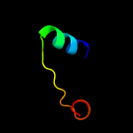
3 d1r0ka3
32.0
38
Fold: FwdE/GAPDH domain-likeSuperfamily: Glyceraldehyde-3-phosphate dehydrogenase-like, C-terminal domainFamily: Dihydrodipicolinate reductase-like
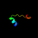
4 d1q0qa3
27.8
25
Fold: FwdE/GAPDH domain-likeSuperfamily: Glyceraldehyde-3-phosphate dehydrogenase-like, C-terminal domainFamily: Dihydrodipicolinate reductase-like
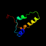
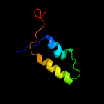
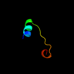
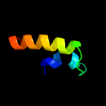
5 d1y82a1
26.6
9
Fold: PIN domain-likeSuperfamily: PIN domain-likeFamily: PIN domain
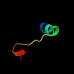
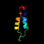
6 c6a7vG_
22.5
19
PDB header: toxin/antitoxinChain: G: PDB Molecule: ribonuclease vapc11;PDBTitle: crystal structure of mycobacterium tuberculosis vapbc11 toxin-2 antitoxin complex
7 c3dboB_
22.2
10
PDB header: toxin/antitoxinChain: B: PDB Molecule: uncharacterized protein;PDBTitle: crystal structure of a member of the vapbc family of toxin-antitoxin2 systems, vapbc-5, from mycobacterium tuberculosis
8 c5l6mC_
22.1
17
PDB header: hydrolaseChain: C: PDB Molecule: ribonuclease vapc;PDBTitle: structure of caulobacter crescentus vapbc1 (vapb1deltac:vapc1 form)
9 c3a14B_
19.2
29
PDB header: oxidoreductaseChain: B: PDB Molecule: 1-deoxy-d-xylulose 5-phosphate reductoisomerase;PDBTitle: crystal structure of dxr from thermotoga maritima, in complex with2 nadph
10 c1r0lD_
17.5
38
PDB header: oxidoreductaseChain: D: PDB Molecule: 1-deoxy-d-xylulose 5-phosphate reductoisomerase;PDBTitle: 1-deoxy-d-xylulose 5-phosphate reductoisomerase from zymomonas mobilis2 in complex with nadph
11 c5kqoA_
14.8
29
PDB header: oxidoreductaseChain: A: PDB Molecule: 1-deoxy-d-xylulose 5-phosphate reductoisomerase;PDBTitle: 1-deoxy-d-xylulose 5-phosphate reductoisomerase from vibrio vulnificus
12 c1k5hB_
14.8
25
PDB header: oxidoreductaseChain: B: PDB Molecule: 1-deoxy-d-xylulose-5-phosphate reductoisomerase;PDBTitle: 1-deoxy-d-xylulose-5-phosphate reductoisomerase
13 c4zn6B_
14.0
25
PDB header: oxidoreductaseChain: B: PDB Molecule: 1-deoxy-d-xylulose 5-phosphate reductoisomerase;PDBTitle: x-ray crystal structure of 1-deoxy-d-xylulose 5-phosphate2 reductoisomerase (ispc) from acinetobacter baumannii
14 c2eghA_
13.5
25
PDB header: oxidoreductaseChain: A: PDB Molecule: 1-deoxy-d-xylulose 5-phosphate reductoisomerase;PDBTitle: crystal structure of 1-deoxy-d-xylulose 5-phosphate reductoisomerase2 complexed with a magnesium ion, nadph and fosmidomycin
15 c3tndC_
13.0
18
PDB header: translation, toxinChain: C: PDB Molecule: trna(fmet)-specific endonuclease vapc;PDBTitle: crystal structure of shigella flexneri vapbc toxin-antitoxin complex
16 c2i5oA_
10.8
25
PDB header: transferaseChain: A: PDB Molecule: dna polymerase eta;PDBTitle: solution structure of the ubiquitin-binding zinc finger2 (ubz) domain of the human dna y-polymerase eta
17 c5fmtB_
9.8
26
PDB header: protein transportChain: B: PDB Molecule: flagellar associated protein;PDBTitle: crift54 ch-domain
18 c3fiaA_
9.2
19
PDB header: protein bindingChain: A: PDB Molecule: intersectin-1;PDBTitle: crystal structure of the eh 1 domain from human intersectin-1 protein.2 northeast structural genomics consortium target hr3646e.
19 c4cu2A_
8.3
83
PDB header: hydrolaseChain: A: PDB Molecule: endolysin;PDBTitle: c-terminal domain of ctp1l endolysin mutant v195p that reduces2 autoproteolysis
20 d2gycm1
8.1
25
Fold: Ribonuclease H-like motifSuperfamily: Translational machinery componentsFamily: Ribosomal protein L18 and S11
21 c4ce4h_
not modelled
7.9
18
PDB header: ribosomeChain: H: PDB Molecule: PDBTitle: 39s large subunit of the porcine mitochondrial ribosome
22 c3mtuE_
not modelled
7.7
24
PDB header: contractile proteinChain: E: PDB Molecule: capsid assembly scaffolding protein,tropomyosin alpha-1PDBTitle: structure of the tropomyosin overlap complex from chicken smooth2 muscle
23 c6nmiB_
not modelled
7.2
23
PDB header: transcriptionChain: B: PDB Molecule: general transcription and dna repair factor iih helicasePDBTitle: cryo-em structure of the human tfiih core complex
24 c3zvkC_
not modelled
7.2
11
PDB header: antitoxin/toxin/dnaChain: C: PDB Molecule: toxin of toxin-antitoxin system;PDBTitle: crystal structure of vapbc2 from rickettsia felis bound to2 a dna fragment from their promoter
25 c4bejB_
not modelled
6.8
13
PDB header: hydrolaseChain: B: PDB Molecule: dynamin 1-like protein;PDBTitle: nucleotide-free dynamin 1-like protein (dnm1l, drp1, dlp1)
26 c4oogA_
not modelled
6.2
20
PDB header: hydrolase/rnaChain: A: PDB Molecule: ribonuclease 3;PDBTitle: crystal structure of yeast rnase iii (rnt1p) complexed with the2 product of dsrna processing
27 c5husA_
not modelled
6.1
9
PDB header: transferaseChain: A: PDB Molecule: trehalose synthase regulatory protein;PDBTitle: structure of candida albicans trehalose synthase regulatory protein c-2 terminal domain
28 c3ke3A_
not modelled
5.9
19
PDB header: transferaseChain: A: PDB Molecule: putative serine-pyruvate aminotransferase;PDBTitle: crystal structure of putative serine-pyruvate aminotransferase2 (yp_263484.1) from psychrobacter arcticum 273-4 at 2.20 a resolution
29 c3bboQ_
not modelled
5.6
17
PDB header: ribosomeChain: Q: PDB Molecule: ribosomal protein l18;PDBTitle: homology model for the spinach chloroplast 50s subunit fitted to 9.4a2 cryo-em map of the 70s chlororibosome
30 c2mx7A_
not modelled
5.3
21
PDB header: protein bindingChain: A: PDB Molecule: synergin gamma;PDBTitle: solution structure of the internal eh domain of gamma-synergin
31 c4pwuC_
not modelled
5.2
17
PDB header: signaling proteinChain: C: PDB Molecule: modulator protein mzra;PDBTitle: crystal structure of a modulator protein mzra (kpn_03524) from2 klebsiella pneumoniae subsp. pneumoniae mgh 78578 at 2.45 a3 resolution