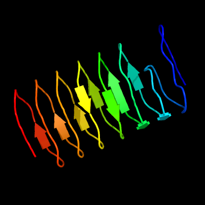
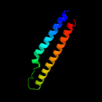
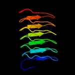
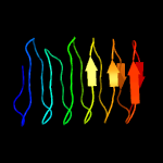
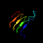
| 1 |
|
PDB 3ult chain B
Region: 271 - 378
Aligned: 108
Modelled: 108
Confidence: 98.5%
Identity: 16%
PDB header:antifreeze protein
Chain: B: PDB Molecule:ice recrystallization inhibition protein-like protein;
PDBTitle: crystal structure of an ice-binding protein from the perennial2 ryegrass, lolium perenne
Phyre2
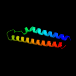
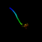
| 2 |
|
PDB 5xfs chain A
Region: 7 - 84
Aligned: 78
Modelled: 78
Confidence: 98.1%
Identity: 50%
PDB header:protein transport
Chain: A: PDB Molecule:pe family protein pe8;
PDBTitle: crystal structure of pe8-ppe15 in complex with espg5 from m.2 tuberculosis
Phyre2
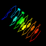
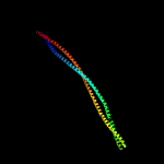
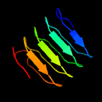
| 3 |
|
PDB 2g38 chain A
Region: 8 - 84
Aligned: 77
Modelled: 77
Confidence: 97.9%
Identity: 36%
PDB header:structural genomics, unknown function
Chain: A: PDB Molecule:pe family protein;
PDBTitle: a pe/ppe protein complex from mycobacterium tuberculosis
Phyre2
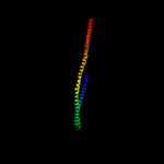
| 4 |
|
PDB 2g38 chain A domain 1
Region: 8 - 84
Aligned: 77
Modelled: 77
Confidence: 97.9%
Identity: 36%
Fold: Ferritin-like
Superfamily: PE/PPE dimer-like
Family: PE
Phyre2
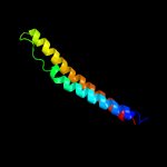
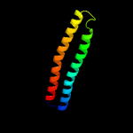
| 5 |
|
PDB 1qu7 chain A
Region: 186 - 369
Aligned: 183
Modelled: 184
Confidence: 97.7%
Identity: 14%
PDB header:signaling protein
Chain: A: PDB Molecule:methyl-accepting chemotaxis protein i;
PDBTitle: four helical-bundle structure of the cytoplasmic domain of a serine2 chemotaxis receptor
Phyre2
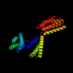
| 6 |
|
PDB 2ch7 chain A
Region: 5 - 369
Aligned: 308
Modelled: 309
Confidence: 97.5%
Identity: 9%
PDB header:chemotaxis
Chain: A: PDB Molecule:methyl-accepting chemotaxis protein;
PDBTitle: crystal structure of the cytoplasmic domain of a bacterial2 chemoreceptor from thermotoga maritima
Phyre2
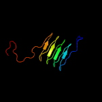
| 7 |
|
PDB 3s6l chain D
Region: 293 - 382
Aligned: 90
Modelled: 90
Confidence: 94.2%
Identity: 19%
PDB header:cell adhesion
Chain: D: PDB Molecule:hep_hag family;
PDBTitle: crystal structure of a yada-like head domain of the trimeric2 autotransporter adhesin boaa from burkholderia pseudomallei solved by3 iodide ion sad phasing
Phyre2
| 8 |
|
PDB 2yo3 chain C
Region: 221 - 361
Aligned: 141
Modelled: 141
Confidence: 94.0%
Identity: 7%
PDB header:membrane protein
Chain: C: PDB Molecule:general control protein gcn4, putative inner membrane
PDBTitle: salmonella enterica sada 1185-1386 fused to gcn4 adaptors (sadak14)
Phyre2
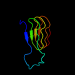
| 9 |
|
PDB 5ic1 chain A
Region: 9 - 369
Aligned: 361
Modelled: 361
Confidence: 93.2%
Identity: 9%
PDB header:peptide binding protein
Chain: A: PDB Molecule:talin-1;
PDBTitle: structural analysis of a talin triple domain module, e1794y, e1797y,2 q1801y mutant
Phyre2
| 10 |
|
PDB 2xqh chain A
Region: 307 - 372
Aligned: 66
Modelled: 66
Confidence: 44.7%
Identity: 17%
PDB header:cell adhesion
Chain: A: PDB Molecule:immunoglobulin-binding protein eibd;
PDBTitle: crystal structure of an immunoglobulin-binding fragment of the2 trimeric autotransporter adhesin eibd
Phyre2
| 11 |
|
PDB 3ntn chain B
Region: 290 - 379
Aligned: 90
Modelled: 90
Confidence: 41.6%
Identity: 9%
PDB header:membrane protein
Chain: B: PDB Molecule:uspa1;
PDBTitle: crystal structure of uspa1 head and neck domain from moraxella2 catarrhalis
Phyre2
| 12 |
|
PDB 4iog chain D
Region: 1 - 79
Aligned: 79
Modelled: 79
Confidence: 9.7%
Identity: 10%
PDB header:unknown function
Chain: D: PDB Molecule:secreted protein esxb;
PDBTitle: the crystal structure of a secreted protein esxb (wild-type, in p212 space group) from bacillus anthracis str. sterne
Phyre2
| 13 |
|
PDB 2yo1 chain A
Region: 297 - 378
Aligned: 82
Modelled: 82
Confidence: 8.9%
Identity: 12%
PDB header:membrane protein
Chain: A: PDB Molecule:general control protein gcn4, putative inner membrane
PDBTitle: salmonella enterica sada 1049-1304 fused to gcn4 adaptors (sadak9-2 cfii)
Phyre2
| 14 |
|
PDB 1p9h chain A
Region: 311 - 377
Aligned: 67
Modelled: 67
Confidence: 7.8%
Identity: 13%
Fold: Single-stranded left-handed beta-helix
Superfamily: Adhesin YadA, collagen-binding domain
Family: Adhesin YadA, collagen-binding domain
Phyre2
| 15 |
|
PDB 1p9h chain A
Region: 311 - 377
Aligned: 67
Modelled: 67
Confidence: 7.8%
Identity: 13%
PDB header:cell adhesion
Chain: A: PDB Molecule:invasin;
PDBTitle: crystal structure of the collagen-binding domain of yersinia adhesin2 yada
Phyre2