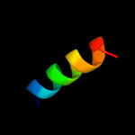
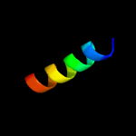
| 1 |
|
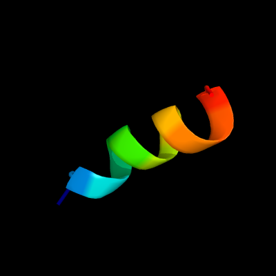
PDB 2mbh chain B
Region: 2 - 15
Aligned: 14
Modelled: 14
Confidence: 19.9%
Identity: 36%
PDB header:transcription
Chain: B: PDB Molecule:krueppel-like factor 1;
PDBTitle: nmr structure of eklf(22-40)/ubiquitin complex
Phyre2
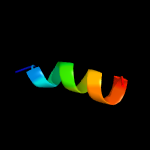
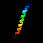
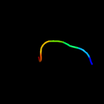
| 2 |
|
PDB 5wbi chain A
Region: 13 - 56
Aligned: 44
Modelled: 44
Confidence: 19.0%
Identity: 27%
PDB header:protein binding
Chain: A: PDB Molecule:regulatory-associated protein of tor 1;
PDBTitle: crystal structure of the arabidopsis thaliana raptor
Phyre2
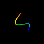
| 3 |
|
PDB 1loi chain A
Region: 6 - 14
Aligned: 9
Modelled: 9
Confidence: 14.4%
Identity: 33%
PDB header:hydrolase
Chain: A: PDB Molecule:cyclic 3',5'-amp specific phosphodiesterase rd1;
PDBTitle: n-terminal splice region of rat c-amp phosphodiesterase,2 nmr, 7 structures
Phyre2
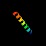
| 4 |
|
PDB 5gpd chain A
Region: 2 - 16
Aligned: 15
Modelled: 15
Confidence: 12.2%
Identity: 27%
PDB header:dna binding protein
Chain: A: PDB Molecule:sterol regulatory element-binding protein 1;
PDBTitle: crystal structure of the binding domain of srebp from fission yeast
Phyre2
| 5 |
|
PDB 2lgz chain A
Region: 7 - 12
Aligned: 6
Modelled: 6
Confidence: 11.0%
Identity: 50%
PDB header:transferase, membrane protein
Chain: A: PDB Molecule:dolichyl-diphosphooligosaccharide--protein
PDBTitle: solution structure of stt3p
Phyre2
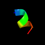
| 6 |
|
PDB 2mom chain C
Region: 34 - 60
Aligned: 27
Modelled: 27
Confidence: 10.3%
Identity: 15%
PDB header:membrane protein
Chain: C: PDB Molecule:lysosome-associated membrane glycoprotein 2;
PDBTitle: structural insights of tm domain of lamp-2a in dpc micelles
Phyre2
| 7 |
|
PDB 2mom chain B
Region: 34 - 60
Aligned: 27
Modelled: 27
Confidence: 9.7%
Identity: 15%
PDB header:membrane protein
Chain: B: PDB Molecule:lysosome-associated membrane glycoprotein 2;
PDBTitle: structural insights of tm domain of lamp-2a in dpc micelles
Phyre2
| 8 |
|
PDB 5ldw chain F
Region: 32 - 47
Aligned: 16
Modelled: 16
Confidence: 9.0%
Identity: 19%
PDB header:oxidoreductase
Chain: F: PDB Molecule:nadh dehydrogenase [ubiquinone] flavoprotein 1,
PDBTitle: structure of mammalian respiratory complex i, class1
Phyre2
| 9 |
|
PDB 5lc5 chain F
Region: 32 - 47
Aligned: 16
Modelled: 16
Confidence: 9.0%
Identity: 19%
PDB header:oxidoreductase
Chain: F: PDB Molecule:nadh dehydrogenase [ubiquinone] flavoprotein 1,
PDBTitle: structure of mammalian respiratory complex i, class2
Phyre2
| 10 |
|
PDB 5ir6 chain B
Region: 4 - 18
Aligned: 15
Modelled: 15
Confidence: 6.6%
Identity: 13%
PDB header:oxidoreductase
Chain: B: PDB Molecule:bd-type quinol oxidase subunit ii;
PDBTitle: the structure of bd oxidase from geobacillus thermodenitrificans
Phyre2
| 11 |
|
PDB 3vu0 chain B
Region: 6 - 12
Aligned: 7
Modelled: 7
Confidence: 6.3%
Identity: 71%
PDB header:transferase
Chain: B: PDB Molecule:putative uncharacterized protein;
PDBTitle: crystal structure of the c-terminal globular domain of2 oligosaccharyltransferase (afaglb-s2, af_0040, o30195_arcfu) from3 archaeoglobus fulgidus
Phyre2
| 12 |
|
PDB 1iie chain A
Region: 11 - 18
Aligned: 8
Modelled: 8
Confidence: 6.1%
Identity: 38%
Fold: Class II MHC-associated invariant chain ectoplasmic trimerization domain
Superfamily: Class II MHC-associated invariant chain ectoplasmic trimerization domain
Family: Class II MHC-associated invariant chain ectoplasmic trimerization domain
Phyre2