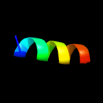
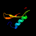
PDB header:structural genomics, unknown function Chain: A: PDB Molecule:bh2414 protein; PDBTitle: the structure of a protein with unknown function from bacillus2 halodurans c
Confidence and coverage
Confidence:
17.8%
Coverage:
46%
28 residues ( 46% of your sequence) have been modelled with 17.8% confidence by the single highest scoring template.
You may wish to submit your sequence to Phyrealarm. This will automatically scan your sequence every week for new potential templates as they appear in the Phyre2 library.
Please note: You must be registered and logged in to use Phyrealarm.
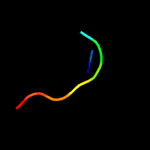
Region: 28 - 55 Aligned: 28 Modelled: 28 Confidence: 17.8% Identity: 14% PDB header:structural genomics, unknown function Chain: A: PDB Molecule:bh2414 protein; PDBTitle: the structure of a protein with unknown function from bacillus2 halodurans c
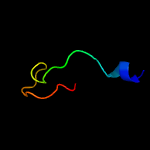
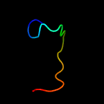
Region: 3 - 26 Aligned: 22 Modelled: 24 Confidence: 7.0% Identity: 32% PDB header:transcription Chain: B: PDB Molecule:heterocyst differentiation control protein; PDBTitle: crystal structure of the hood domain of anabaena hetr in complex with2 the hexapeptide ergsgr derived from pats