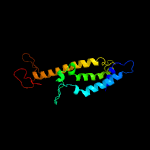
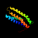
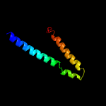
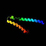
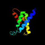
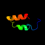
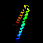
1 c6hwhb_
79.9
16
PDB header: electron transportChain: B: PDB Molecule: ubiquinol-cytochrome c reductase iron-sulfur subunit;PDBTitle: structure of a functional obligate respiratory supercomplex from2 mycobacterium smegmatis
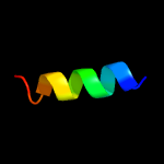
2 c6ajjA_
66.5
10
PDB header: membrane protein, hydrolaseChain: A: PDB Molecule: drug exporters of the rnd superfamily-like protein,PDBTitle: crystal structure of mycolic acid transporter mmpl3 from mycobacterium2 smegmatis complexed with ica38
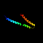
3 c3nd0A_
51.9
19
PDB header: transport proteinChain: A: PDB Molecule: sll0855 protein;PDBTitle: x-ray crystal structure of a slow cyanobacterial cl-/h+ antiporter
4 d1otsa_
47.2
20
Fold: Clc chloride channelSuperfamily: Clc chloride channelFamily: Clc chloride channel
5 c4tkrB_
46.3
22
PDB header: membrane proteinChain: B: PDB Molecule: thiamine transporter thia;PDBTitle: native-sad phasing for thit from listeria monocytogenes serovar.
6 d1iqva_
41.7
40
Fold: Ribosomal protein S7Superfamily: Ribosomal protein S7Family: Ribosomal protein S7
7 c2zkqg_
41.6
35
PDB header: ribosomal protein/rnaChain: G: PDB Molecule: rna helix;PDBTitle: structure of a mammalian ribosomal 40s subunit within an 80s complex2 obtained by docking homology models of the rna and proteins into an3 8.7 a cryo-em map
8 c5xyiF_
36.1
40
PDB header: ribosomeChain: F: PDB Molecule: 40s ribosomal protein s5-b, putative;PDBTitle: small subunit of trichomonas vaginalis ribosome
9 c3izbF_
35.6
40
PDB header: ribosomeChain: F: PDB Molecule: 40s ribosomal protein rps5 (s7p);PDBTitle: localization of the small subunit ribosomal proteins into a 6.1 a2 cryo-em map of saccharomyces cerevisiae translating 80s ribosome
10 c5khnB_
35.4
11
PDB header: membrane proteinChain: B: PDB Molecule: rnd transporter;PDBTitle: crystal structures of the burkholderia multivorans hopanoid2 transporter hpnn
11 c2xzmG_
34.0
40
PDB header: ribosomeChain: G: PDB Molecule: ribosomal protein s7 containing protein;PDBTitle: crystal structure of the eukaryotic 40s ribosomal2 subunit in complex with initiation factor 1. this file3 contains the 40s subunit and initiation factor for4 molecule 1
12 c3zey2_
33.1
33
PDB header: ribosomeChain: 2: PDB Molecule: 40s ribosomal protein s5, putative;PDBTitle: high-resolution cryo-electron microscopy structure of the trypanosoma2 brucei ribosome
13 c1s1hG_
30.1
40
PDB header: ribosomeChain: G: PDB Molecule: 40s ribosomal protein s5;PDBTitle: structure of the ribosomal 80s-eef2-sordarin complex from yeast2 obtained by docking atomic models for rna and protein components into3 a 11.7 a cryo-em map. this file, 1s1h, contains 40s subunit. the 60s4 ribosomal subunit is in file 1s1i.
14 c1p58E_
30.0
22
PDB header: virusChain: E: PDB Molecule: envelope protein m;PDBTitle: complex organization of dengue virus membrane proteins as revealed by2 9.5 angstrom cryo-em reconstruction
15 d1husa_
29.6
47
Fold: Ribosomal protein S7Superfamily: Ribosomal protein S7Family: Ribosomal protein S7
16 d1rssa_
29.6
47
Fold: Ribosomal protein S7Superfamily: Ribosomal protein S7Family: Ribosomal protein S7
17 d2qalg1
28.5
47
Fold: Ribosomal protein S7Superfamily: Ribosomal protein S7Family: Ribosomal protein S7
18 c6coyB_
28.2
10
PDB header: transport proteinChain: B: PDB Molecule: chloride channel protein 1;PDBTitle: human clc-1 chloride ion channel, transmembrane domain
19 c3gtyS_
27.2
33
PDB header: chaperone/ribosomal proteinChain: S: PDB Molecule: 30s ribosomal protein s7;PDBTitle: promiscuous substrate recognition in folding and assembly activities2 of the trigger factor chaperone
20 c2ht2B_
26.6
20
PDB header: membrane proteinChain: B: PDB Molecule: h(+)/cl(-) exchange transporter clca;PDBTitle: structure of the escherichia coli clc chloride channel2 y445h mutant and fab complex
21 c3j6vG_
not modelled
26.4
40
PDB header: ribosomeChain: G: PDB Molecule: 28s ribosomal protein s7, mitochondrial;PDBTitle: cryo-em structure of the small subunit of the mammalian mitochondrial2 ribosome
22 c2lowA_
not modelled
25.7
39
PDB header: membrane proteinChain: A: PDB Molecule: apelin receptor;PDBTitle: solution structure of ar55 in 50% hfip
23 c5wsnD_
not modelled
24.7
24
PDB header: virusChain: D: PDB Molecule: m protein;PDBTitle: structure of japanese encephalitis virus
24 c5o5jG_
not modelled
24.6
47
PDB header: ribosomeChain: G: PDB Molecule: 30s ribosomal protein s7;PDBTitle: structure of the 30s small ribosomal subunit from mycobacterium2 smegmatis
25 c3bbnG_
not modelled
23.4
33
PDB header: ribosomeChain: G: PDB Molecule: ribosomal protein s7;PDBTitle: homology model for the spinach chloroplast 30s subunit fitted to 9.4a2 cryo-em map of the 70s chlororibosome.
26 c6rdr8_
not modelled
23.3
30
PDB header: proton transportChain: 8: PDB Molecule: mitochondrial atp synthase subunit asa8;PDBTitle: cryo-em structure of polytomella f-atp synthase, rotary substate 1d,2 monomer-masked refinement
27 c6rdi8_
not modelled
23.3
30
PDB header: proton transportChain: 8: PDB Molecule: mitochondrial atp synthase subunit asa8;PDBTitle: cryo-em structure of polytomella f-atp synthase, rotary substate 1a,2 monomer-masked refinement
28 c3j0xJ_
not modelled
22.7
47
PDB header: ribosomeChain: J: PDB Molecule: 30s ribosomal protein s7;PDBTitle: structural characterization of mrna-trna translocation intermediates2 (30s ribosome of class 4b of the six classes)
29 c6hhkB_
not modelled
22.6
14
PDB header: viral proteinChain: B: PDB Molecule: gp105;PDBTitle: structure of gp105 of listeria bacteriophage a511
30 c1vs5G_
not modelled
21.8
47
PDB header: ribosomeChain: G: PDB Molecule: 30s ribosomal protein s7;PDBTitle: crystal structure of the bacterial ribosome from escherichia coli in2 complex with the antibiotic kasugamyin at 3.5a resolution. this file3 contains the 30s subunit of one 70s ribosome. the entire crystal4 structure contains two 70s ribosomes and is described in remark 400.
31 c5tr1A_
not modelled
20.8
10
PDB header: transport proteinChain: A: PDB Molecule: chloride channel protein;PDBTitle: cryo-electron microscopy structure of a bovine clc-k chloride channel,2 alternate (class 2) conformation
32 c3j2pD_
not modelled
19.5
22
PDB header: viral proteinChain: D: PDB Molecule: small envelope protein m;PDBTitle: cryoem structure of dengue virus envelope protein heterotetramer
33 c6hwhX_
not modelled
19.5
11
PDB header: electron transportChain: X: PDB Molecule: cytochrome c oxidase polypeptide 4;PDBTitle: structure of a functional obligate respiratory supercomplex from2 mycobacterium smegmatis
34 d1kpla_
not modelled
19.0
20
Fold: Clc chloride channelSuperfamily: Clc chloride channelFamily: Clc chloride channel
35 c2k9pA_
not modelled
17.5
33
PDB header: membrane proteinChain: A: PDB Molecule: pheromone alpha factor receptor;PDBTitle: structure of tm1_tm2 in lppg micelles
36 c6qvcB_
not modelled
17.5
10
PDB header: membrane proteinChain: B: PDB Molecule: chloride channel protein 1;PDBTitle: cryoem structure of the human clc-1 chloride channel, cbs state 1
37 c2losA_
not modelled
15.6
24
PDB header: membrane proteinChain: A: PDB Molecule: transmembrane protein 14c;PDBTitle: backbone structure of human membrane protein tmem14c
38 c2k9yB_
not modelled
14.7
15
PDB header: transferaseChain: B: PDB Molecule: ephrin type-a receptor 2;PDBTitle: epha2 dimeric structure in the lipidic bicelle at ph 5.0
39 c6eyuA_
not modelled
14.3
20
PDB header: membrane proteinChain: A: PDB Molecule: bacteriorhodopsin;PDBTitle: crystal structure of the inward h(+) pump xenorhodopsin
40 c2mpnA_
not modelled
14.2
23
PDB header: membrane proteinChain: A: PDB Molecule: inner membrane protein ygap;PDBTitle: 3d nmr structure of the transmembrane domain of the full-length inner2 membrane protein ygap from escherichia coli
41 c2mpnB_
not modelled
14.2
23
PDB header: membrane proteinChain: B: PDB Molecule: inner membrane protein ygap;PDBTitle: 3d nmr structure of the transmembrane domain of the full-length inner2 membrane protein ygap from escherichia coli
42 c3e37B_
not modelled
13.7
16
PDB header: transferaseChain: B: PDB Molecule: protein farnesyltransferase subunit beta;PDBTitle: protein farnesyltransferase complexed with bisubstrate2 ethylenediamine scaffold inhibitor 5
43 c5ireD_
not modelled
13.5
22
PDB header: virusChain: D: PDB Molecule: m protein;PDBTitle: the cryo-em structure of zika virus
44 c1p58F_
not modelled
13.3
22
PDB header: virusChain: F: PDB Molecule: envelope protein m;PDBTitle: complex organization of dengue virus membrane proteins as revealed by2 9.5 angstrom cryo-em reconstruction
45 c3dinD_
not modelled
13.3
18
PDB header: membrane protein, protein transportChain: D: PDB Molecule: preprotein translocase subunit sece;PDBTitle: crystal structure of the protein-translocation complex formed by the2 secy channel and the seca atpase
46 c6dmoA_
not modelled
13.1
11
PDB header: protein bindingChain: A: PDB Molecule: protein patched homolog 1;PDBTitle: cryo-em structure of human ptch1 with three mutations2 l282q/t500f/p504l
47 c6fosK_
not modelled
12.3
47
PDB header: photosynthesisChain: K: PDB Molecule: photosystem i reaction center subunit x;PDBTitle: cyanidioschyzon merolae photosystem i
48 c5z62N_
not modelled
11.8
28
PDB header: electron transportChain: N: PDB Molecule: cytochrome c oxidase subunit ndufa4;PDBTitle: structure of human cytochrome c oxidase
49 c2k9yA_
not modelled
11.3
16
PDB header: transferaseChain: A: PDB Molecule: ephrin type-a receptor 2;PDBTitle: epha2 dimeric structure in the lipidic bicelle at ph 5.0
50 c5zghK_
not modelled
11.3
47
PDB header: photosynthesisChain: K: PDB Molecule: psak;PDBTitle: cryo-em structure of the red algal psi-lhcr
51 c3q7aB_
not modelled
10.6
19
PDB header: transferase/transferase inhibitorChain: B: PDB Molecule: farnesyltransferase beta subunit;PDBTitle: cryptococcus neoformans protein farnesyltransferase in complex with2 fpp and l-778,123
52 c4ydeB_
not modelled
10.2
22
PDB header: transferaseChain: B: PDB Molecule: protein farnesyltransferase/geranylgeranyltransferase type-PDBTitle: crystal structure of candida albicans protein farnesyltransferase2 binary complex with the isoprenoid farnesyldiphosphate
53 c4px7A_
not modelled
9.7
18
PDB header: hydrolaseChain: A: PDB Molecule: phosphatidylglycerophosphatase;PDBTitle: crystal structure of lipid phosphatase e. coli pgpb
54 c6f46A_
not modelled
9.1
32
PDB header: apoptosisChain: A: PDB Molecule: bcl-2-like protein 1;PDBTitle: structure of the transmembrane helix of bclxl in phospholipid2 nanodiscs
55 c5n1cA_
not modelled
8.8
26
PDB header: dna binding proteinChain: A: PDB Molecule: probable transcriptional regulatory protein;PDBTitle: iodinated form of the mycobacterium tuberculosis repressor ethr2
56 c3aygA_
not modelled
8.7
14
PDB header: oxidoreductaseChain: A: PDB Molecule: nitric oxide reductase;PDBTitle: crystal structure of nitric oxide reductase complex with hqno
57 c4cdiC_
not modelled
8.5
43
PDB header: membrane proteinChain: C: PDB Molecule: predicted protein;PDBTitle: crystal structure of acrb-acrz complex
58 c6igzL_
not modelled
8.5
36
PDB header: plant proteinChain: L: PDB Molecule: psal;PDBTitle: structure of psi-lhci
59 d2soba_
not modelled
8.4
20
Fold: OB-foldSuperfamily: Staphylococcal nucleaseFamily: Staphylococcal nuclease
60 c4nj7P_
not modelled
8.2
19
PDB header: protein bindingChain: P: PDB Molecule: auxin response factor 7;PDBTitle: pb1 domain of atarf7 - semet derivative
61 c4mbgB_
not modelled
8.0
27
PDB header: transferaseChain: B: PDB Molecule: caax farnesyltransferase beta subunit ram1;PDBTitle: crystal structure of aspergillus fumigatus protein farnesyltransferase2 binary complex with farnesyldiphosphate
62 c6f0kD_
not modelled
7.8
19
PDB header: membrane proteinChain: D: PDB Molecule: actd;PDBTitle: alternative complex iii
63 d2h6fb1
not modelled
7.7
15
Fold: alpha/alpha toroidSuperfamily: Terpenoid cyclases/Protein prenyltransferasesFamily: Protein prenyltransferases
64 c4y7jE_
not modelled
6.9
16
PDB header: membrane protein,transport proteinChain: E: PDB Molecule: large conductance mechanosensitive channel protein,PDBTitle: structure of an archaeal mechanosensitive channel in expanded state
65 c4cbfB_
not modelled
6.7
25
PDB header: virusChain: B: PDB Molecule: m protein;PDBTitle: near-atomic resolution cryo-em structure of dengue serotype 4 virus
66 c5fruB_
not modelled
6.5
27
PDB header: transcriptionChain: B: PDB Molecule: positive phenol-degradative gene regulator;PDBTitle: crystal structure of the phenol-responsive sensory domain2 of the transcription activator poxr
67 d2osoa1
not modelled
6.4
33
Fold: Ligand-binding domain in the NO signalling and Golgi transportSuperfamily: Ligand-binding domain in the NO signalling and Golgi transportFamily: MJ1460-like
68 c2v5iA_
not modelled
6.0
43
PDB header: viral proteinChain: A: PDB Molecule: salmonella typhimurium db7155 bacteriophage det7 tailspike;PDBTitle: structure of the receptor-binding protein of bacteriophage det7: a2 podoviral tailspike in a myovirus
69 c5kbhB_
not modelled
5.9
36
PDB header: transcriptionChain: B: PDB Molecule: mopr;PDBTitle: crystal structure of the aromatic sensor domain of mopr in complex2 with 3-chloro-phenol
70 c2j5dA_
not modelled
5.9
47
PDB header: membrane proteinChain: A: PDB Molecule: bcl2/adenovirus e1b 19 kda protein-interacting protein 3;PDBTitle: nmr structure of bnip3 transmembrane domain in lipid bicelles
71 d1kf6c_
not modelled
5.4
16
Fold: Heme-binding four-helical bundleSuperfamily: Fumarate reductase respiratory complex transmembrane subunitsFamily: Succinate dehydrogenase/Fumarate reductase transmembrane subunits (SdhC/FrdC and SdhD/FrdD)
72 c3ofgA_
not modelled
5.1
11
PDB header: chaperoneChain: A: PDB Molecule: boca/mesd chaperone for ywtd beta-propeller-egf protein 1;PDBTitle: structured domain of caenorhabditis elegans bmy-1
73 c4m0qB_
not modelled
5.1
26
PDB header: viral proteinChain: B: PDB Molecule: membrane-associated protein vp24;PDBTitle: ebola virus vp24 structure
74 c1mowG_
not modelled
5.1
36
PDB header: hydrolase/dnaChain: G: PDB Molecule: chimera of homing endonuclease i-dmoi and dna endonucleasePDBTitle: e-drei


